- 原文:CNS tumor with BCOR internal tandem duplication。
- 作者:Jared T. Ahrendsen, M.D., Ph.D.。
- 编辑部最后更新 2026-09-14(更新进行中)。
定义 / 概述
- 中枢神经系统(CNS)的恶性肿瘤。
- 存在累及 BCOR 基因第 15 外显子的内部串联重复(ITD)。
要点
- 中枢神经系统恶性肿瘤。
- 形态均一、实性至梭形的肿瘤细胞主要呈实性生长,位于致密毛细血管网中,并形成假菊形团。
- 存在累及第 15 外显子的 BCOR ITD。
- 对于未能明确的病变,采用 CNS 肿瘤伴 BCOR ITD 的 DNA 甲基化谱。
术语
- 不推荐使用:CNS 高级别神经上皮肿瘤伴 BCOR 内部串联重复;CNS 原始神经上皮肿瘤(CNS PNET)。
ICD 编码
- ICD-O:9500/3 — CNS 肿瘤伴 BCOR 内部串联重复。
- ICD-11:2A00.1Y — 其他特指的脑胚胎性肿瘤。
流行病学
- 主要发生于儿童,中位年龄 3.2 岁;无性别倾向(Brain Pathol 2020;30:46;J Pathol Transl Med 2025;59:273)。
- 无特定的环境或遗传危险因素(J Pathol Transl Med 2025;59:273)。
部位
- 最常见于大脑或小脑半球;罕见累及基底节、脑干或脊髓(Cell 2016;164:1060;Brain Pathol 2020;30:46)。
病理生理
- BCOR 位于染色体 Xq26.2,是表观遗传和转录调控因子(Genes Dev 2000;14:1810;Nat Cell Biol 2009;11:1002)。
- 体细胞杂合性 BCOR ITD 很可能产生功能获得性(激活性)突变,在肿瘤发生中发挥重要作用,但细胞机制仍不清楚(Cell 2016;164:1060)。
- 常观察到 WNT 信号通路激活,可能参与肿瘤发生(Cell 2016;164:1060)。
临床特征
- 症状和临床表现取决于肿瘤部位,通常与颅内压增高相关,如头痛、恶心、呕吐。
- 部分患者表现为局灶性神经功能缺损或癫痫发作。
- 参考文献:Brain Pathol 2020;30:46。
诊断
- 脑影像检查,首选 MRI。
- 对手术切除组织进行组织病理学和分子分析,以作出最终诊断。
影像描述
- MRI 显示大型、境界清楚的轴内肿块,常紧贴硬脑膜,可见坏死、钙化或出血区域(AJNR Am J Neuroradiol 2022;43:151)。
- T1 低信号,呈不均匀强化;T2 / FLAIR 高信号。
- 弥散加权成像 / 表观弥散系数(DWI / ADC)显示弥散受限。
影像图
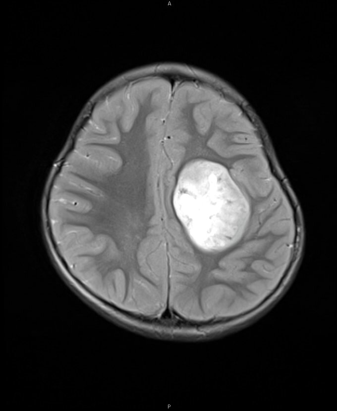
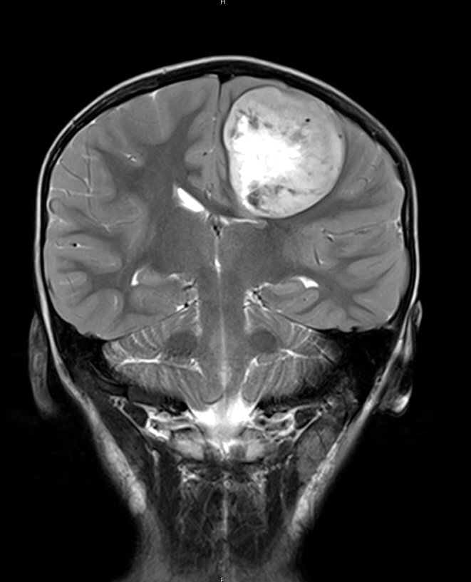
影像贡献:Jared T. Ahrendsen, M.D., Ph.D.;Nitin Wadhwani, M.D.。原文另有外部托管的 MRI 特征图,本站未收录。
预后因素
- 预后通常较差,许多患者出现复发,5 年总生存率约 50%(Brain Pathol 2020;30:46)。
- 一项研究汇总 46 例已发表病例,报告中位生存期为 1.7 年(Pathol Res Pract 2022;236:153995)。
- 有少数长期生存(超过 10 年)的病例报告(Brain Pathol 2020;30:46)。
病例报告
- 18 个月男孩,斜颈和呕吐(Childs Nerv Syst 2023;39:321)。
- 5 岁男孩,左额叶肿瘤(NMC Case Rep J 2023;10:343)。
- 6 岁女孩和 3 岁男孩,CNS 肿瘤伴 BCOR ITD(Pathol Res Pract 2022;236:153995)。
治疗
- 最大限度安全手术切除,随后化疗,并常接受放疗(Brain Pathol 2020;30:46)。
冰冻切片描述
- 涂片:圆形至卵圆形肿瘤细胞,染色质疏松、胞质嗜酸性,常有纤细的纤维状细胞突起。
- 冰冻:致密生长,与邻近脑组织分界清楚,可见血管周围假菊形团;坏死常见(Cytopathology 2024;35:561)。
术中冰冻 / 涂片细胞学图像
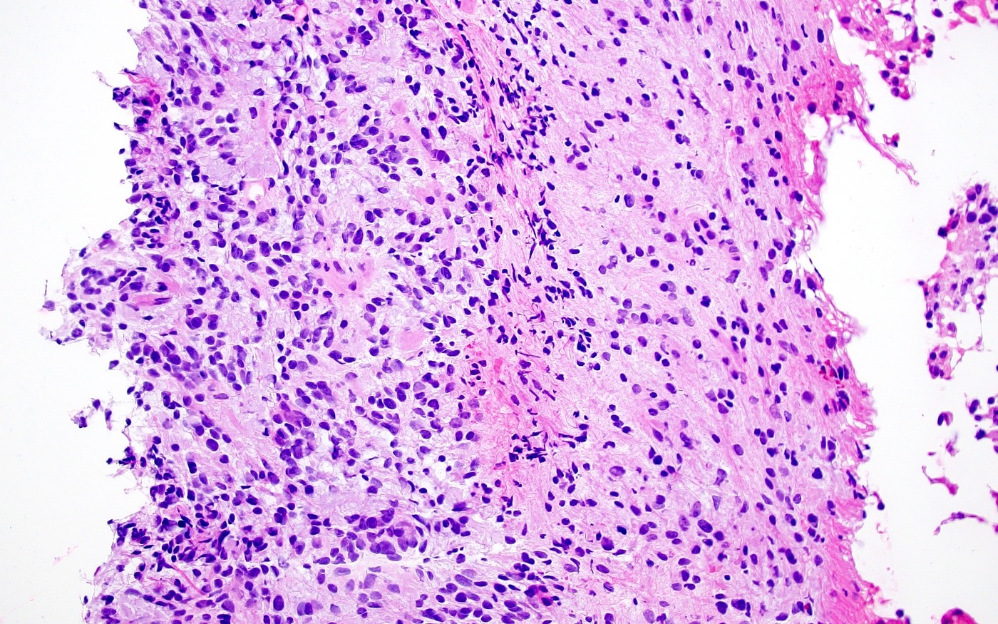
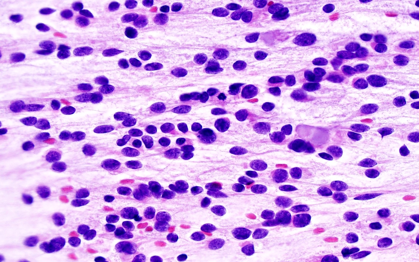
镜下(组织学)描述
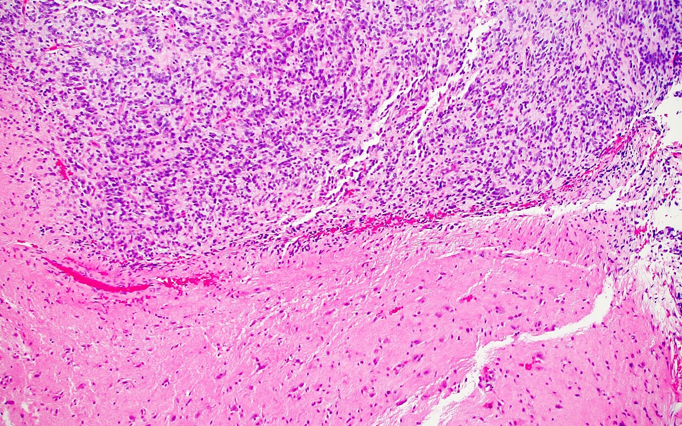
- 与邻近脑组织分界清楚,呈实性生长,但可有局灶浸润。
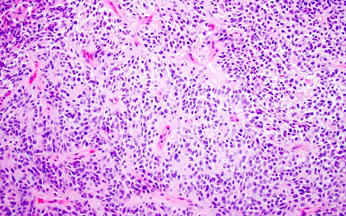
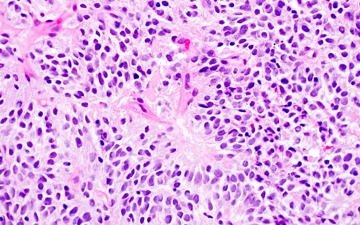
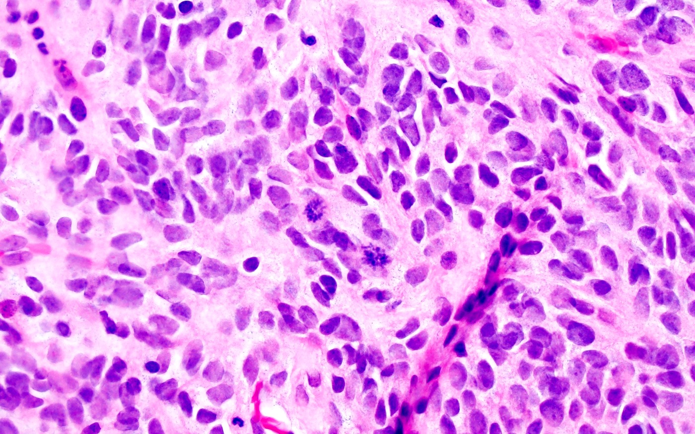
- 形态均一的梭形至卵圆形细胞,染色质细腻。
- 丰富的分支状毛细血管网。
- 坏死常见,核分裂活性不等。
- 常见胶质瘤样纤维性结构和血管周围假菊形团;肿瘤细胞偶尔形成菊形团样结构。
- 其他可见特征包括:黏液样 / 微囊性改变、微钙化、玻璃样变间质,以及细胞密集并伴活跃核分裂。
- 参考文献:Brain Pathol 2020;30:46;Brain Pathol 2018;28:710;Acta Neuropathol Commun 2020;8:105。
镜下(组织学)图像
细胞学描述
- 圆形至卵圆形肿瘤细胞,染色质疏松,胞质嗜酸性,常有纤细的纤维状细胞突起(Cytopathology 2024;35:561)。
细胞学图像
原文该栏目未提供独立图像。
阳性染色
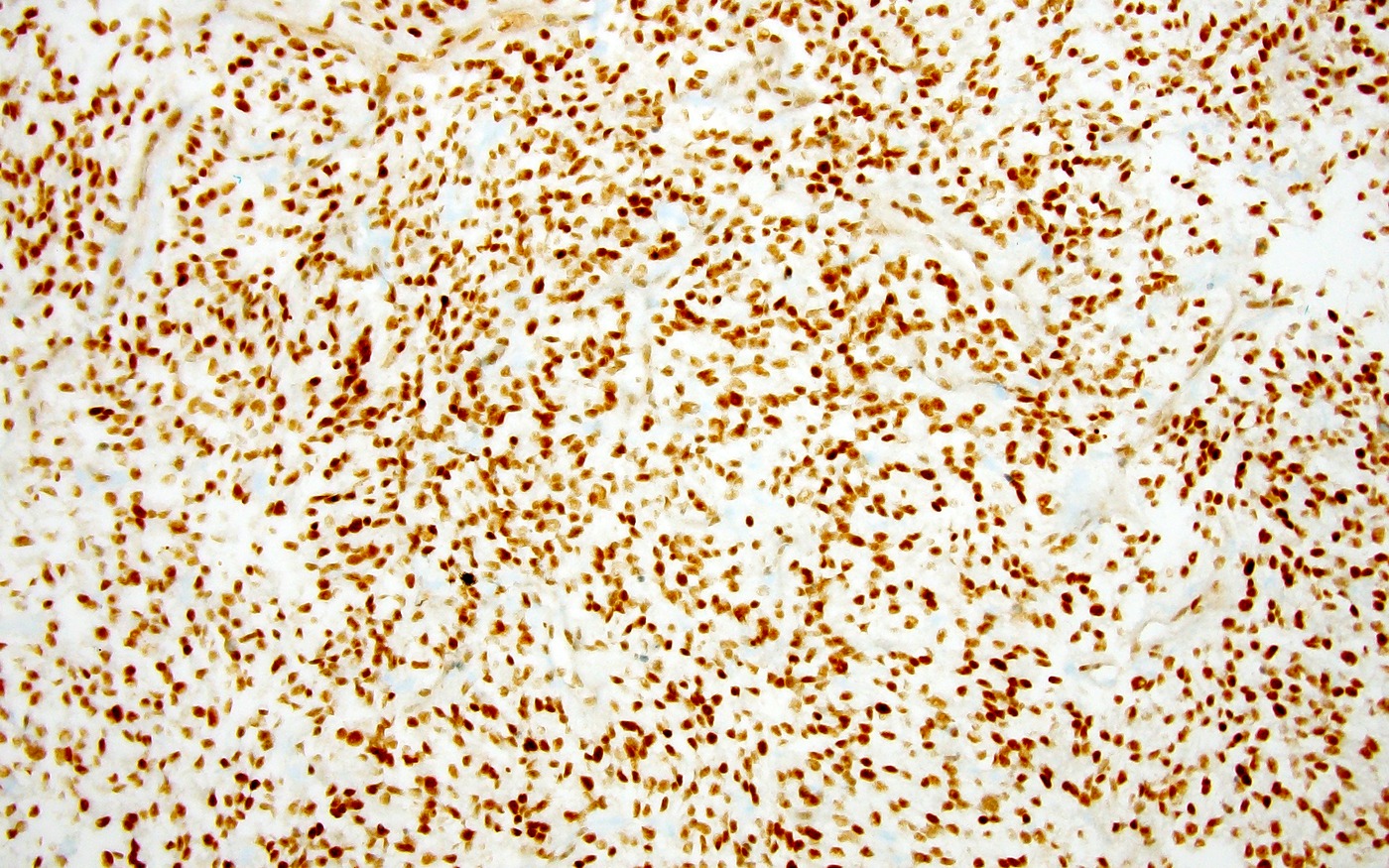
- 波形蛋白、CD56 和 BCOR 一致阳性。
- 多数肿瘤 Olig2 和 NeuN 呈不同程度阳性。
- Ki67:15–60%。
- 参考文献:Brain Pathol 2020;30:46。
阴性染色
- GFAP 和突触素主要阴性。
- EMA 阴性,或呈微弱的胞质颗粒状染色。
- 参考文献:Brain Pathol 2020;30:46。
电镜描述
- 不用于常规诊断。
- 原始细胞含丰富粗面内质网(Brain Pathol 2020;30:46)。
电镜图像
原文超微结构特征图托管于其他服务器;本站未收录。
分子 / 细胞遗传学描述
- 以 BCOR 第 15 外显子存在杂合性 ITD 为定义(Cell 2016;164:1060)。
- 这一改变也见于肾透明细胞肉瘤和婴儿原始黏液样间叶性肿瘤(Nat Genet 2015;47:861;Am J Surg Pathol 2016;40:1009)。
- 其他遗传改变不常见,但少数病例可同时存在 TP53、ATRX 或 CDKN2A / CDKN2B 改变(Brain Pathol 2020;30:46)。
- DNA 甲基化谱与 CNS 肿瘤伴 BCOR ITD 相符(Nature 2018;555:469)。
病理报告示例
- 脑,左额叶肿瘤,切除:CNS 肿瘤伴 BCOR 内部串联重复,CNS WHO 4 级(见评注)。
- 评注:组织学显示胚胎性肿瘤,呈实性生长,具有均一的卵圆形核及分支状毛细血管网。可见坏死和增高的核分裂活性,偶见血管周围假菊形团。肿瘤细胞 Olig2 呈不同程度阳性,GFAP 和突触素阴性。分子检测发现累及 BCOR 基因第 15 外显子的内部串联重复,DNA 甲基化分析以高置信度匹配 CNS 肿瘤伴 BCOR ITD 甲基化类别。
鉴别诊断
室管膜瘤
- 沿脑室或位于脑室附近。
- 免疫组化 GFAP 弥漫阳性,EMA 核周点状阳性;Olig2 主要阴性。
- 具有不同的分子病理特征,包括幕上肿瘤的基因融合以及后颅窝肿瘤的表观遗传特征(后颅窝室管膜瘤 A 组 / B 组)。
非典型畸胎样 / 横纹肌样肿瘤(AT / RT)
- 通常见于婴儿的恶性胚胎性肿瘤。
- 肿瘤细胞分化差,可不同程度出现横纹肌样形态。
- 免疫组化 INI1 表达缺失。
- SMARCB1 或罕见的 SMARCA4 双等位基因失活。
髓母细胞瘤
- 发生于小脑 / 第四脑室。
- 是 CNS 典型的小圆蓝细胞肿瘤。
- 肿瘤细胞突触素阳性,GFAP 可不同程度阳性。
伴多层菊形团的胚胎性肿瘤(ETMR)
- 恶性胚胎性细胞形成多层菊形团。
- LIN28 强、弥漫胞质阳性。
- 存在 C19MC 遗传改变,或罕见的 DICER1 突变。
儿童型高级别胶质瘤
- 弥漫浸润性生长。
- 通常 GFAP 和 Olig2 均阳性。
- 儿童大脑半球肿瘤可有 H3 G34 改变。
- DNA 甲基化谱匹配弥漫性儿童型高级别胶质瘤或其他高级别胶质瘤类别。
- 鉴别诊断参考文献:Am J Surg Pathol 2017;41:1254;Brain Pathol 2020;30:46;Acta Neuropathol Commun 2020;8:105。
补充参考文献
原文该栏目未列出条目。
练习题 1
一名 4 岁儿童,右额叶浅表部出现巨大脑肿块。肿瘤切除后的组织学表现如图。分子检测发现 BCOR 基因第 15 外显子的内部串联重复(ITD)。该肿瘤最可能的免疫表型是什么?
- GFAP 阴性,Olig2 阴性,NeuN 阴性
- GFAP 阴性,Olig2 阳性,NeuN 阳性
- GFAP 阳性,Olig2 阴性,NeuN 阳性
- GFAP 阳性,Olig2 阳性,NeuN 阴性
- GFAP 阳性,Olig2 阳性,NeuN 阳性
练习题 1 答案
B. GFAP 阴性,Olig2 阳性,NeuN 阳性。本病例描述的是 CNS 肿瘤伴 BCOR ITD。典型免疫表型为 Olig2 和 NeuN 阳性,GFAP 和突触素阴性。A、C、D、E 不正确,因为它们不符合 CNS 肿瘤伴 BCOR ITD 的免疫表型。练习题 2
一名 3 岁儿童,小脑出现巨大脑肿块。切除后组织病理学显示恶性肿瘤,呈致密生长,卵圆形细胞形态均一,在毛细血管丰富的背景中偶见假菊形团,并有坏死区。免疫组化显示肿瘤细胞 Olig2 呈不同程度阳性,NeuN、ATRX 和 INI1 阳性;GFAP、突触素、IDH1 R132H 和 H3 K27M 阴性。该肿瘤最可能存在下列哪项分子改变?
- BCOR 第 15 外显子内部串联重复(ITD)
- SMARCB1 双等位基因丢失
- EGFR 基因扩增
- H3 G34R 突变
- KIAA1549::BRAF 融合
练习题 2 答案
A. BCOR 第 15 外显子内部串联重复(ITD)。病例描述了 CNS 肿瘤伴 BCOR ITD 的经典组织学和免疫表型。该 CNS 恶性肿瘤以 BCOR 基因第 15 外显子 ITD 为定义。B 不正确:SMARCB1 双等位基因丢失是非典型畸胎样 / 横纹肌样肿瘤的特征,而本例保留 INI1。C、D 不正确:EGFR 基因扩增和 H3 G34R 突变更常见于高级别胶质瘤,分别如胶质母细胞瘤和弥漫性大脑半球胶质瘤。E 不正确:KIAA1549::BRAF 融合是毛细胞型星形细胞瘤(尤其小脑肿瘤)的特征,后者是低级别胶质肿瘤。Definition / general
- Malignant neoplasm of the central nervous system (CNS)
- Internal tandem duplication (ITD) involving exon 15 of the BCOR gene
Essential features
- Malignant neoplasm of the CNS
- Predominantly solid growth of uniform appearing, solid to spindled tumor cells in a dense capillary network with pseudorosette formation
- BCOR ITD involving exon 15
- For unresolved lesions, DNA methylation profile of CNS tumor with BCOR ITD
Terminology
- Not recommended
- CNS high grade neuroepithelial tumor with BCOR internal tandem duplication
- CNS primitive neuroepithelial tumor (CNS PNET)
ICD coding
- ICD-O: 9500/3 - CNS tumor with BCOR internal tandem duplication
- ICD-11: 2A00.1Y - other specified embryonal tumors of brain
Epidemiology
- Primarily affects children (median age of 3.2 years); no sex predilection (Brain Pathol 2020;30:46, J Pathol Transl Med 2025;59:273)
- No specific environmental or genetic risk factors (J Pathol Transl Med 2025;59:273)
Sites
- Most commonly in the cerebral or cerebellar hemispheres; rarely involves the basal ganglia, brainstem or spinal cord (Cell 2016;164:1060, Brain Pathol 2020;30:46)
Pathophysiology
- BCOR (chromosome Xq26.2) is an epigenetic and transcriptional regulator (Genes Dev 2000;14:1810, Nat Cell Biol 2009;11:1002)
- Somatic heterozygous BCOR ITD, likely producing a gain of function (activating) mutation, plays an important role in tumorigenesis but cellular mechanisms remain unclear (Cell 2016;164:1060)
- WNT signaling activation is frequently observed, which may contribute to tumorigenesis (Cell 2016;164:1060)
Clinical features
- Symptoms and clinical presentation are based on tumor location and are usually related to increased intracranial pressure (e.g., headache, nausea, vomiting)
- Some patients present with focal neurologic deficits or seizures
- Reference: Brain Pathol 2020;30:46
Diagnosis
- Brain imaging, preferably MRI
- Histopathologic and molecular analyses of surgically resected tissue to arrive at final diagnosis
Radiology description
- By MRI: large, well defined, intra-axial masses, often abutting the dura, with areas of necrosis, calcification or hemorrhage (AJNR Am J Neuroradiol 2022;43:151)
- T1 hypointense with heterogeneous enhancement
- T2 / FLAIR hyperintense
- Diffusion weighted imaging / apparent diffusion coefficient (DWI / ADC): reduced diffusivity
Radiology images
Contributed by Jared T. Ahrendsen, M.D., Ph.D.; Nitin Wadhwani, M.D. Additional MRI feature images hosted on other servers were not archived locally.
Prognostic factors
- Prognosis is generally poor, with many patients experiencing disease recurrence and a 5 year overall survival rate of ~50% (Brain Pathol 2020;30:46)
- One study reported a median survival of 1.7 years among 46 published cases (Pathol Res Pract 2022;236:153995)
- Rare cases of long term survival (> 10 years) have been reported (Brain Pathol 2020;30:46)
Case reports
- 18 month old boy with torticollis and vomiting (Childs Nerv Syst 2023;39:321)
- 5 year old boy with a left frontal tumor (NMC Case Rep J 2023;10:343)
- 6 year old girl and 3 year old boy with CNS tumors with BCOR ITD (Pathol Res Pract 2022;236:153995)
Treatment
- Maximal safe surgical resection followed by chemotherapy and often radiation therapy (Brain Pathol 2020;30:46)
Frozen section description
- Smear preparation: round to oval tumor cells with open chromatin, eosinophilic cytoplasm and often with delicate fibrillary cellular processes
- Frozen: compact growth, well demarcated from adjacent brain, with perivascular pseudorosettes; necrosis is common (Cytopathology 2024;35:561)
Intraoperative frozen / smear cytology images
Microscopic (histologic) description
- Well demarcated from adjacent brain with solid growth, though focal areas of infiltration can occur
- Uniform appearing, spindled to oval cells with fine chromatin
- Abundant branching capillary network
- Necrosis is common with variable mitotic activity
- Glioma-like fibrillarity and perivascular pseudorosettes are common; tumor cells occasionally form rosette-like structures
- Other features that are variably encountered: myxoid / microcystic changes, microcalcifications, hyalinized stroma and dense cellularity with brisk mitotic activity
- References: Brain Pathol 2020;30:46, Brain Pathol 2018;28:710, Acta Neuropathol Commun 2020;8:105
Microscopic (histologic) images
Cytology description
- Round to oval tumor cells with open chromatin, eosinophilic cytoplasm and often with delicate fibrillary cellular processes (Cytopathology 2024;35:561)
Cytology images
No separate images were provided in this section of the original topic.
Positive stains
- Uniformly positive for vimentin, CD56 and BCOR
- Most tumors are variably positive for Olig2 and NeuN
- Ki67: 15 - 60%
- Reference: Brain Pathol 2020;30:46
Negative stains
- GFAP and synaptophysin are predominantly negative
- EMA is either negative or faint granular cytoplasmic staining
- Reference: Brain Pathol 2020;30:46
Electron microscopy description
- Not used for routine diagnosis
- Primitive cells with abundant rough endoplasmic reticulum (Brain Pathol 2020;30:46)
Electron microscopy images
Ultrastructural feature images from the original topic were hosted on other servers and were not archived locally.
Molecular / cytogenetics description
- Defined by the presence of heterozygous ITD in exon 15 of BCOR (Cell 2016;164:1060)
- This is the same alteration seen in clear cell sarcoma of kidney and primitive myxoid mesenchymal tumor of infancy (Nat Genet 2015;47:861, Am J Surg Pathol 2016;40:1009)
- Additional genetic alterations are uncommon but there are rare cases with co-occurring TP53, ATRX or CDKN2A / CDKN2B alterations (Brain Pathol 2020;30:46)
- Demonstrates a DNA methylation profile that aligns with CNS tumor with BCOR ITD (Nature 2018;555:469)
Sample pathology report
- Brain, left frontal tumor, resection:
- CNS tumor with BCOR internal tandem duplication, CNS WHO grade 4 (see comment)
- Comment: Histology reveals an embryonal neoplasm with solid growth, uniform oval nuclei and branching capillary network. Necrosis and elevated mitotic activity are observed and there are occasional perivascular pseudorosettes. The tumor cells are variably positive for Olig2 and negative for GFAP and synaptophysin. Molecular studies reveal an internal tandem duplication involving exon 15 of the BCOR gene and DNA methylation profiling matched with high confidence to methylation class CNS tumor with BCOR ITD.
Differential diagnosis
Ependymoma
- Located along / near ventricle
- IHC positive for GFAP (diffuse) and EMA (perinuclear dot-like)
- IHC predominantly negative for Olig2
- Distinct molecular pathology, including gene fusions for supratentorial tumors and epigenetic signature for posterior fossa tumors (posterior fossa ependymoma group A / group B)
Atypical teratoid / rhabdoid tumor (AT / RT)
- Malignant embryonal neoplasm generally encountered in infancy
- Poorly differentiated tumor cells with variably present rhabdoid morphology
- Loss of INI1 expression by immunohistochemistry
- Biallelic inactivation of SMARCB1 or (rarely) SMARCA4
Medulloblastoma
- Occur in the cerebellum / fourth ventricle
- Prototypical small round blue cell tumor of the CNS
- Tumor cells are positive for synaptophysin and (variably) GFAP
Embryonal tumor with multilayered rosettes (ETMR)
- Multilayered rosettes of malignant embryonal cells
- Strong, diffuse cytoplasmic reactivity for LIN28
- C19MC genetic alteration or (rarely) DICER1 mutation
Pediatric type high grade glioma
- Diffusely infiltrative growth pattern
- Usually positive for both GFAP and Olig2
- Hemispheric tumors in children may harbor H3 G34 alteration
- DNA methylation profile aligns with diffuse pediatric type high grade glioma or other high grade glioma classes
- References: Am J Surg Pathol 2017;41:1254, Brain Pathol 2020;30:46, Acta Neuropathol Commun 2020;8:105
Additional references
No entries listed in this section of the original topic.
Practice question #1
A 4 year old child presented with a large brain mass located in the superficial right frontal lobe. The tumor was resected and histology findings are shown in the image above. Molecular testing reveals an internal tandem duplication (ITD) of exon 15 in the BCOR gene. What is the most likely immunophenotype of this tumor?
- GFAP negative, Olig2 negative, NeuN negative
- GFAP negative, Olig2 positive, NeuN positive
- GFAP positive, Olig2 negative, NeuN positive
- GFAP positive, Olig2 positive, NeuN negative
- GFAP positive, Olig2 positive, NeuN positive
Practice answer #1
B. GFAP negative, Olig2 positive, NeuN positive. The clinical vignette describes a CNS tumor with BCOR ITD. The typical immunophenotype for this tumor is positive expression for Olig2 and NeuN and negative expression for GFAP and synaptophysin. Answers A, C, D and E are incorrect because they are not the immunophenotype for CNS tumor with BCOR ITD.Practice question #2
A 3 year old child presented with a large brain mass located in the cerebellum. The mass was resected and histopathologic evaluation reveals a malignant neoplasm with compact growth pattern, uniform appearing oval cells with occasional pseudorosettes in a capillary rich background and areas of necrosis. By immunohistochemistry, the tumor cells are variably positive for Olig2 and positive for NeuN, ATRX and INI1; the tumor cells are negative for GFAP, synaptophysin, IDH1 R132H and H3 K27M. Which molecular alteration is most likely present in this tumor?
- BCOR exon 15 internal tandem duplication (ITD)
- Biallelic loss of SMARCB1
- EGFR gene amplification
- H3 G34R mutation
- KIAA1549::BRAF fusion
Practice answer #2
A.BCOR exon 15 internal tandem duplication (ITD). The vignette describes classic histology and immunophenotype of CNS tumor with BCOR ITD. The malignant CNS tumor is defined by the presence of ITD of exon 15 in the BCOR gene. Answer B is incorrect because biallelic loss of SMARCB1 is characteristic of atypical teratoid / rhabdoid tumor; however, the tumor here has retained INI1. Answers C and D are incorrect because EGFR gene amplification and H3 G34R mutation are more commonly observed in high grade gliomas, such as glioblastoma or diffuse hemispheric glioma, respectively. Answer E is incorrect because KIAA1549::BRAF gene fusion is characteristic of pilocytic astrocytoma (especially those in the cerebellum), which is a low grade glial neoplasm.Cite this page: Ahrendsen JT. CNS tumor with BCOR internal tandem duplication. PathologyOutlines.com website. https://www.pathologyoutlines.com/topic/cnstumorbcor.html. Accessed September 15th, 2026.
English text and images © contributors and PathologyOutlines.com. For personal study only; not medical advice.