可在临床和影像上模拟膀胱癌的少见炎症;诊断重点为膀胱壁嗜酸性粒细胞浸润,以及急性期与慢性期的差异。
定义 / 概述
- 膀胱的炎症性疾病,反复出现尿频、排尿困难、肉眼血尿和排尿时耻骨上疼痛。
- 与朗格汉斯细胞组织细胞增生症无关。
要点
- 引起膀胱刺激症状的少见间质性膀胱炎。
- 症状多样,与尿路感染(UTI)相似。
- 影像学上可模拟膀胱恶性病变。
术语
过敏性膀胱炎:许多病例与潜在过敏反应或显著过敏史相关,因而使用这一名称。
ICD 编码
ICD-10:N30.80 — 其他不伴血尿的膀胱炎。
流行病学
- 少见,已报道约 200 例。
- 各年龄组均有报道,女性明显多见。
- 可见于有过敏性疾病及外周血嗜酸性粒细胞增多的女性或儿童,以及有前列腺 / 膀胱疾病或寄生虫感染的老年男性。
- 20% 发生于儿童;症状往往自行消退(Arch Dis Child 2001;84:344)。
部位
膀胱壁全层炎症,见于 90% 的病例(Ann Med Surg (Lond) 2021;68:102626)。
病因
- 通常不明;诱因包括过敏、局部创伤(双 J 管、膀胱手术)、药物(青霉素、卡介苗[BCG]、丝裂霉素 C)、感染(结核分枝杆菌、血吸虫)或潜在全身性疾病。
- 可有反复尿路感染、嗜酸细胞性肠炎、长期留置导尿管病史(Acad Pathol 2023;10:100078;Int Urol Nephrol 2004;36:159)。
病理生理
- 抗原暴露导致抗原–抗体复合物形成。
- IgE 介导的肥大细胞脱颗粒募集嗜酸性粒细胞,触发炎症及组织损伤(J Pediatr Urol 2013;9:738)。
临床特征
- 尿频、尿急、排尿困难(BMJ Case Rep 2014;2014:bcr2014205708)。
- 耻骨上 / 盆腔疼痛。
- 尿潴留。
- 血尿(肉眼或镜下)及脓尿。
诊断
- 结合组织病理学(活检)、临床表现(症状、尿液分析、过敏试验)和影像学(静脉尿路造影、超声、膀胱造影、CT)诊断。
- 可行软性膀胱镜检查;膀胱活检可确诊。
实验室检查
- 尿液分析:血尿(肉眼或镜下)、脓尿、亚硝酸盐阳性、尿培养阳性(26%)。
- 血液检查:外周血嗜酸性粒细胞增多(43%)(Urol Case Rep 2020;33:101379)。
影像学描述
- 膀胱黏膜水肿 / 红斑 / 溃疡。
- 膀胱壁增厚。
- 膀胱肿块 / 出血性病灶。
- 肾积水(Urol Ann 2013;5:50)。
- 罕见膀胱输尿管反流、输尿管口狭窄及膀胱颈挛缩(BMJ Case Rep 2014;2014:bcr2014205708)。
影像图
预后因素
- 经保守药物治疗并去除诱因(药物、寄生虫),总体预后极佳(Ann Med Surg (Lond) 2021;68:102626)。
- 有明显特应性疾病史(如哮喘、环境或食物过敏)及全身性疾病(高嗜酸性粒细胞综合征、Churg–Strauss 综合征)的患者易慢性反复发作(Heliyon 2024;10:e32993)。
- 不影响生存,但影响生活质量(Urol Case Rep 2020;33:101379)。
病例报告
- 4、10、15 岁男孩及 11、15 岁女孩患嗜酸细胞性膀胱炎(Case Rep Urol 2026;2026:2379059)。
- 14 岁男孩和 69 岁男性接受美泊利珠单抗治疗(J Investig Allergol Clin Immunol 2024;34:202)。
- 44 岁男性,病变模拟膀胱肿瘤(Urol Case Rep 2024;54:102714)。
- 49 岁女性,病变模拟出血性膀胱炎(Int Cancer Conf J 2022;11:298)。
- 66 岁男性,难治性病变采用贝那利珠单抗治疗(Front Allergy 2023;3:1055129)。
- 72 岁男性,误诊为膀胱肿瘤(Medicine (Baltimore) 2024;103:e36668)。
治疗
- 尚无标准治疗;识别并去除诱发因素。
- 对症治疗包括口服和膀胱内使用糖皮质激素、非甾体抗炎药(NSAIDs)及抗组胺药。
- 激素无效时可用环孢素或硫唑嘌呤(J Indian Assoc Pediatr Surg 2021;26:51)。
- 重度难治病例可接受手术(膀胱部分切除、经尿道膀胱肿瘤切除术[TURBT])(J Urol 2001;165:805)。
- 白细胞介素 5 通路拮抗治疗:贝那利珠单抗可显著改善生活质量;美泊利珠单抗用于特发性病例(Urol Case Rep 2020;33:101379;J Investig Allergol Clin Immunol 2024;34:202)。
- 仍有复发风险,需要长期泌尿外科随访(Int J Clin Pract 2005;59:356)。
大体描述
黏膜水肿、发红,伴息肉样增生,类似鼻中隔的过敏性息肉。
镜下(组织学)描述
- 组织病理表现可分为急性期和慢性期。
- 急性期:膀胱全层炎症伴显著嗜酸性粒细胞浸润(Yamada 和 Taguchi 标准:每 5 个 20× 视野至少 20 个嗜酸性粒细胞),伴水肿,偶见肌层坏死;可有夏科–莱登结晶(Arch Pathol Lab Med 2009;133:289)。
- 慢性期:嗜酸性粒细胞减少,肥大细胞更显著,可见肌层坏死及纤维化 / 瘢痕形成。
镜下(组织学)图像
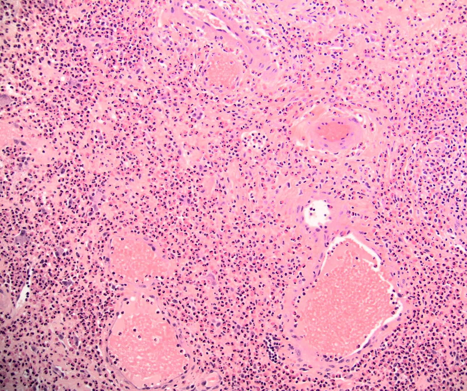
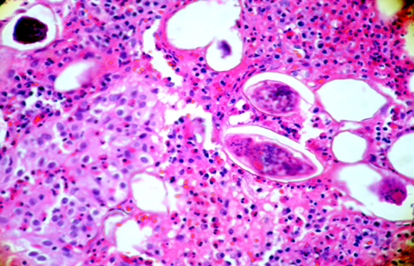
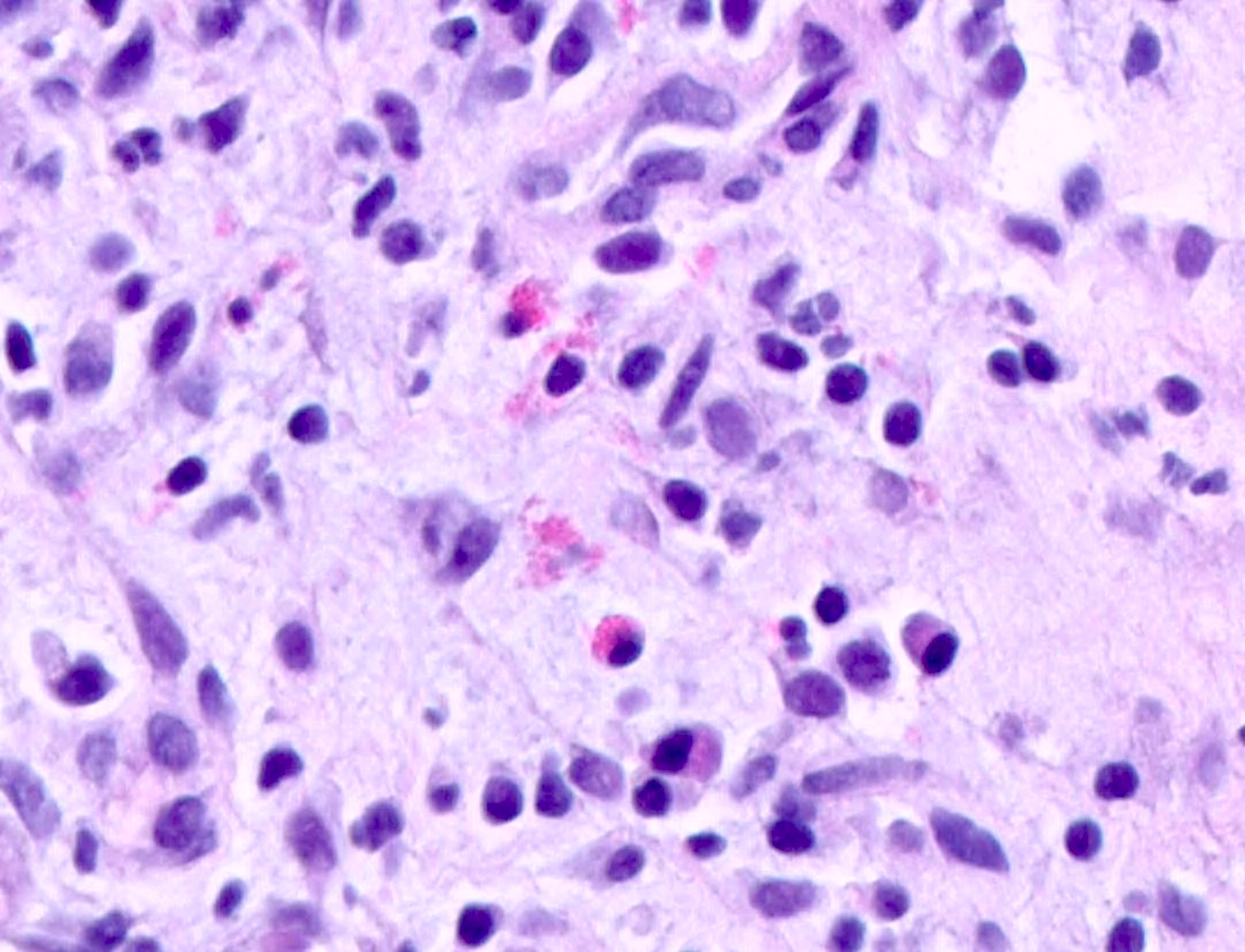
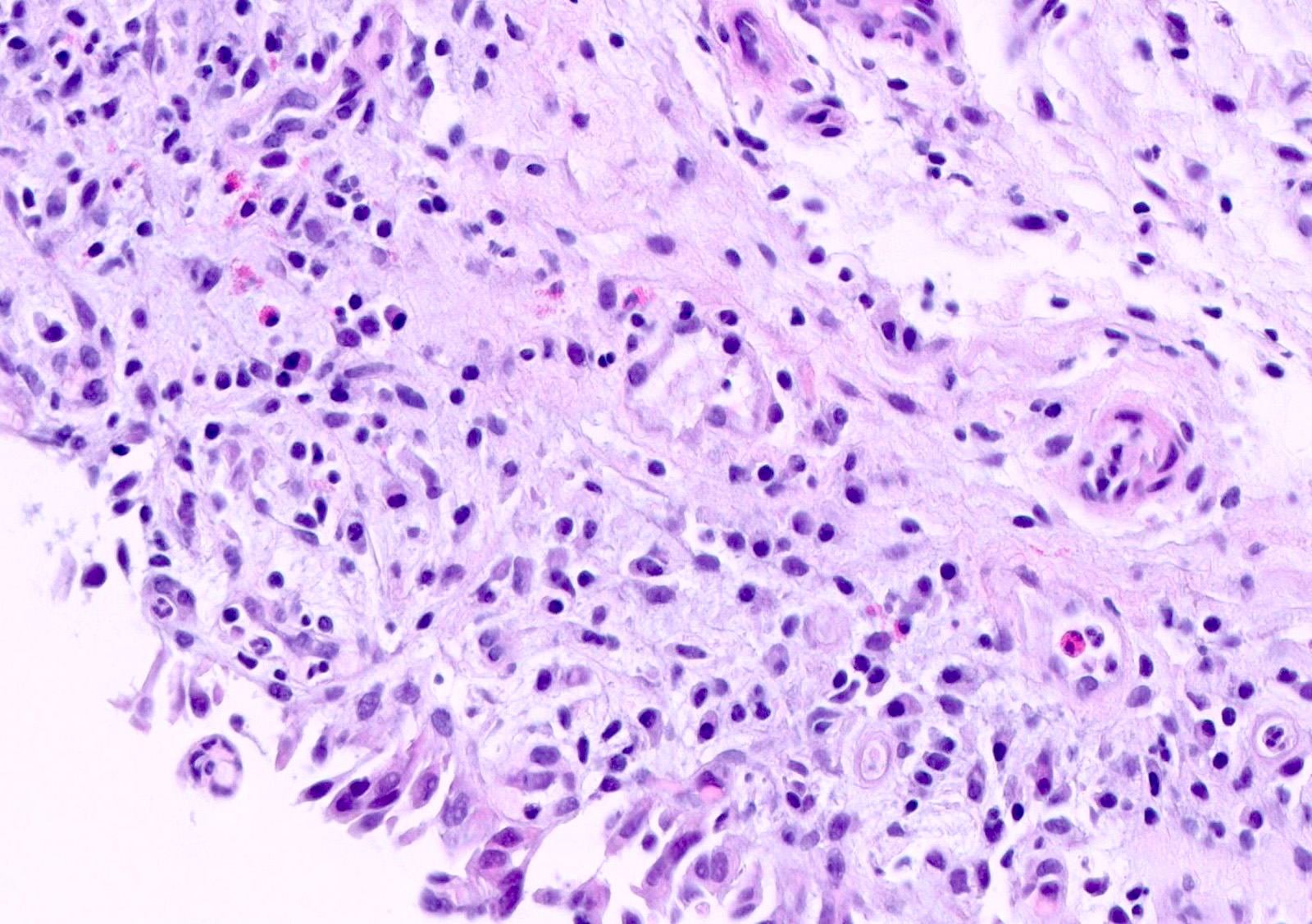
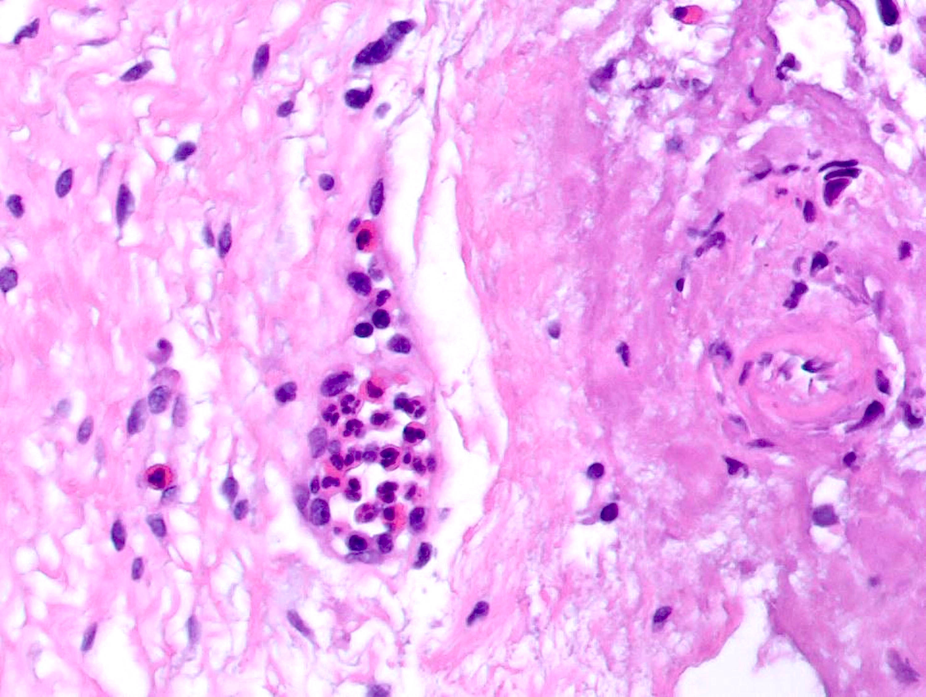
阳性染色
类胰蛋白酶(Tryptase)。
阴性染色
CK7、CK20、p53、Ki67、GATA3、PAX8(BMC Urol 2021;21:116)。
病理报告示例
- 膀胱病变,活检:嗜酸细胞性膀胱炎(见评注)。
- 评注:镜下可见膀胱壁全层炎症,伴显著嗜酸性粒细胞浸润。未见核异型或核分裂象。形态符合嗜酸细胞性膀胱炎。
鉴别诊断
间质性膀胱炎
- 上皮下淋巴细胞和浆细胞浸润,伴淋巴组织聚集。
- 尿路上皮脱离 / 剥脱(J Urol 1982;127:132)。
放射性膀胱炎
- 有盆腔放疗史。
- 非肿瘤性尿路上皮增生,可呈假浸润特征。
- 尿路上皮衬覆区域可见水肿、出血,固有层有以中性粒细胞和淋巴细胞为主的混合炎性浸润。
- 扩张的毛细血管伴纤维素样坏死及血管内纤维蛋白沉积。
浸润性尿路上皮癌
- 尿路上皮异型,缺乏显著的急、慢性炎症特征。
- 肿瘤细胞侵犯固有肌层(J Clin Diagn Res 2013;7:2282;Medicine (Baltimore) 2024;103:e36668)。
软斑病
- 圆形组织细胞(von Hansemann 细胞)浸润,胞质内有特征性嗜碱性包涵体(Michaelis–Gutmann 小体)。
- 黏膜完整的膀胱息肉样肿块。
其他参考文献
Nephron Extra 2013;3:30;Br J Radiol 2010;83:e122。
练习题 1
45 岁男性,排尿困难、耻骨上不适及间歇性无痛性血尿,有哮喘史。膀胱镜见膀胱顶部广基红斑样病变,临床疑为尿路上皮癌。活检显示固有层及固有肌层弥漫嗜酸性粒细胞浸润,伴显著水肿,无细胞异型或浸润性癌。最可能的诊断是什么?
- 嗜酸细胞性膀胱炎
- 间质性膀胱炎
- 浸润性尿路上皮癌
- 息肉样膀胱炎
练习题 1 答案
A.嗜酸细胞性膀胱炎。这是一种少见的炎症性疾病,常表现为膀胱肿块,临床及影像均可模拟恶性肿瘤。组织学见致密嗜酸性粒细胞浸润累及固有肌层,且无肿瘤性尿路上皮细胞,可支持诊断。B 错误:间质性膀胱炎通常为慢性炎症和肉芽组织,并非致密的全层嗜酸性粒细胞浸润。C 错误:尿路上皮癌可见浸润性恶性尿路上皮细胞,伴细胞异型和核分裂活动。D 错误:息肉样膀胱炎常与长期导尿或刺激相关,组织学表现为宽大水肿的乳头,伴血管充血和慢性炎症。
练习题 2
48 岁女性,尿频、排尿困难及间歇性肉眼血尿。膀胱镜见侧壁 2.5 cm 息肉样病变,疑为尿路上皮癌。经尿道切除组织学如上图。下一步最恰当的处理是什么?
- 开始使用贝那利珠单抗
- 开始膀胱内卡介苗(BCG)治疗
- 因恶性转化风险高而行根治性膀胱切除术
- 开始糖皮质激素治疗,并评估潜在过敏或继发原因
练习题 2 答案
D.开始糖皮质激素治疗,并评估潜在过敏或继发原因。嗜酸细胞性膀胱炎是良性炎症性疾病,初始治疗通常保守,包括糖皮质激素、抗组胺药及治疗或去除可识别的病因。B 错误:BCG 用于非肌层浸润性膀胱癌(NMIBC),不用于治疗嗜酸细胞性膀胱炎。C 错误:手术通常用于难治性疾病,而非由于恶性潜能。A 错误:贝那利珠单抗是针对嗜酸性粒细胞及嗜碱性粒细胞所表达 IL5Rα 的单克隆抗体,适用于去除诱因后仍对激素和抗组胺药无效的患者。
A rare inflammatory condition that can mimic bladder malignancy clinically and radiologically.
Definition / general
- Inflammatory condition of the urinary bladder, with recurrent episodes of urinary frequency, dysuria, gross hematuria and suprapubic pain during micturition
- Not related to Langerhans cell histiocytosis
Essential features
- Rare form of interstitial cystitis causing irritative bladder symptoms
- Variable symptoms similar to urinary tract infection (UTI)
- Can mimic malignant bladder lesions on imaging
Terminology
- Allergic cystitis: used because many cases are linked to underlying allergic reactions or a strong history of allergies
ICD coding
- ICD-10: N30.80 - other cystitis without hematuria
Epidemiology
- Rare (~200 reported cases)
- Reported across all age groups with striking predominance in female patients
- Women or children with allergic disorders and peripheral eosinophilia, older men with prostate / bladder disorders or parasitic infestation
- 20% occur in children; symptoms tend to disappear spontaneously (Arch Dis Child 2001;84:344)
Sites
- Transmural inflammation of bladder wall (90% cases) (Ann Med Surg (Lond) 2021;68:102626)
Etiology
- Often unknown; triggered by allergies, local trauma (double J stent, bladder surgery), medications (penicillin, bacillus Calmette-Guérin [BCG], mitomycin C), infections (Mycobacterium tuberculosis, schistosomiasis) or underlying systemic disorders
- History of recurrent UTI, eosinophilic enteritis, long term catheterization (Acad Pathol 2023;10:100078, Int Urol Nephrol 2004;36:159)
Pathophysiology
- Antigen exposure causes formation of the antigen - antibody complexes
- IgE mediated mast cell degranulation recruits eosinophils, triggering inflammation and tissue damage (J Pediatr Urol 2013;9:738)
Clinical features
- Increase in frequency, urgency, dysuria (BMJ Case Rep 2014;2014:bcr2014205708)
- Suprapubic / pelvic pain
- Urine retention
- Hematuria (gross or microscopic), pyuria
Diagnosis
- Diagnosed using histopathology (biopsy), clinical findings (symptoms, urinalysis, allergy testing), imaging (IV urography, ultrasonography, cystography, CT scan)
- Flexible cystoscopy; bladder biopsy confirms the diagnosis
Laboratory
- Urinalysis; hematuria (gross or microscopic), pyuria, positive nitrite, positive urine culture (26% cases)
- Blood investigation; peripheral eosinophilia (43% cases) (Urol Case Rep 2020;33:101379)
Radiology description
- Edema / erythema / ulcer of the bladder mucosa
- Thickened bladder wall
- Bladder mass / hemorrhagic lesions
- Hydronephrosis (Urol Ann 2013;5:50)
- Rarely, vesicoureteral reflux, ureteral orifice stenosis, bladder neck contracture (BMJ Case Rep 2014;2014:bcr2014205708)
Radiology images
Images hosted on other servers: Thickened bladder wall; Bladder ulcer on cystoscopy
Prognostic factors
- Overall prognosis is excellent with conservative medical therapy and elimination of triggers (medications, parasites) (Ann Med Surg (Lond) 2021;68:102626)
- Patients with a strong history of atopic conditions (e.g., asthma, environmental or food allergies) and systemic conditions (hypereosinophilic syndrome, Churg-Strauss syndrome) are prone to chronic recurring flares (Heliyon 2024;10:e32993)
- No impact on survival but impacts quality of life (Urol Case Rep 2020;33:101379)
Case reports
- 4, 10 and 15 year old boys and 11 and 15 year old girls with eosinophilic cystitis (Case Rep Urol 2026;2026:2379059)
- 14 year old boy and 69 year old man with eosinophilic cystitis treated with mepolizumab (J Investig Allergol Clin Immunol 2024;34:202)
- 44 year old man with eosinophilic cystitis mimicking bladder tumor (Urol Case Rep 2024;54:102714)
- 49 year old woman with eosinophilic cystitis mimicking hemorrhagic cystitis (Int Cancer Conf J 2022;11:298)
- 66 year old man with refractory eosinophilic cystitis treated with benralizumab (Front Allergy 2023;3:1055129)
- 72 year old man with eosinophilic cystitis misdiagnosed as bladder tumor (Medicine (Baltimore) 2024;103:e36668)
Treatment
- No standard treatment available
- Identification and removal of the precipitating factor
- Symptomatic management consists of oral and intravesical corticosteroids, nonsteroidal anti-inflammatory drugs (NSAIDs), antihistamines
- Cyclosporine and azathioprine in case of nonresponsiveness to steroids (J Indian Assoc Pediatr Surg 2021;26:51)
- Surgical management (partial cystectomy, transurethral resection of bladder tumor [TURBT]) in severe refractory cases (J Urol 2001;165:805)
- Interleukin 5 antagonists: benralizumab has caused significant improvement in quality of life and mepolizumab in cases of idiopathic eosinophilic cystitis (Urol Case Rep 2020;33:101379, J Investig Allergol Clin Immunol 2024;34:202)
- Risk of recurrence remains, requiring long term urologic follow up (Int J Clin Pract 2005;59:356)
Gross description
- Edematous and erythematous mucosa with polypoid growths resembling allergic polyps of nasal septum
Microscopic (histologic) description
- Histopathological findings can be divided in acute and chronic phase
- Acute phase: transmural inflammation of the bladder with prominent eosinophilic infiltrate (Yamada and Taguchi criteria are 20 or more eosinophils per 5 20x fields) with edema and occasional muscle necrosis; Charcot-Leyden crystals may be present (Arch Pathol Lab Med 2009;133:289)
- Chronic phase: fewer eosinophils but more prominent mast cells, muscle necrosis and fibrosis / scarring
Microscopic (histologic) images
Positive stains
Negative stains
Sample pathology report
- Bladder lesion, biopsy:
- Eosinophilic cystitis (see comment)
- Comment: Microscopic examination reveals transmural inflammation of bladder wall with prominent eosinophilic infiltration. There is no nuclear atypia or mitoses. The morphology is consistent with eosinophilic cystitis.
Differential diagnosis
- Includes other types of cystitis:
- Interstitial cystitis (J Urol 1982;127:132):
- Subepithelial lymphoplasmacytic inflammatory infiltrate with lymphoid aggregates
- Urothelial detachment / denudation
- History of pelvic irradiation
- Nonneoplastic urothelial proliferation with pseudoinfiltrative features
- Urothelial lining showing edema, hemorrhage and mixed inflammatory infiltrate composed predominantly of neutrophils and lymphocytes in the lamina propria
- Telangiectatic vessels with fibrinoid necrosis and intravascular fibrin deposition
- Urothelial atypia without any prominent features of acute and chronic inflammation
- Invasion of muscularis propria by tumor cells
- Round histiocytic infiltrate (von Hansemann cells) with characteristic cytoplasmic basophilic inclusions (Michaelis-Gutmann bodies)
- Polypoid bladder mass with intact mucosa
Additional references
Practice question #1
A 45 year old man presented with dysuria, suprapubic discomfort and intermittent painless hematuria. He had a history of asthma. Cystoscopy revealed a broad based erythematous lesion involving the bladder dome, clinically suspicious for urothelial carcinoma. Biopsy showed diffuse eosinophilic infiltration of the lamina propria and muscularis propria with marked edema, without cytologic atypia or invasive carcinoma. Which of the following is the most likely diagnosis?- Eosinophilic cystitis
- Interstitial cystitis
- Invasive urothelial carcinoma
- Polypoid cystitis
Practice answer #1
A. Eosinophilic cystitis. It is a rare inflammatory condition that often presents as a bladder mass and can mimic malignancy both clinically and radiologically. Histologic identification of dense eosinophilic infiltration, involving the muscularis propria, in the absence of neoplastic urothelial cells is diagnostic. Answer B is incorrect because histology typically shows chronic inflammation and granulation tissue and not dense transmural eosinophilic infiltrates. Answer C is incorrect because urothelial carcinoma shows infiltrating malignant urothelial cells with cytologic atypia and mitotic activity. Answer D is incorrect because polypoid cystitis is commonly associated with chronic catheterization or irritation and demonstrates broad edematous papillae with vascular congestion and chronic inflammation on histology.Reference: Eosinophilic cystitis
Practice question #2
A 48 year old woman presented with urinary frequency, dysuria and intermittent gross hematuria. Cystoscopy revealed a 2.5 cm polypoid lesion on the lateral bladder wall concerning for urothelial carcinoma. Transurethral resection histology is demonstrated in the image above. Which of the following is the most appropriate next step in management?
- Initiate benralizumab
- Initiate intravesical bacillus Calmette-Guérin (BCG) therapy
- Proceed with radical cystectomy because of the high risk of malignant transformation
- Start corticosteroid therapy and evaluate for potential underlying allergic or secondary causes
Practice answer #2
D. Start corticosteroid therapy and evaluate for potential underlying allergic or secondary causes. Eosinophilic cystitis is a benign inflammatory disorder. Initial management is typically conservative and includes corticosteroids, antihistamines and treatment or removal of any identifiable underlying cause. Answer B is incorrect because BCG is indicated for nonmuscle invasive bladder cancer (NMIBC) and has no role in the treatment of eosinophilic cystitis. Answer C is incorrect because surgical intervention is generally reserved for refractory disease rather than malignant potential. Answer A is incorrect because benralizumab is a monoclonal antibody against IL5Rα expressed on eosinophils and basophils and is indicated when the patient is refractory to steroids and antihistamines and after the removal of triggers.Reference: Eosinophilic cystitis