- 38 岁女性:夜间可疑全面强直–阵挛发作;右额白质 / 中央前回交界区无强化弥漫性肿块,拟低级别胶质瘤。
- 形态兼有少突与星形细胞特征;分子同时具备少突(IDH1 R132H、TERT、1p/19q 共缺失)与星形(致病性 ATRX + 亚克隆 TP53)信号。
- 诊断:双基因型 IDH 突变胶质瘤,符合少突星形细胞瘤 NEC(WHO CNS 2021),主体 2 级,局灶核分裂升高可疑向 3 级演进。
虚拟玻片
数字玻片(Box 1):https://isn-slidearchive.org/?&col=ISN&fol=Archive&file=ID-7213631.svs
临床病史与影像
本例为 38 岁女性,既往无明显病史。近 6 个月内发生两次无人目睹的夜间发作,伴舌咬伤与尿失禁,高度可疑全面强直–阵挛发作。神经系统查体其余未见异常。
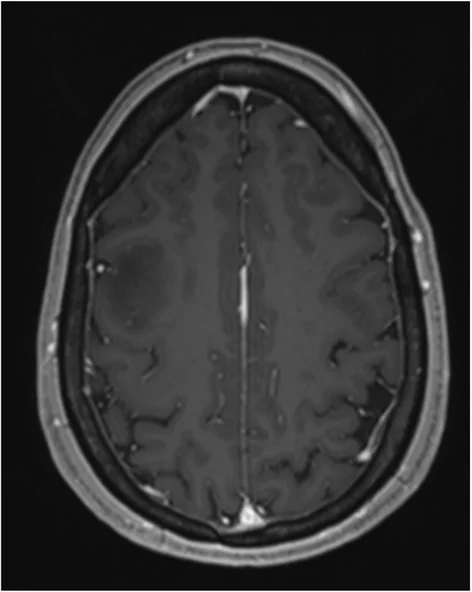
脑 MRI 显示位于右额叶的 T2 / FLAIR 高信号、T1 低信号弥漫性肿块,病灶位于白质并向后延伸至中央前回。未见对比剂强化、弥散受限或高级别生物学 / 转化征象,影像符合低级别胶质瘤(图 1)。患者接受了最大限度安全切除。
病理所见
组织学显示弥漫浸润性胶质肿瘤,累及皮层及深部白质(Box 1 虚拟玻片)。肿瘤中等细胞密度,由中等大小、核轻度不规则的细胞组成(图 2A)。相当一部分肿瘤细胞胞质透亮并具核周空晕,符合少突胶质细胞形态;其间混杂更小、形态偏星形细胞谱系的细胞。背景显著纤维性。未见坏死或微血管增生,血管亦缺乏典型「鸡爪样」构型。核分裂少(1 个 / 10 HPF;2 个 / 2.6 mm²),Ki67 约 5%–8%。极局灶见一增殖性结节,核分裂可高达 14 个 / 10 HPF,Ki67 约 20%(图 2B)。
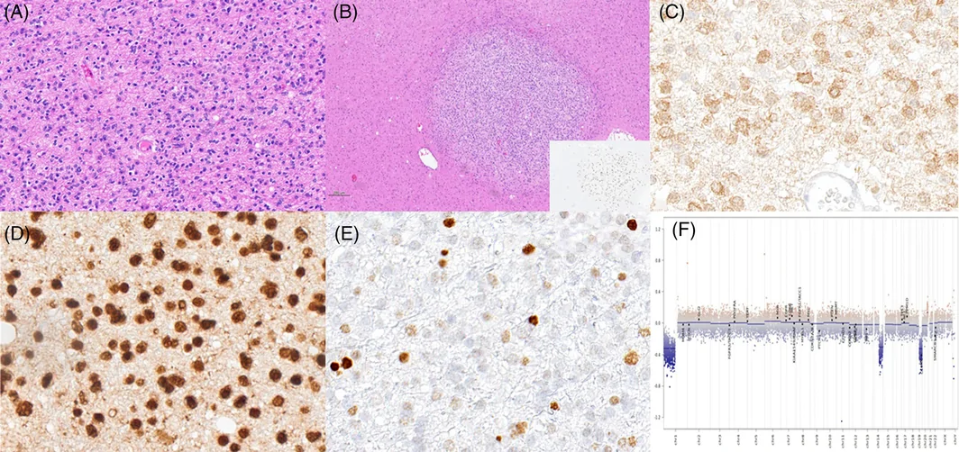
免疫组化:GFAP 强阳性;OLIG2 部分阳性(>60% 肿瘤细胞);IDH1 R132H 阳性(图 2C);ATRX 表达保留;p53 约 5% 阳性(图 2D、E);H3K27me3 保留;波形蛋白阴性。
基于 DNA 的二代测序检出致病性 IDH1 R132H,并共存 TERT 启动子与 ATRX 致病性变异。三者变异等位基因频率相近,分别为 33%、32% 与 36%。另见亚克隆致病性 TP53 变异(等位基因频率 5%)。FISH 示 1p/19q 共缺失:分析细胞中 1p36 与 19p13 缺失比例分别为 72% 与 75%。未见纯合或杂合 CDKN2A/B 缺失。
DNA 甲基化分型(Heidelberg 脑肿瘤分类器 v12.8)归类为「弥漫性胶质瘤,IDH 突变」高置信(0.99);亚类提示「少突胶质细胞瘤,IDH 突变且 1p/19q 共缺失」(0.65),「星形细胞瘤,IDH 突变,较低级别」评分较低(0.33)。拷贝数分析证实 1p/19q 共缺失(图 2F)。
最终诊断
双基因型 IDH 突变胶质瘤,符合少突星形细胞瘤,未另作分类(NEC);按 WHO CNS 2021 主体为 2 级,但极局灶核分裂活性增高,可疑向 3 级演进。
讨论
双基因型少突星形细胞瘤罕见,目前并非 WHO CNS 2021 分类与 cIMPACT-NOW 建议中的独立实体。尽管 IDH 突变少突胶质细胞瘤与星形细胞瘤在形态上可有重叠,二者鉴别现主要依据分子特征。本例支持少突胶质细胞瘤、IDH 突变的证据包括致病性 IDH1 R132H 与 TERT 变异以及 1p/19q 共缺失。然而,同时存在致病性 IDH1、ATRX 与 TP53 变异通常被认为与 1p/19q 共缺失少突胶质细胞瘤相互排斥,反而更支持星形细胞瘤、IDH 突变。上述重叠分子特征促成了双基因型少突星形细胞瘤的诊断。有趣的是,尽管检出致病性变异,本例肿瘤细胞 ATRX 免疫组化仍弥漫保留;该变异在蛋白近 C 端引入提前终止密码子,可能解释异常蛋白仍可检出。
既往形态学定义的少突星形细胞瘤研究曾报道空间分隔或弥漫混杂的少突与星形细胞群体;但其免疫组化与分子谱并不重叠,分别归入 1p/19q 共缺失、ATRX 保留的少突成分,与 1p/19q 完整、ATRX 及 TP53 突变的星形成分。本例免疫组化未见可分离的两群;IDH1、ATRX 与 TERT 变异等位基因频率相近,且多数细胞存在 1p/19q 共缺失。亚克隆 TP53 变异或可来自混杂星形成分和 / 或增殖性结节,但免疫组化未能证实。分子检测基于全切片提取 DNA,故无法判定各改变是否局限于特定组织学成分或为各成分共有。
无坏死与微血管增生支持 WHO CNS 2 级;但局灶核分裂与增殖指数升高令人担忧向 WHO CNS 3 级进展。cIMPACT-NOW update 12 亦承认少突胶质细胞瘤中 ≥6 个核分裂 / 10 HPF(0.24 mm²)的阈值;而如本例所示,MRI 无强化似与更长生存相关。
总之,本例说明即便形态与免疫组化看似典型,详细分子研究(含 NGS 与甲基化分型)仍可在 IDH 突变胶质瘤中揭示额外、意料之外的发现。
Virtual glass slide
Box 1. Access at https://isn-slidearchive.org/?&col=ISN&fol=Archive&file=ID-7213631.svs.
Clinical history and imaging
A 38-year-old woman without significant clinical history experienced two unwitnessed nocturnal episodes over the past 6 months, characterized by tongue biting and urinary incontinence, highly suspicious for tonic–clonic-generalized seizures. Neurological examination was otherwise unremarkable.
MRI of the brain revealed a T2- and FLAIR-hyperintense, T1-hypointense diffuse mass lesion, located in the right frontal lobe. The lesion was located in the white matter, extending posteriorly into the precentral gyrus. No contrast enhancement, diffusion restriction, or signs of high-grade biology or transformation were observed. The findings were consistent with a low-grade glioma (Figure 1). The patient underwent a maximally safe resection.
Findings
Histological examination revealed a diffusely infiltrating glial tumor involving the cortex and underlying white matter (Box 1). The tumor was moderately cellular and composed of medium-sized cells with mildly irregular nuclei (Figure 2A). A substantial proportion of tumor cells exhibited abundant clear cytoplasm with a perinuclear halo, consistent with oligodendroglial morphology, while intermingled smaller cells showed a morphology reminiscent of astrocytic lineage. The background was prominently fibrillary. No necrosis or microvascular proliferation was observed, and the vasculature lacked a characteristic “chicken-wire” pattern. The mitotic count was low (1 mitosis/10 high power fields (HPF), 2 mitoses/2.6 mm²) and proliferation index (Ki67) in the tumor ranged between 5% and 8%. Very focally, a proliferative nodule was identified, showing increased mitotic activity of up to 14 mitoses per 10 HPF and an increased Ki67 index of approximately 20% (Figure 2B).
Immunohistochemical analysis demonstrated strong positivity for GFAP and partial positivity for OLIG2 (>60% of tumor cells). IDH1 R132H immunostaining was positive (Figure 2C). ATRX expression was retained, and p53 was positive in approximately 5% of tumor cells (Figure 2D,E). H3K27me3 expression was retained. Tumor cells were negative for Vimentin.
DNA-based next-generation sequencing revealed an IDH1 R132H pathogenic variant with coexisting pathogenic variants in TERT promoter and ATRX. Notably, the allele frequency of the IDH1, TERT, and ATRX pathogenic variants was similar, being 33%, 32% and 36%, respectively. A subclonal pathogenic variant in TP53 was identified at 5% allele frequency. Fluorescence in situ hybridization demonstrated 1p/19q co-deletion, with 72% and 75% of analyzed cells showing loss of 1p36 and 19p13, respectively. No homozygous or heterozygous CDKN2A/B deletion was detected.
DNA methylation profiling (Heidelberg Brain Tumor Classifier v12.8) classified the tumor as a “diffuse glioma, IDH-mutant” with a high confidence score (0.99), with a subclassification suggestive of “oligodendroglioma, IDH-mutant and 1p/19q-codeleted” (score 0.65), and a lower score for “astrocytoma, IDH-mutant, lower grade” (score 0.33). Copy number variation analysis confirmed 1p/19q co-deletion (Figure 2F).
Final diagnosis
Dual-genotype IDH-mutant glioma, consistent with an oligoastrocytoma, not elsewhere classified (NEC), WHO CNS 2021 predominantly Grade 2, but with a very focal area of increased mitotic activity suspicious for evolution toward Grade 3.
Discussion
Dual-genotype oligoastrocytomas are rare and currently not recognized as a distinct entity by the WHO CNS 2021 classification and cIMPACT-NOW recommendations. Although morphological overlap between IDH-mutant oligodendrogliomas and astrocytomas is recognized, distinction between these two entities is now primarily based on molecular features. The diagnosis of oligodendroglioma, IDH-mutant in our case was supported by pathogenic variants in IDH1 R132H and TERT, and 1p/19q co-deletion. However, the presence of concurrent pathogenic variants in IDH1, ATRX, and TP53 is considered mutually exclusive with 1p/19q-codeleted oligodendrogliomas, and instead favor a diagnosis of astrocytoma, IDH-mutant. The coexistence of these overlapping molecular features led to a diagnosis of a dual-genotype oligoastrocytoma. Interestingly, ATRX was diffusely retained in tumor cells in our patient despite the detection of a pathogenic variant. This variant induced a premature stop codon located at the C-terminal end of the protein, potentially explaining maintained aberrant protein expression.
Previous studies of morphologically defined oligoastrocytomas have reported spatially distinct or diffusely intermixed oligodendroglial and astrocytic populations. However, immunohistochemical and molecular profiles did not overlap, segregating into 1p19q-co-deleted, ATRX retained oligodendroglial, and 1p19q-intact, ATRX and TP53 mutant astrocytic populations. In the present case, immunohistochemically no separate populations were present, variant allele frequencies were similar for IDH1, ATRX, and TERT, and 1p19q co-deletion was present in the majority of cells. Although the subclonal TP53 variant may originate from the intermixed astrocytic component and/or the proliferative nodule, this could not be confirmed by immunohistochemistry. Given that molecular profiling was performed on DNA extracted from the whole tissue section, it could not be established whether the identified molecular alterations were restricted to specific histological components or shared across them.
The absence of necrosis and microvascular proliferation supports WHO CNS Grade 2; however, the presence of a focal area with increased mitotic activity and an elevated proliferative index raised concern for progression toward WHO CNS Grade 3. While this has also been acknowledged by c-IMPACT-NOW update 12, which suggests a cut-off of ≥6 mitoses/10 HPF of 0.24 mm² in oligodendrogliomas, lack of contrast enhancement on MRI, as in our patient, appears to be associated with prolonged survival.
In conclusion, this case highlights how detailed molecular studies, including NGS and methylation profiling, may disclose additional unexpected findings in IDH-mutant gliomas, with an otherwise typical morphological and immunohistochemical profile.