- 53 份淋巴结活检涵盖 NLPHL-OL、cHL-LR 及典型 NLPHL、其他亚型 cHL 对照,以双重 κ/λ mRNA ISH 评估轻链表达。
- 典型弥漫胞质模式见于全部 NLPHL 和多数 NLPHL-OL;cHL 为阴性或非典型核周/颗粒状模式,不能将任何阳性信号都作为 NLPHL 的依据。
- 重叠病例仍需结合组织结构、B 细胞标志物、MEF2B 与 EBV 状态;非典型信号的生物学基础尚待验证。
摘要
结节性淋巴细胞为主型霍奇金/B 细胞淋巴瘤(NLPHL/BL)与经典霍奇金淋巴瘤(cHL)是生物学上不同的实体,分别表现为 B 细胞分化及免疫球蛋白(IG)转录的保留与丧失。然而,NLPHL 与富于淋巴细胞的 cHL(cHL-LR)之间具有重叠形态及免疫表型特征的病例,本文称为 NLPHL-OL,其生物学性质仍不明确。我们采用一种新型高敏感双重 κ/λ mRNA 原位杂交检测,评估这一谱系中的 IG 轻链表达。分析 53 份淋巴结活检,包括 NLPHL-OL 15 例、cHL-LR 11 例,并以典型 NLPHL 13 例和其他亚型 cHL 14 例为对照。系统评估肿瘤细胞 κ/λ mRNA 表达的分布、强度及模式,并与组织学、结构、免疫表型及 EB 病毒(EBV)状态关联。所有 NLPHL(13/13)及多数 NLPHL-OL(12/15,80%)的肿瘤细胞均检出弥漫、强的胞质轻链 mRNA 表达,即典型模式。相反,6/11(55%)cHL-LR 及 4/14(28%)其他亚型 cHL 检出微弱的核周或胞质颗粒状信号,即非典型模式。因此,典型 κ/λ 表达仅见于 NLPHL 及 NLPHL-OL,证实后者应归为具有异常特征的 NLPHL;cHL 则阴性或仅呈非典型模式。3/15(20%)NLPHL-OL 也出现非典型染色,其中 2 例 EBV 阳性。NLPHL 与 NLPHL-OL 中罕见的异常轻链谱包括单细胞 κ/λ 共表达,以及分别互斥表达 κ、λ 的不同 LP 细胞群。总之,κ/λ mRNA ISH 可细化 NLPHL–cHL 谱系的诊断分类,尤其适用于特征重叠病例,并揭示可能反映 B 细胞程序完整性的典型、非典型及阴性染色模式,将这些病例置于一个生物学连续谱中。
引言
经典霍奇金淋巴瘤(cHL)和结节性淋巴细胞为主型霍奇金/B 细胞淋巴瘤(NLPHL/BL)是 WHO 第 5 版及 ICC 2022 分类认可的两个实体[1(第 5 版),2]。历史上共同使用“霍奇金”一词反映了形态相似性:大型异型肿瘤细胞散布于致密炎症背景,该背景既塑造组织结构,也为肿瘤细胞群提供关键生存信号。然而,cHL 与 NLPHL 在临床、组织学、表型及分子方面明显不同,被认为是生物学上不同的疾病。cHL 的 B 细胞转录程序受破坏,反映一种“功能残缺”的凋亡前 B 细胞状态;NLPHL 则具有生发中心 B 细胞的表型和基因表达谱。二者的关键区别被认为是是否存在免疫球蛋白转录;缺失该转录会阻碍生理性的生发中心后轻链成熟[3–7]。部分 NLPHL 病例的 B 细胞受体可识别卡他莫拉菌等常见细菌病原体的抗原,因而被推测由抗原驱动,这支持淋巴细胞为主型(LP)细胞的免疫球蛋白仍具功能的观点[8]。
尽管生物学不同,NLPHL 与 cHL 之间仍存在形态及免疫表型灰区,包括标志物谱异常的 NLPHL 和 cHL 的富于淋巴细胞亚型(cHL-LR)。cHL-LR 在形态上可能非常类似 NLPHL,且表达 OCT2、BOB.1、BCL6 等 B 细胞转录因子的频率高于其他 cHL 亚型[9]。此外,多达 50% 的 cHL-LR 可见滤泡辅助性 T 细胞(TFH)微环境,PD1+ 或 CD57+ T 细胞形成花环[10,11]。反过来,NLPHL 可能表达 CD30 或 CD15、不同程度下调 B 细胞标志物,或出现 EBV 感染,这些通常更常见于 cHL[12–15]。同时发生的 cHL 与 NLPHL 之间已有克隆相关性记录,也支持该灰区存在[16]。
虽然理论上 NLPHL 与 cHL 的免疫球蛋白转录应有区别,特征重叠病例却很少见,研究不足。流式细胞术虽能有效评估 B 细胞淋巴瘤中的免疫球蛋白轻链(IGL)表达,但由于 Hodgkin/Reed–Sternberg(HRS)细胞及 LP 细胞稀少而脆弱,并不适合 cHL 和 NLPHL[17]。同样,在 FFPE 组织中,背景染色使免疫组化评估轻链限制性表达具有挑战[18]。
κ/λ mRNA 原位杂交(κ/λ mRNA ISH)可能是评估 B 细胞淋巴瘤轻链限制性表达的有效方法,诊断准确性接近流式细胞术[19–24]。既往研究表明,该方法有助于区分 cHL 与 NLPHL,因为 NLPHL 的 LP 细胞保留功能性 B 细胞分化,因而可检出 κ/λ mRNA 信号[25–27]。然而,在 cHL–NLPHL 特征重叠病例中,IGL mRNA 表达尚未得到系统研究[28,29]。借助近期可用的高敏感双重 κ/λ mRNA ISH,本研究旨在:(i)验证既往报道的 cHL 与 NLPHL 间 IGL 表达差异;(ii)表征轻链限制性表达模式;(iii)探索这一新技术对 cHL–NLPHL 特征重叠疑难病例的分类价值。
病例选择与诊断复核
回顾性收集德国蒂宾根大学医院病理研究所于 2010–2025 年诊断为 NLPHL 与 cHL 特征重叠或 cHL-LR 的淋巴结切除及粗针活检,包括会诊病例。若有代表性 FFPE 组织块或未染色切片可供 κ/λ mRNA ISH,则纳入研究。特征重叠病例(NLPHL-OL)定义为具有常规 NLPHL 不典型形态和/或免疫表型的诊断困难病例,包括具有 cHL 样特征的 NLPHL,如 CD30 和/或 CD15 表达、B 细胞表面标志物和/或转录因子(CD20、CD79a、PAX5、OCT2、BOB.1)部分丧失、EBV 阳性、生发中心结构保留、纯滤泡间生长或显著硬化。另外,为覆盖完整 NLPHL–cHL 谱系,纳入诊断为 cHL-LR 的病例;该亚型已知可见提示中间表型的特征,如生发中心进行性转化(PTGC)样形态、B 细胞标志物保留或异常强表达、PD1+ T 细胞花环及明显 TFH 背景[10,11]。
另纳入形态与表型典型的 NLPHL 及其他亚型 cHL 作为对照。由 3 名血液病理医师(JBS、FF、LQM)依据 WHO 第 5 版及 ICC 2022 标准复核全部诊断,并按现行体系进一步分类(cHL 亚型、Fan 等的模式、NLPHL 的 ICC 分级)[1,2,30,31]。临床数据来自病理申请单和机构病历。研究遵循《赫尔辛基宣言》,并获当地伦理审查委员会批准。
免疫组化
若初始检查尚未包括相关检测,则按厂商方案,在 2–3 µm FFPE 切片上使用 Ventana Ultra 自动染色系统(Ventana Medical Systems,美国亚利桑那州图森)及 Ventana 试剂进行免疫组化。完整抗体组合见补充方法。NLPHL-OL 病例的染色强度半定量评分为阴性(0)、弱(1)、中等(2)或强(3),分歧通过共同复核解决。
κ/λ mRNA 原位杂交
κ/λ mRNA ISH 的详细方法与数据解释见补充方法。按照厂商说明,采用 VENTANA Kappa and Lambda Dual ISH mRNA Probe Cocktail(Ventana Medical Systems,Roche Diagnostics),在 BenchMark ULTRA 平台对 FFPE 切片进行显色双重原位杂交,评估 κ、λ 轻链 mRNA 表达。根据表达 κ 或 λ mRNA 的肿瘤细胞比例判断轻链限制性。作为同片外部阳性质控的反应性扁桃体套区 B 细胞,在成熟反应性 B 细胞中具有最低的 IGL mRNA 表达;背景浆细胞与 B 淋巴细胞作为内部阳性对照。
多名观察者结合扫描数字切片和常规玻片光学显微镜,评估阳性 LP 或 HRS 细胞比例,以及胞质 κ/λ mRNA ISH 信号的强度、轻链 mRNA 类型和染色模式。仅对明确可识别的肿瘤细胞内无疑义的胞质信号评分;分歧通过共同镜下复核解决。信号强度分为阴性(0)、弱(1)、中等(2)或强(3)。轻链 mRNA 表达分为 κ 限制型、λ 限制型或异常型。染色模式分为:(1)组织学界定的肿瘤细胞中较高比例呈弥漫胞质染色,即典型模式;(2)不同比例肿瘤细胞出现弱核周环状和/或微弱胞质颗粒点状染色,即非典型模式;(3)阴性。每例可归入一种或多种模式。所有非典型模式病例均重复 κ/λ mRNA ISH 以确认。
结果
临床病理特征
NLPHL-OL、cHL-LR、NLPHL 及 cHL 各组的详细临床病理特征见表 1 和补充表 S1–S4。
| 特征 | NLPHL-OL | cHL-LR | NLPHL | cHL | 合计 |
|---|---|---|---|---|---|
| 病例数 | 15 | 11 | 13 | 14 | 53 |
| 中位年龄,岁(范围) | 47 (19–84) | 41 (15–80) | 37 (4–82) | 35.5 (15–81) | 41 (4–84) |
| 性别,男/女 | 男 > 女 | 男 > 女 | 男 > 女 | 男 > 女 | 2:1 |
| 最常见部位 | 腋窝 | 颈部 | 腋窝 | 颈部 | — |
| CD20 表达 | 15/15 (100%) | 1/9 ∗ (11%) | 13/13 (100%) | 1/14 (7%) | 30/53 (57%) |
| CD30 表达 | 12/15 (80%) | 11/11 (100%) | 0/13 (0%) | 14/14 (100%) | 37/53 (70%) |
| CD15 表达 | 2/15 (13%) | 9/11 (82%) | 0/13 (0%) | 14/14 (100%) | 23/53 (43%) |
| EBV 阳性 | 5/15 (33%) | 4/11 (36%) | 0/13 (0%) | 6/14 (43%) | 15/53 (28%) |
| κ/λ mRNA ISH 阳性 | 15/15 (100%) | 6/11 (55%) | 13/13 (100%) | 4/14 (29%) | 38/53 (72%) |
| 典型 κ/λ 模式 | 12/15 (80%) | 0/6 (0%) | 13/13 (100%) | 0/4 (0%) | 25/53 (47%) |
| 非典型 κ/λ 模式 | 3/15 (20%) | 6/6 (100%) | 0/13 (0%) | 4/4 (100%) | 13/53 (25%) |
∗)以可获得的临床资料为准。
共分析 53 份淋巴结活检,主要为切除标本,包括 NLPHL-OL 15 例、cHL-LR 11 例及 NLPHL 13 例、cHL 14 例对照。中位年龄 41 岁(4–84 岁),总体男性为主,男女比约 2:1。
NLPHL-OL 队列(n = 15)男性略多,年龄跨度大,中位 47 岁(19–84 岁)。有临床资料的大多数病例就诊时为早期(9/10)。NLPHL-OL 涵盖广泛 Fan 模式,包括 A 型以及结合低风险与较高风险特征的复合模式。1 例无法按 Fan 等的体系分类。2 例(NLPHL-OL-4、NLPHL-OL-7)既往有 cHL 诊断或接受过针对 cHL 的治疗。NLPHL-OL-7 的既往诊断修订为 NLPHL;NLPHL-OL-4 的既往活检无法获取复核。
cHL-LR 组(n = 11)男性为主,中位年龄 41 岁(15–80 岁)。最常见受累部位为颈部(7/11)。6 例有临床资料者中 2 例纵隔受累。1 例患者 HIV 阳性。
NLPHL 对照队列(n = 13)采用非连续病例,以覆盖 NLPHL 模式的形态谱。患者明显以男性为主,中位年龄 37 岁(4–82 岁),多数为早期。约半数主要表现为 Fan A 型(6/13,46%),单独出现或合并其他低风险模式。较高风险模式,尤其 Fan D 或 E 型(6/13,46%),与脾受累及复发相关。所有病例均强表达 CD20、OCT2、MEF2B,并有 PD1+ T 细胞花环。1 例(NLP-13)IgD 阳性。
cHL 对照队列(n = 14)男性为主,中位年龄 35.5 岁(15–81 岁)。结节硬化型(cHL-NS)最多见(8 例),其次为混合细胞型(cHL-MC,3 例)。早期与晚期均有病例(各 5/10)。纵隔受累常见(8/11),尤其在 cHL-NS 中(6/6);脾与骨髓受累主要见于晚期。全部 cHL-MC 病例 EBV 阳性,cHL-NS 偶见阳性(3/8,38%)。3 例未进一步分亚型。全部 14 例呈典型 cHL 免疫表型:RS 细胞 CD30、MUM1 阳性,CD15 表达不一,PAX5 弱表达,CD20 缺失。
NLPHL-OL 与 cHL-LR 队列的组织学和免疫表型特征
NLPHL-OL 队列表现出广泛的 NLPHL 与 cHL 重叠结构、细胞学及表型特征。各例具体重叠特征见表 1、补充表 S4 和 S5。PTGC 样改变常见(8/15,56%),滤泡结构保留相对少见且常为局灶(6/15,40%)。少数病例硬化(3/15,20%),其中 2 例为 Fan D 型[32]。全部病例均见 T 细胞花环及 LP 细胞(15/15,100%)。13/15(87%)和 9/15(60%)分别可见 Hodgkin 样及 Reed–Sternberg 样细胞,木乃伊样细胞仅见于少数病例(4/15,27%)。
免疫表型方面,NLPHL-OL 保留 B 细胞分化,全部表达 CD20(15/15),OCT2(14/14)和 MEF2B(12/12)以强阳性为主,PAX5 则多数弱阳性。CD79a、BOB.1 表达不一;全部可评估病例 BCL6 阳性(12/12),强度不一。多数表达 CD30(12/15,80%),通常弱至中等;CD15 阳性少见(2/15,13%)。5/15(33%)EBV 阳性,均表达 LMP1(5/5,100%),EBNA2 阴性。尽管存在非典型形态及免疫表型,CD20、OCT2 的强表达联合 MEF2B 阳性,使所有病例均倾向诊断 NLPHL。
cHL-LR 队列中,1/11(9%)见 PTGC 样改变,2/11(18%)有富于 TFH 的微环境,6/11(55%)见 T 细胞花环。1 例有木乃伊样细胞。所有病例均强表达 CD30(11/11,100%),CD15 阳性常见(9/11,82%),B 细胞标志物丧失或明显下调,仅有弱 PAX5 染色(9/9),几乎所有病例不表达 CD20(8/9,89%)。2 例(18%)OCT2 强表达。MUM1 强阳性亦常见(9/9),8 例可评估者中 5 例 BCL6 弱阳性。4/11(36%)EBV 阳性。
NLPHL-OL、cHL-LR、NLPHL 及 cHL 中的 κ/λ mRNA ISH 表达
详细 κ/λ ISH 特征汇总于表 1 和补充表 S6–S9,构成下述分析的框架。κ/λ mRNA ISH 阳性病例的 B 细胞免疫表型详见表 2。
| 病例编号 | κ/λ mRNA ISH 类别 | CD20 | PAX5 | OCT2 | BOB.1 | CD79a | BCL6 | MEF2B |
|---|---|---|---|---|---|---|---|---|
| NLPHL-OL-1 | 典型 | 3 | 2 | 2 | – | – | 3 | – |
| NLPHL-OL-2 | 典型 | 3 | 1 | 3 | 3 | 0 | 2 | – |
| NLPHL-OL-3 | 典型 | 3 | 2 | 3 | 0 | – | – | 3 |
| NLPHL-OL-4 | 典型 | 3 | 1 | 3 | 3 | 2 | – | 3 |
| NLPHL-OL-5 | 非典型 | 3 | 1 | 3 | 2 | 0 | 1 | 3 |
| NLPHL-OL-6 | 非典型 | 3 | 1 | 3 | 1 | 1 | 3 | 3 |
| NLPHL-OL-7 | 典型 | 3 | 1 | 3 | 2 | 1 | 2 | 3 |
| NLPHL-OL-8 | 典型 | 3 | 1 | 3 | – | 2 | 3 | 3 |
| NLPHL-OL-9 | 典型 | 3 | 3 | 3 | – | – | 1 | 2 |
| NLPHL-OL-10 | 典型 | 2 | 2 | 3 | 1 | 2 | 1 | 2 |
| NLPHL-OL-11 | 典型 | 3 | 2 | 2 | 2 | – | 2 | – |
| NLPHL-OL-12 | 典型 | 3 | 3 | 3 | 2 | 3 | 2 | 3 |
| NLPHL-OL-13 | 典型 | 3 | 1 | 3 | 3 | 1 | 1 | 3 |
| NLPHL-OL-14 | 典型 | 3 | – | – | – | – | – | 3 |
| NLPHL-OL-15 | 非典型 | 2 | 1 | 3 | 0 | 0 | 2 | 3 |
| cHL-LR-3 | 非典型 | 1 | 1 | 3 | – | – | 0 | – |
| cHL-LR-4 | 非典型 | 0 | 1 | 1 | 1 | 0 | 1 | 1 |
| cHL-LR-6 | 非典型 | 0 | 1 | 1 | 0 | 0 | 0 | 0 |
| cHL-LR-7 | 非典型 | 0 | 1 | 1 | 0 | 0 | 1 | 1 |
| cHL-LR-9 | 非典型 | 0 | 1 | 0 | – | – | 0 | 0 |
| cHL-LR-11 | 非典型 | 0 | 1 | 1 | 0 | 0 | 1 | 0 |
| cHL-1 | 非典型 | 1 | 1 | 0 | 0 | 0 | 0 | 0* |
| cHL-3 | 非典型 | 0 | 1 | 1 | 0 | 0 | 0 | 0 |
| cHL-4 | 非典型 | 0 | 1 | 0 | 0 | 0 | 1 | 0 |
| cHL-9 | 非典型 | 0 | 1 | 0 | 0 | – | 1 | 0 |
大型异型细胞的免疫组化染色强度半定量评分为 0(阴性)、1(弱)、2(中等)或 3(强)。“–”表示未评估或无法评估;* 表示罕见单个细胞弱阳性。注意:尽管呈非典型 κ/λ mRNA ISH 模式,NLPHL-OL 病例的 B 细胞标志物表达仍强于 cHL-LR 及 cHL。
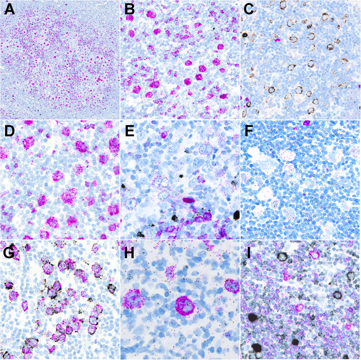
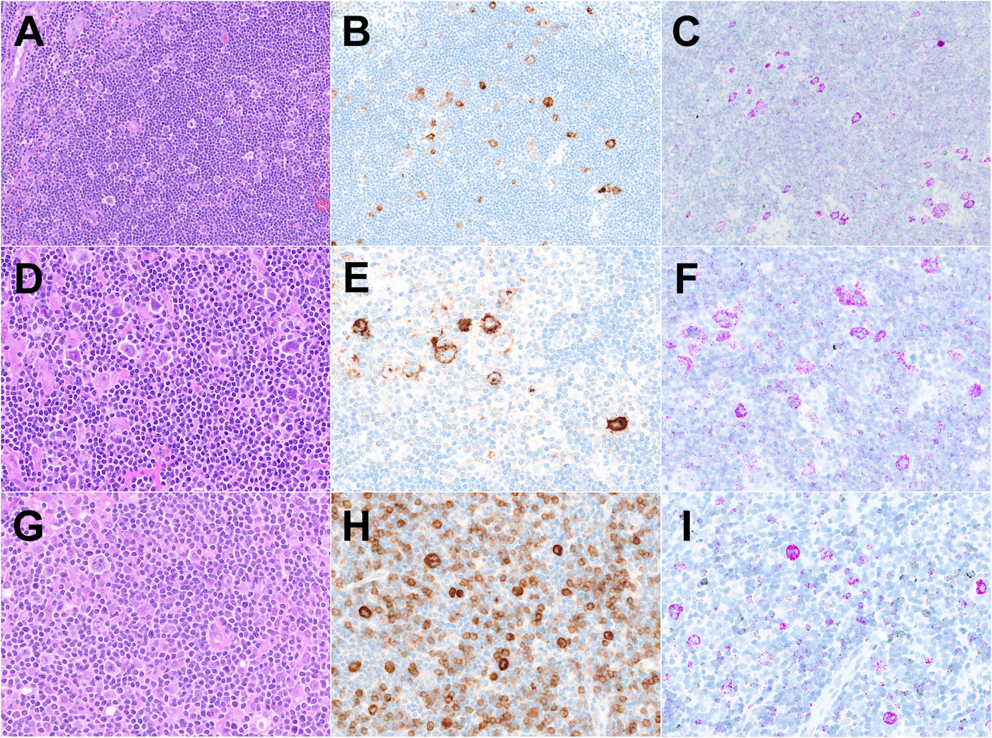
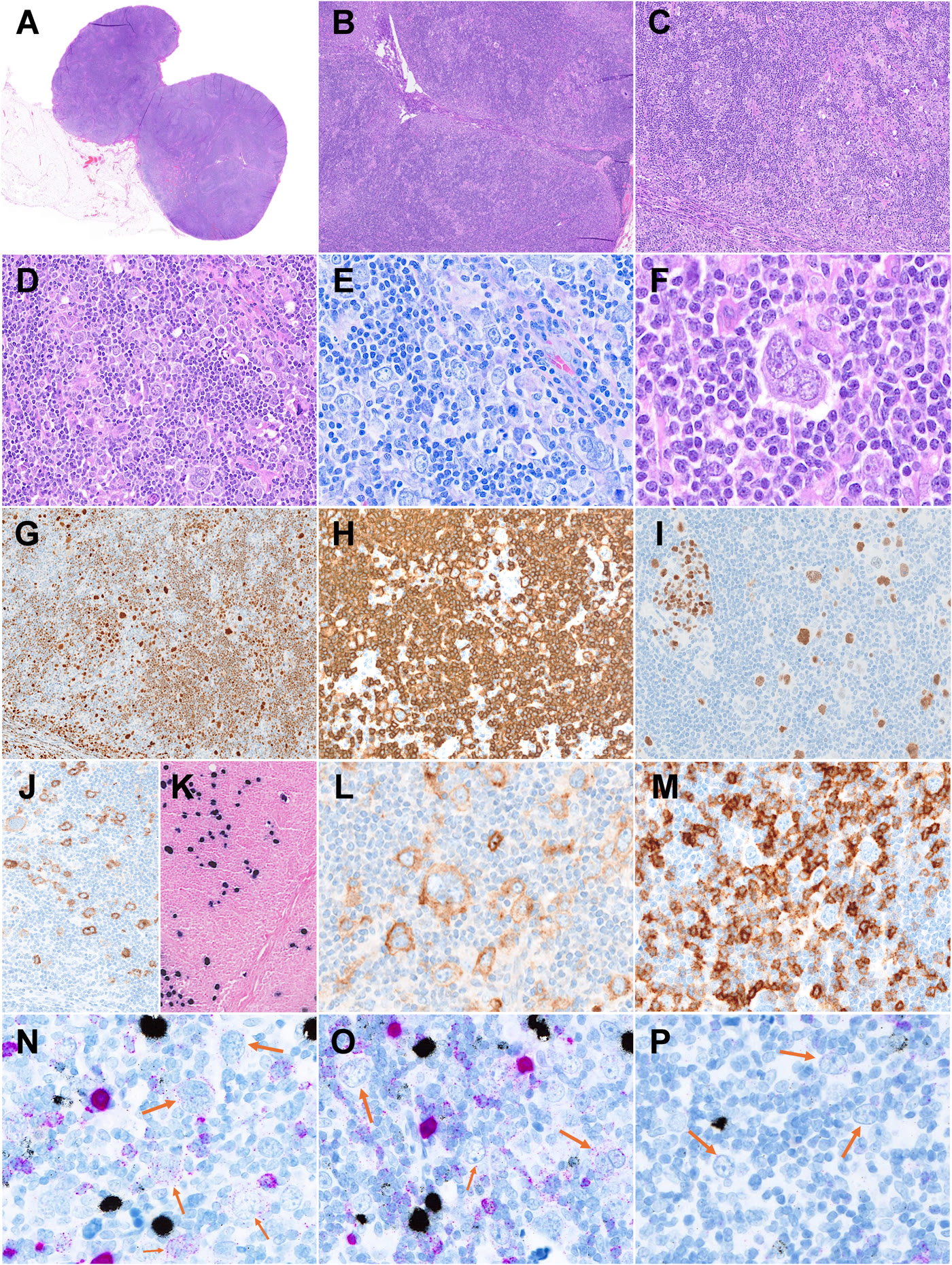
所有 NLPHL-OL、cHL-LR 及 NLPHL、cHL 对照均可评估 κ/λ mRNA ISH。全部 NLPHL(13/13)及 NLPHL-OL(15/15)呈 κ/λ ISH 阳性(图 1)。较高比例 LP 细胞表达 κ/λ mRNA,阳性比例中位数分别为 NLPHL 的 100% 及 NLPHL-OL 的 90%。两组主要染色模式均为弥漫胞质型(典型),分别见于 13/13 和 12/15(80%),显示两组 κ/λ mRNA 表达高度一致。CD30 或 CD15 阳性的 NLPHL-OL,其 mRNA 表达谱与常规 NLPHL 无差异(图 2)。3 例 NLPHL-OL(20%)呈非典型染色,其中 2 例 EBV 阳性(NLPHL-OL-5、-6)。NLPHL-OL-5 因多项异常特征与 cHL-LR 重叠而难以分类(图 3)。NLPHL-OL-7 亦为 EBV 阳性,仅 50% LP 细胞表达 κ/λ mRNA,但肿瘤细胞仍呈弥漫/典型模式。较高风险或复合模式仍保留典型阳性,但偶见异质性,包括部分肿瘤细胞非典型染色及强度不一。
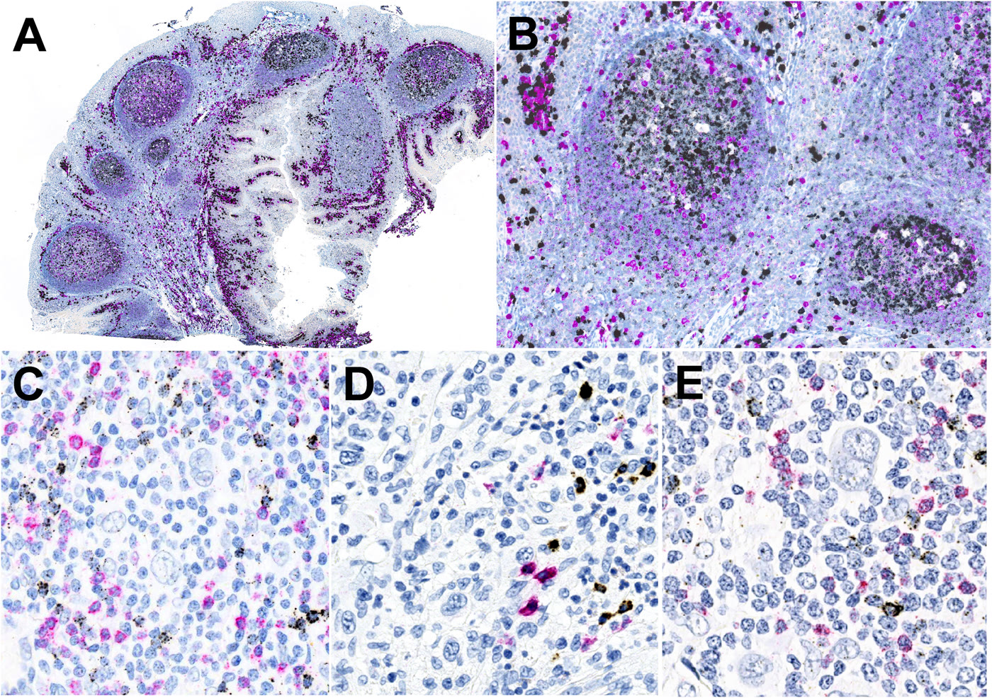
cHL-LR 中 6/11(55%)κ/λ mRNA 阳性,但均为非典型染色。相反,多数 cHL(10/14,71%)κ/λ mRNA ISH 完全阴性,包括 EBV 阳性 cHL(5/6,83%)(图 4)。cHL-NS 与 cHL-MC 阳性频率低于 cHL-LR,仅部分病例表达 κ/λ mRNA,分别为 3/8(38%)及 1/3(33%)。总体而言,cHL 中 κ/λ mRNA ISH 阳性肿瘤细胞比例的变异更大,且始终呈非典型模式,包括胞质颗粒点状和/或核周环状信号,未见典型模式。所有非典型模式病例重复 ISH 后结果相同。
Dunn 事后检验的组间比较显示,NLPHL 与 NLPHL-OL(校正后 p = 0.312)、cHL 与 cHL-LR(校正后 p = 0.440)之间无显著差异(图 5)。相反,NLPHL 与 cHL 的 κ/λ mRNA ISH 阳性存在显著差异(校正后 p = 2.88 × 10⁻⁶)。NLPHL-OL 与 cHL-LR 的差异亦显著(校正后 p = 0.0037)。全部病例结果汇总于图 6。
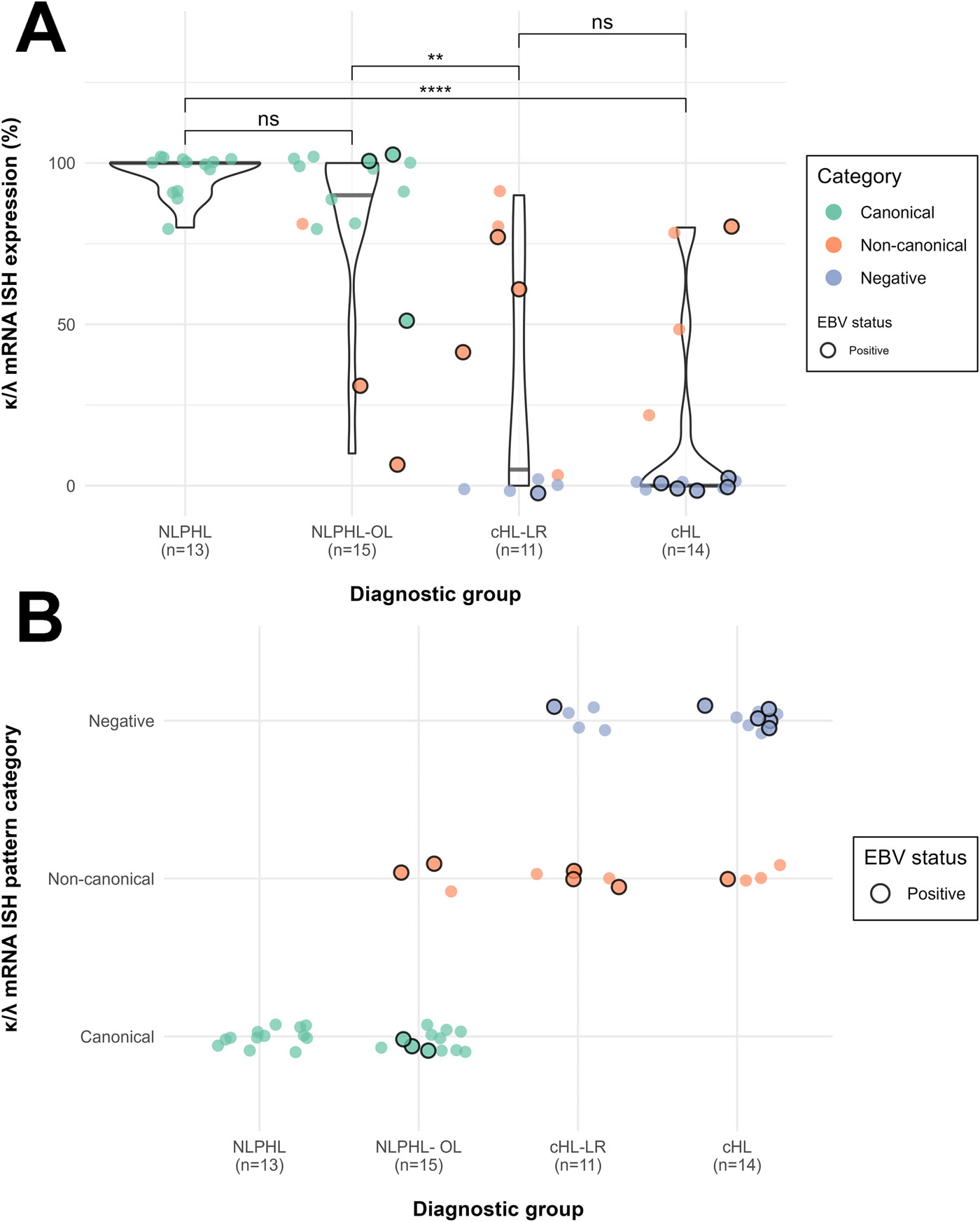
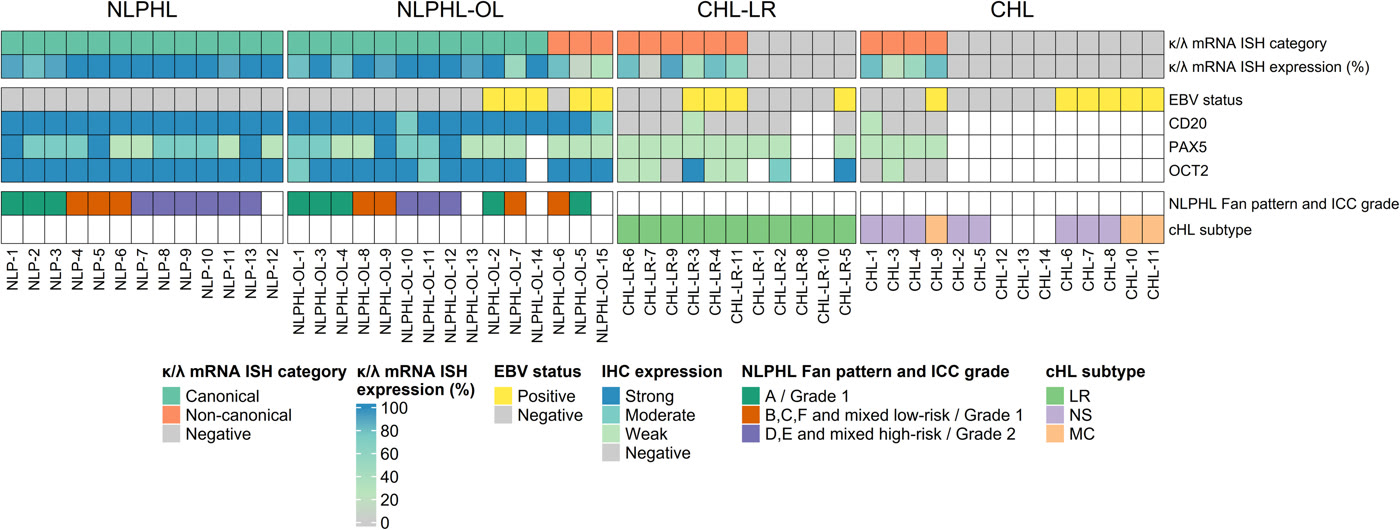
所有队列中,κ 轻链限制性表达占主导(32/38,84%),λ 轻链表达少见(4/38,11%),且仅存在于 NLPHL 和 NLPHL-OL。还发现异常双重 κ/λ mRNA ISH 表达谱,包括 1 例 EBV 阳性病例中分别表达 κ 或 λ 的不同 LP 细胞群(补充图 S1),以及 1 例 LP 细胞内 κ/λ 共表达。4 例 κ 阳性肿瘤细胞出现局灶核内颗粒状 λ 信号,反映 IGLL5 转录本标记(图 2G–I)。最后,背景 B 淋巴细胞评估未发现第二个克隆性细胞群;但少数有 PTGC 样改变的滤泡显示偏向 κ 轻链 mRNA 表达,尚无明确轻链限制性证据。
讨论
本研究考察 κ/λ mRNA 表达作为 NLPHL 与 cHL 中 IG 轻链位点功能性转录活性的替代标志,重点关注特征重叠的 NLPHL 及 cHL-LR,旨在细化这些疑难病例的分类。结果表明,新型超敏 κ/λ mRNA ISH 能可靠区分典型 NLPHL 与 cHL,并为重叠病例提供额外诊断支持。此外,在相当一部分 cHL 中发现意义尚不明确的减弱核周及颗粒点状染色,本文称为非典型模式。这些模式可能反映 B 细胞转录程序不同程度的保留或改变。
既往采用其他检测方法的研究已表明,κ/λ mRNA ISH 可通过 NLPHL 一致阳性、cHL 阴性来区分二者[25–27]。与之相符,本研究也显示明确区分:NLPHL(包括特征重叠病例)100% 阳性,而 cHL 完全不出现典型染色。这证实其内在 B 细胞转录程序存在显著差异,并提供进一步诊断支持。与保留的 B 细胞程序一致,典型 NLPHL 始终呈典型弥漫胞质染色。NLPHL 的结构变异与异质性增加相关,但典型模式大体保留。
与近期报道所有 cHL 均阴性的研究不同,本系列中约半数 cHL-LR 及少数但占一定比例的 cHL 表现有限的 κ/λ mRNA ISH 阳性[25]。虽然尚不完全清楚这些非典型染色是否代表肿瘤细胞内低水平免疫球蛋白 mRNA 表达,识别它们具有实际诊断意义。迄今文献未专门讨论这些模式[25,26]。既往研究未见该模式,可能反映检测性能与敏感性差异,包括本研究试剂可能检出其他平台无法捕捉的低水平轻链信号。
NLPHL-OL 病例持续存在轻链 mRNA 表达,证实其属于具有异常特征的 NLPHL,其中 3 例呈非典型模式,1 例表达减少;这几例中有 3 例 EBV 阳性。尽管异常 CD30/CD15 共表达或部分 B 细胞标志物丧失等形态、免疫表型特征增加了诊断难度,κ/λ mRNA ISH 仍获得稳健且可重复的结果。相反,cHL-LR 仅呈非典型阳性(55%)或阴性(45%)。cHL-LR 的 κ/λ mRNA ISH 阳性比常规 cHL 更常见(55% 对 29%),支持其与 NLPHL 具有更近的生物学关系,以及既往研究所示的 B 细胞程序部分保留[9]。
然而,这种阳性完全属于非典型模式,更提示免疫球蛋白转录失调,而非 NLPHL 中的生理性轻链表达[6]。它可能反映免疫球蛋白转录减少或残余转录,也可能来自无义介导的 mRNA 降解增强,导致异常转录本被更强地清除;该机制是一种去除错误 mRNA 的细胞监控机制[33,34]。
NLPHL 与 NLPHL-OL、cHL 与 cHL-LR 之间均未见显著差异。但 NLPHL-OL 与 cHL-LR 在典型/非典型模式及 κ/λ mRNA ISH 阳性百分比方面存在显著差异。从诊断角度,非典型 κ/λ mRNA ISH 模式不应被解读为完整 B 细胞身份的证据,也不应作为诊断 NLPHL 的依据,但可能反映 IG 转录活性的连续谱。在 NLPHL–cHL-LR 灰区,这一区别尤其重要:常规形态与免疫表型标准可能无法定论,而不同实体治疗策略显著不同,最终诊断直接影响治疗。在此类病例中,仅典型 κ/λ mRNA 表达能提供额外诊断信息,但仍需结合结构、B 细胞标志物、MEF2B 表达及 EBV 状态[35]。
与 EBV 阴性 NLPHL 相比,EBV 阳性 NLPHL-OL 更常表现为 κ/λ mRNA ISH 表达减少或非典型染色。EBV 阳性 NLPHL 已在各年龄组得到认识,常表达 CD30,通常为 II 型潜伏感染,虽也有罕见 I 型潜伏感染报道[15,36,37]。在本研究这类 EBV 均一阳性病例中,EBV 可能是早期驱动事件;但这些病变中 LP 细胞继发性/亚克隆性 EBV 感染的意义仍不明确[38,39]。在此背景下,本研究提示 EBV 可能调节 LP 细胞内 κ/λ 转录。cHL 中 EBV 相关的 B 细胞程序扰动已有充分记录[38,40,41]。
机制上,LMP2A、LMP1 等 EBV 潜伏蛋白可通过影响 PAX5 等关键转录调控因子,并诱导 ID2 等 B 细胞分化抑制因子,干扰 B 细胞分化程序[42–44]。其他 EBV 驱动的 B 细胞淋巴瘤也已证实这一现象[45,46];这可能解释为何 EBV 阳性 NLPHL-OL 虽保留 B 细胞标志物表达,包括强 CD20、OCT2,仍出现非典型 κ/λ 谱。
κ/λ mRNA ISH 还揭示了据我们所知尚未在 LP 细胞中描述的非常规表达模式。4 例 NLPHL 的 κ 阳性 LP 细胞内检出核内颗粒状 λ 信号,与已知的 IGLL5 交叉反应一致。IGLL5 是一种与 IGL 恒定区共享 2 个外显子的 λ 样假基因,其转录本通常位于细胞核,可在淋巴及非淋巴细胞中检出[19–21,25,26,47]。识别这一现象是避免误判为 λ 轻链 mRNA 表达的关键。
另有 2 例罕见异常模式:NLP-10 的同一 LP 细胞中 κ 和 λ 呈强胞质共表达。其他 B 细胞恶性肿瘤中罕有同一细胞真正双等位轻链表达的描述;在单型轻链表达的慢性淋巴细胞白血病(CLL)中,也曾发现两种轻链的功能性 mRNA 转录本,提示等位排斥可发生于翻译层面[26,47,48]。NLPHL-OL-2 是一例复发 EBV 阳性 NLPHL-OL,有两个分别表达 κ、λ 的不同 LP 细胞群,未见单细胞共表达证据。其究竟是真正双克隆,还是同一克隆内发生轻链转换,需要包括单细胞分析在内的更高分辨率方法。
本研究有若干局限。固定与标本保存时间可能影响 RNA 保存,并引入分析前变异。尽管严格评估了内、外对照的阳性,固定欠佳区域或背景染色异质性仍可能降低特定病例的敏感性。此外,虽然非典型 κ/λ mRNA ISH 模式可重复,且与诊断类别和 EBV 状态呈明确的非随机关联,但其生物学基础无法在本研究范围内完全阐明;非特异性背景染色虽不太可能,仍无法彻底排除。需要更高分辨率或单细胞方法,确认这些信号是否反映低水平 IG 转录,并研究 EBV 感染对 LP 细胞轻链转录本的影响。最后,对照病例相对较少,可能无法反映典型 NLPHL 与 cHL 轻链 mRNA 表达的完整谱系,强调了更大规模多中心队列验证的必要性。
总之,高敏感 κ/λ mRNA ISH 可可靠区分 NLPHL 与 cHL,并通过提供 IG 转录读出,成为 NLPHL–cHL-LR 灰区病例分类的有价值辅助手段。除二元结果外,EBV 阳性 NLPHL 及部分 cHL,尤其 cHL-LR 中的不同非典型表达模式,可能反映低水平 IG mRNA 转录,但确认这一假设仍需更先进的方法。
- Dual κ/λ mRNA ISH assessed light-chain expression in 53 lymph node biopsies spanning NLPHL-OL, cHL-LR, typical NLPHL, and other cHL subtypes.
- Canonical diffuse cytoplasmic staining occurred in all NLPHL and most NLPHL-OL cases; cHL was negative or showed non-canonical perinuclear/granular patterns, so positivity alone does not establish NLPHL.
- Overlap cases still require integration with architecture, B-cell markers, MEF2B, and EBV status; the biological basis of non-canonical signals requires validation.
Abstract
Nodular lymphocyte-predominant Hodgkin/B-cell lymphoma (NLPHL/BL) and classic Hodgkin lymphoma (cHL) are biologically distinct entities, exhibiting preserved versus lost B-cell differentiation and immunoglobulin (IG) transcription. However, cases with overlapping morphologic and immunophenotypic features between NLPHL and lymphocyte-rich cHL (cHL-LR), here designated as NLPHL-OL, remain biologically ambiguous. We applied a novel, highly sensitive dual κ/λ mRNA in situ hybridization assay to assess IG light-chain expression across this spectrum. We analyzed 53 lymph node biopsies, including NLPHL-OL cases (n =15), cHL-LR (n = 11), along with typical NLPHL (n = 13) and cHL of other subtypes (n = 14) as controls. Distribution, intensity, and patterns of κ/λ mRNA expression in neoplastic cells was systematically evaluated and correlated with histology, architecture, immunophenotype, and Epstein-Barr virus (EBV) status. Diffuse, strong cytoplasmic light chain mRNA expression, representing the canonical pattern, was detected in the tumor cells of all NLPHL (13/13) and the majority of NLPHL-OL cases (12/15, 80%). In contrast, faint perinuclear or granular cytoplasmic signals (non-canonical patterns) were detected in 6/11 (55%) cHL-LR and 4/14 (28%) of other cHL subtypes. Canonical κ/λ expression was thus restricted to NLPHL and NLPHL-OL, confirming their classification as NLPHL with aberrant features, whereas cHL was negative or displayed exclusively non-canonical patterns. Non-canonical staining was also seen in 3/15 (20%) NLPHL-OL, including two EBV-positive cases. Rare aberrant light chain profiles in NLPHL and NLPHL-OL cases included single-cell κ/λ coexpression, and distinct LP-cell populations with mutually exclusive κ and λ expression. In summary, κ/λ mRNA ISH refines diagnostic classification across the NLPHL-cHL spectrum, is particularly useful in cases with overlapping features, and reveals canonical, non-canonical, and negative staining patterns that may reflect B-cell program integrity, placing these cases along a biological continuum.
Introduction
Classic Hodgkin lymphoma (cHL) and nodular lymphocyte-predominant Hodgkin/B-cell lymphoma (NLPHL/BL) are two entities recognized in the WHO 5 th Edition and ICC 2022 classifications. 1(p5),2 The historical use of the shared term “Hodgkin” reflects their morphological resemblance - large, atypical tumor cells embedded in a dense inflammatory milieu that both shapes tissue architecture and provides critical survival signaling cues to the neoplastic population. However, cHL and NLPHL show marked clinical, histologic, phenotypic and molecular differences and are regarded as biologically distinct. cHL exhibits a disrupted B-cell transcriptional program reflecting a “crippled” pre-apoptotic B cell, whereas NLPHL has the phenotype and gene expression profile of a germinal center B cell. The key distinction is considered the presence or absence of immunoglobulin transcription; the latter precludes physiologic post-germinal center light-chain maturation. 3–7 Evidence that a subset of NLPHL cases expresses B-cell receptors recognizing antigens of common bacterial pathogens such as Moraxella catarrhalis and thus are putatively antigen-driven supports the notion that lymphocyte-predominant (LP) cell immunoglobulin remains functional. 8
Despite their contrasting biology, a morphological and immunophenotypic gray zone exists between NLPHL and cHL, encompassing NLPHL with aberrant marker profile and the lymphocyte-rich subtype of cHL (cHL-LR). cHL-LR may closely resemble NLPHL morphologically and expresses B-cell transcription factors such as OCT2, BOB.1, and BCL6 more frequently than other cHL subtypes. 9 In addition, a follicular helper T-cell (TFH) microenvironment with rosetting by PD1+ or CD57+ T-cells can be seen in up to 50% of cHL-LR cases. 10 , 11 Conversely, NLPHL may express CD30 or CD15, exhibit variable downregulation of B-cell markers, or show Epstein-Barr virus (EBV) infection, features more typically associated with cHL. 12–15 Support for this gray zone also comes from cases with documented clonal relationships between synchronous cHL and NLPHL. 16
Although NLPHL and cHL are conceptually expected to differ in their immunoglobulin transcription, cases with overlapping features are rare and poorly studied. Flow cytometry, despite being an effective method for assessing immunoglobulin light-chain (IGL) expression in B-cell lymphomas, is poorly suited for cHL and NLPHL due to the scarcity and fragility of Hodgkin/Reed-Sternberg (HRS) and LP cells. 17 Likewise, in formalin-fixed paraffin-embedded (FFPE) tissue, assessment of light-chain restriction with immunohistochemistry is challenging because of background staining. 18
κ/λ mRNA in situ hybridization (κ/λ mRNA ISH) may provide a useful approach for assessing light-chain restriction in B-cell lymphomas, with diagnostic accuracy approaching that of flow cytometry. 19–24 Previous studies have shown that κ/λ mRNA ISH can help in distinguishing cHL from NLPHL, since LP cells in NLPHL retain functional B-cell differentiation and therefore show detectable κ/λ mRNA signals. 25–27 However, the expression of IGL mRNA has not been systematically explored in the context of cases with overlapping cHL-NLPHL features. 28 , 29 Making use of a recently available, highly sensitive dual κ/λ mRNA ISH, we specifically aimed to (i) validate previously reported differences in IGL expression between cHL and NLPHL; (ii) characterize light-chain restriction patterns; and (iii) explore the use of this novel technology for classifying challenging cases with overlapping cHL-NLPHL features.
Case Selection / Diagnostic Review
We retrospectively collected excisional lymph node and core needle biopsies, including cases submitted for second opinion, diagnosed either as showing overlapping features between NLPHL and cHL or as cHL-LR at the Institute of Pathology, University Hospital Tuebingen, Germany, between 2010 and 2025. Cases were included if representative FFPE tissue blocks or unstained slides were available for κ/λ mRNA ISH. Cases with overlapping features (NLPHL-OL) were defined as diagnostically challenging cases showing morphologic and/or immunophenotypic features atypical for conventional NLPHL. These included NLPHL with cHL-like features, such as CD30 and/or CD15 expression, partial loss of B-cell surface markers and/or transcription factors (CD20, CD79a, PAX5, OCT2, BOB.1), EBV positivity, preservation of germinal center architecture, purely interfollicular growth, or marked sclerosis. Additionally, to cover the full NLPHL–cHL spectrum, we included cases classified as cHL-LR, in which features suggestive of an intermediate phenotype - such as progressive transformation of germinal centers (PTGC)-like morphology, retention or unusually strong expression of B-cell markers, PD1+ T-cell rosettes, and a prominent TFH background - are well recognized. 10 , 11
Furthermore, cases of morphologically and phenotypically typical NLPHL and cHL of other subtypes were included as controls. All diagnoses were reassessed by three hematopathologists (JBS, FF, LQM) according to WHO 5 th Edition and ICC 2022 criteria. Cases were also subclassified according to current systems (cHL subtype, Fan et al. patterns, ICC grade in NLPHL). 1 , 2 , 30 , 31 Clinical data were retrieved from pathology request forms and institutional medical records. The study was conducted in accordance with the Declaration of Helsinki and approved by the local Ethics Review Committee.
Immunohistochemistry
Immunohistochemical studies were performed (if they had not already been part of the initial workup) on 2-3 μm FFPE sections using the Ventana Ultra automated staining system (Ventana Medical Systems, Tucson, AZ, USA) and Ventana reagents, following the manufacturer’s protocols. The complete antibody panel is provided in the Supplementary Methods. Staining intensity was semiquantitatively scored as negative (0), weak (1), moderate (2), or strong (3) in NLPHL-OL cases, and discrepant results were resolved by joint review.
κ/λ mRNA In Situ Hybridization
A detailed description of κ/λ mRNA ISH methodology and data interpretation is presented in the Supplementary Methods. Kappa and lambda light-chain mRNA expression was assessed by chromogenic dual in situ hybridization on FFPE tissue sections using the VENTANA Kappa and Lambda Dual ISH mRNA Probe Cocktail (Ventana Medical Systems, Roche Diagnostics), according to the manufacturer’s instructions, on a BenchMark ULTRA platform. Light-chain restriction was determined by evaluating the proportion of tumor cells expressing kappa or lambda mRNA. Mantle zone B-cells of a reactive on-slide tonsil, which show the lowest expression of IGL mRNA among mature reactive B-cells, served as external positive quality controls, while background plasma cells and B lymphocytes served as internal positive controls.
The percentage of positive LP or HRS cells, as well as intensity, light-chain mRNA type, and staining pattern of cytoplasmic κ/λ mRNA ISH signals were evaluated by multiple observers using both scanned digital slides and conventional glass-slide light microscopy. Only unequivocal cytoplasmic signals in clearly identifiable tumor cells were scored, and discordances were resolved through joint microscopic review. Signal intensity was scored as negative (0), weak (1), moderate (2), or strong (3). Light-chain mRNA expression was classified as kappa-restricted, lambda-restricted, or atypical. Staining patterns were categorized as (1) diffuse cytoplasmic in a high proportion of histologically defined tumor cells / canonical, (2) weak perinuclear rimming and/or faint granular punctate cytoplasmic in variable numbers of tumor cells / non-canonical, or (3) negative. Each case was classified into one or more of these patterns. κ/λ mRNA ISH was repeated in all cases with a non-canonical pattern for confirmation.
Results
Clinicopathologic Characteristics
Detailed clinicopathologic characteristics of the NLPHL-OL, cHL-LR, NLPHL and cHL groups are provided in Table 1 and Supplementary Tables S1-S4 .
| Characteristic | NLPHL-OL | cHL-LR | NLPHL | cHL | Total |
|---|---|---|---|---|---|
| No. of cases | 15 | 11 | 13 | 14 | 53 |
| Median age, years (range) | 47 (19–84) | 41 (15–80) | 37 (4–82) | 35.5 (15–81) | 41 (4–84) |
| Sex, M/F | M > F | M > F | M > F | M > F | 2:1 |
| Most common site | Axillary | Cervical | Axillary | Cervical | — |
| CD20 expression | 15/15 (100%) | 1/9 ∗ (11%) | 13/13 (100%) | 1/14 (7%) | 30/53 (57%) |
| CD30 expression | 12/15 (80%) | 11/11 (100%) | 0/13 (0%) | 14/14 (100%) | 37/53 (70%) |
| CD15 expression | 2/15 (13%) | 9/11 (82%) | 0/13 (0%) | 14/14 (100%) | 23/53 (43%) |
| EBV positivity | 5/15 (33%) | 4/11 (36%) | 0/13 (0%) | 6/14 (43%) | 15/53 (28%) |
| κ/λ mRNA ISH positive | 15/15 (100%) | 6/11 (55%) | 13/13 (100%) | 4/14 (29%) | 38/53 (72%) |
| Canonical κ/λ pattern | 12/15 (80%) | 0/6 (0%) | 13/13 (100%) | 0/4 (0%) | 25/53 (47%) |
| Non-canonical κ/λ pattern | 3/15 (20%) | 6/6 (100%) | 0/13 (0%) | 4/4 (100%) | 13/53 (25%) |
∗ ) of available clinical data
A total of 53 lymph node biopsies were analyzed, predominantly excisional specimens, including NLPHL-OL (n = 15) and cHL-LR (n = 11) cases, as well as NLPHL (n = 13) and cHL (n = 14) controls. The median age was 41 years (range, 4-84), with an overall male predominance (male-to-female ratio approximately 2:1).
The NLPHL-OL cohort (n = 15) showed slight male predominance and a wide age range (median 47 years, range 19–84). The majority of cases with available clinical data presented in early stages (9/10). NLPHL-OL showed a broad range of Fan patterns, including pattern A and composite patterns combining low- and higher-risk features. In one case, classification according to Fan et al. was not feasible. Two cases (NLPHL-OL-4 and NLPHL-OL-7) had a history of prior cHL diagnosis or cHL-directed therapy. In NLPHL-OL-7, the prior diagnosis was revised to NLPHL; in NLPHL-OL-4, the previous biopsy was unavailable for review.
The cHL-LR group (n = 11) showed male predominance and a median age of 41 years (range, 15–80). The most frequent site of involvement was the cervical region (7/11). Mediastinal involvement was present in 2 of 6 cases with available clinical data. One patient was HIV-positive.
The NLPHL control cohort (n = 13) comprised non-consecutive cases selected to cover the morphological spectrum of NLPHL patterns. Patients showed marked male predominance and a median age of 37 years (range, 4–82). Most cases presented at early stages. Half of them predominantly displayed Fan pattern A (6/13, 46%), either alone or in combination with other low-risk patterns. Higher-risk patterns, particularly Fan D or E (6/13, 46%), were associated with splenic involvement and relapses. All cases showed strong CD20, OCT2, and MEF2B expression and PD1+ T-cell rosettes. One case (NLP-13) was positive for IgD.
The cHL control cohort (n = 14) showed male predominance and a median age of 35.5 years (range, 15–81). Nodular sclerosis (cHL-NS) was most frequent (8 cases), followed by 3 cases of mixed cellularity (cHL-MC). Both early- and advanced-stage disease was represented (5/10 each). Mediastinal involvement was common (8/11), particularly in cHL-NS (6/6), whereas splenic and bone marrow involvement were mainly seen in advanced disease. EBV positivity was present in all cHL-MC cases and occasionally in cHL-NS (3/8, 38%). Three cases were not subclassified. All 14 cases showed a typical cHL immunophenotype, with CD30-positive, MUM1-positive RS cells showing variable CD15 expression, weak PAX5 expression, and lack of CD20.
Histological and Immunophenotypic Features of NLPHL-OL and cHL-LR Cohort
The NLPHL-OL cohort showed a broad spectrum of architectural, cytological and phenotypical features overlapping NLPHL and cHL. The specific overlapping features of each case are detailed in Table 1 and Supplementary Tables S4 and S5 . PTGC-like changes were frequent (8/15, 56%), whereas preserved follicular architecture was less common and often focal (6/15, 40%). Sclerosis was present in a minority (3/15, 20%), two of the cases showing Fan pattern D. 32 T-cell rosettes and LP cells were seen in all cases (15/15, 100%). Hodgkin-like and Reed-Sternberg-like cells were present in 13/15 cases (87%) and 9/15 cases (60%) respectively, whereas mummified cells were present in a minority of cases (4/15, 27%).
Immunophenotypically, NLPHL-OL retained B-cell differentiation, with constant CD20 expression (15/15) and predominantly strong OCT2 (14/14) and MEF2B (12/12) positivity, whereas PAX5 was mostly weakly positive. CD79a and BOB.1 showed variable expression, while BCL6 was positive in all evaluable cases (12/12), with variable intensity. CD30 was expressed in the majority of cases (12/15, 80%), typically weak to moderate, and CD15 positivity was rare (2/15, 13%). EBV positivity was observed in 5/15 cases (33%); all cases expressed LMP1 (5/5, 100%) and were negative for EBNA2. Despite the unusual morphologic and immunophenotypic features, the strong CD20 and OCT2 expression together with MEF2B positivity favored the diagnosis of NLPHL in all cases.
The cHL-LR cohort showed PTGC-like changes in 1/11 (9%), a TFH-rich microenvironment in 2/11 (18%), and T-cell rosettes in 6/11 cases (55%). Mummified cells were present in 1 case. All cases uniformly showed strong CD30 expression (11/11, 100%), frequent CD15 positivity (9/11, 82%), and loss or marked downregulation of B-cell markers, with only weak PAX5 staining (9/9) and absence of CD20 expression in almost all cases (8/9, 89%). OCT2 was strongly expressed in 2 cases (18%). Strong MUM1 positivity was also common (9/9), whereas BCL6 was weakly positive in 5/8 evaluable cases. EBV positivity was observed in 4/11 cases (36%).
κ/λ mRNA ISH expression in NLPHL-OL, cHL-LR, NLPHL, and cHL
Detailed κ/λ ISH characteristics are summarized in Table 1 and Supplementary Tables S6-S9 and provide the framework for the analyses described below. The B-cell immunophenotypic profile of κ/λ mRNA ISH-positive cases is detailed in Table 2 .
| Case ID | κ/λ mRNA ISH category | CD20 | PAX5 | OCT2 | BOB.1 | CD79a | BCL6 | MEF2B |
|---|---|---|---|---|---|---|---|---|
| NLPHL-OL-1 | Canonical | 3 | 2 | 2 | – | – | 3 | – |
| NLPHL-OL-2 | Canonical | 3 | 1 | 3 | 3 | 0 | 2 | – |
| NLPHL-OL-3 | Canonical | 3 | 2 | 3 | 0 | – | – | 3 |
| NLPHL-OL-4 | Canonical | 3 | 1 | 3 | 3 | 2 | – | 3 |
| NLPHL-OL-5 | Non-canonical | 3 | 1 | 3 | 2 | 0 | 1 | 3 |
| NLPHL-OL-6 | Non-canonical | 3 | 1 | 3 | 1 | 1 | 3 | 3 |
| NLPHL-OL-7 | Canonical | 3 | 1 | 3 | 2 | 1 | 2 | 3 |
| NLPHL-OL-8 | Canonical | 3 | 1 | 3 | – | 2 | 3 | 3 |
| NLPHL-OL-9 | Canonical | 3 | 3 | 3 | – | – | 1 | 2 |
| NLPHL-OL-10 | Canonical | 2 | 2 | 3 | 1 | 2 | 1 | 2 |
| NLPHL-OL-11 | Canonical | 3 | 2 | 2 | 2 | – | 2 | – |
| NLPHL-OL-12 | Canonical | 3 | 3 | 3 | 2 | 3 | 2 | 3 |
| NLPHL-OL-13 | Canonical | 3 | 1 | 3 | 3 | 1 | 1 | 3 |
| NLPHL-OL-14 | Canonical | 3 | – | – | – | – | – | 3 |
| NLPHL-OL-15 | Non-canonical | 2 | 1 | 3 | 0 | 0 | 2 | 3 |
| cHL-LR-3 | Non-canonical | 1 | 1 | 3 | – | – | 0 | – |
| cHL-LR-4 | Non-canonical | 0 | 1 | 1 | 1 | 0 | 1 | 1 |
| cHL-LR-6 | Non-canonical | 0 | 1 | 1 | 0 | 0 | 0 | 0 |
| cHL-LR-7 | Non-canonical | 0 | 1 | 1 | 0 | 0 | 1 | 1 |
| cHL-LR-9 | Non-canonical | 0 | 1 | 0 | – | – | 0 | 0 |
| cHL-LR-11 | Non-canonical | 0 | 1 | 1 | 0 | 0 | 1 | 0 |
| cHL-1 | Non-canonical | 1 | 1 | 0 | 0 | 0 | 0 | 0* |
| cHL-3 | Non-canonical | 0 | 1 | 1 | 0 | 0 | 0 | 0 |
| cHL-4 | Non-canonical | 0 | 1 | 0 | 0 | 0 | 1 | 0 |
| cHL-9 | Non-canonical | 0 | 1 | 0 | 0 | – | 1 | 0 |
Immunohistochemical staining intensity in large atypical cells was scored semiquantitatively as 0 (negative), 1 (weak), 2 (moderate), or 3 (strong). “–” indicates not assessed or not evaluable. * indicates rare single cells with weak positivity. Note that, despite non-canonical κ/λ mRNA ISH patterns, NLPHL-OL cases show stronger B-cell marker expression than cHL-LR and cHL cases.
κ/λ mRNA ISH was evaluable in all NLPHL-OL and cHL-LR, as well as in the control cohorts of NLPHL and cHL cases. Positive κ/λ ISH expression was observed in all NLPHL (13/13) and NLPHL-OL cases (15/15) ( Figure 1 ). κ/λ mRNA expression was identified in a high proportion of LP cells with a median of 100% in NLPHL and 90% in NLPHL-OL. In both groups the predominant staining pattern was diffuse cytoplasmic (canonical) - observed in 13/13 and 12/15 (80%), respectively - showing close alignment of κ/λ mRNA expression between these groups. NLPHL-OL cases with CD30 or CD15 positivity did not differ in their mRNA expression profile from conventional NLPHL ( Figure 2 ). Three cases of NLPHL-OL (20%) showed non-canonical staining, two of which were EBV-positive (NLPHL-OL-5 and -6). NLPHL-OL-5 was difficult to classify due to several aberrant features overlapping with cHL-LR ( Figure 3 ). NLPHL-OL-7 was also EBV-positive and showed κ/λ mRNA expression in only 50% of LP-cells; nevertheless, the tumor cells still had a diffuse/canonical pattern. Higher-risk or composite patterns retained canonical positivity but occasionally showed heterogeneity, including non-canonical staining in a subset of tumor cells and variable intensity.
In cHL-LR, 6/11 cases (55%) were positive for κ/λ mRNA; however, all of these showed a non-canonical staining pattern. By contrast, κ/λ mRNA ISH signals were completely negative in the majority of cHL (10/14, 71%), including cHL EBV-positive cases (5/6, 83%) ( Figure 4 ). cHL-NS and cHL-MC were less frequently positive than cHL-LR, with κ/λ mRNA expression observed only in a subset (3/8, 38% and 1/3, 33%, respectively). Overall, κ/λ mRNA ISH in cHL showed greater variability in the percentage of tumor cells and consistently displayed non-canonical staining patterns, including granular punctate and/or perinuclear rimming cytoplasmic signals; a canonical pattern was not observed. All cases with non-canonical patterns underwent repeat ISH and gave identical results.
Group comparisons using Dunn’s post hoc test showed no significant differences between NLPHL and NLPHL-OL (adjusted p = 0.312) or between cHL and cHL-LR (adjusted p = 0.440) ( Figure 5 ). In contrast, κ/λ mRNA ISH positivity differed significantly between NLPHL and cHL (adjusted p = 2.88 × 10 -6 ). The comparison between NLPHL-OL and cHL-LR was also significant (adjusted p = 0.0037). A summary of the findings in all cases is presented in Figure 6 .
Across all cohorts, κ light-chain restriction predominated (32/38, 84%), whereas λ light-chain expression was uncommon (4/38, 11%) and only present in NLPHL and NLPHL-OL. Atypical double κ/λ mRNA ISH profiles were also identified, including one EBV+ case with distinct LP-cell populations each expressing either κ or λ ( Supplementary Figure S1 ), and one case with κ/λ coexpression in LP cells. Four cases of κ-positive tumor cells revealed focal intranuclear granular λ signals, reflecting labeling of IGLL5 transcripts ( Figure 2 G-I ). Finally, assessment of the background B lymphocytes showed no secondary clonal population; however, rare follicles with PTGC-like changes showed skewing toward kappa light-chain mRNA expression, without definite evidence of light chain restriction.
Discussion
In this study, we investigated the expression of κ/λ mRNA as a surrogate marker for the functional transcriptional activity of the IG light chain locus in NLPHL and cHL, with particular focus on NLPHL with overlapping features and cHL-LR, aiming to refine the classification of these challenging cases. Our results demonstrate that the novel ultra-sensitive κ/λ mRNA ISH reliably distinguishes typical NLPHL from cHL and provides additional diagnostic support in overlap cases. In addition, we identified attenuated perinuclear and granular punctate staining patterns of currently unknown significance, referred to here as non-canonical patterns in a substantial subset of cases of cHL. These patterns may reflect varying degrees of preservation/alteration of the B-cell transcriptional program.
Previous studies using other assays have demonstrated that κ/λ mRNA ISH differentiates NLPHL from cHL, with uniform positivity in NLPHL and negativity in cHL. 25–27 In line with these findings, our results also indicate a clear distinction with 100% positivity in NLPHL, including cases with overlapping features, and complete absence of canonical staining in cHL. This distinction confirms the significant differences in the underlying B-cell transcriptional programs and offers further diagnostic support. In line with their preserved B-cell program, typical NLPHL revealed invariably canonical diffuse cytoplasmic staining. Architectural variation in NLPHL correlated with increased heterogeneity, although the canonical pattern was largely maintained.
Of note, in contrast to a recent study reporting negativity in all cHL cases, half of cHL-LR and a significant minority of cHL cases in our series showed limited κ/λ mRNA ISH positivity. 25 Although it is not completely clear whether these non-canonical staining patterns represent low-level immunoglobulin mRNA expression in the tumor cells, their recognition is of practical diagnostic relevance. To date, these patterns have not been specifically addressed in the literature. 25 , 26 The absence of this non-canonical pattern in previous studies may reflect differences in assay performance and sensitivity, including the possibility that the reagents used here may detect low-level light-chain signals not captured by other platforms.
Importantly, the constant expression of light chain mRNA in NLPHL-OL cases confirms their classification as NLPHL with aberrant features, with three cases displaying non-canonical patterns and one case showing reduced expression. Notably, three of these cases were EBV-positive. Despite morphological and immunophenotypic features that may complicate diagnosis, such as aberrant CD30/CD15 co-expression or partial loss of B-cell markers, κ/λ mRNA ISH yielded robust, reproducible results. In contrast, cHL-LR exhibited only non-canonical positivity (55%) or negativity (45%). κ/λ mRNA ISH positivity occurred more frequently in cHL-LR than in conventional cHL (55% vs. 29%), supporting a closer biological relationship to NLPHL and partial preservation of the B-cell program as indicated in previous studies. 9
However, the exclusively non-canonical nature of this positivity points more to a dysregulated immunoglobulin transcription than to a physiologic light chain expression as in NLPHL. 6 This may reflect reduced or residual immunoglobulin transcription, but could also result from enhanced degradation of aberrant transcripts through nonsense-mediated mRNA decay, a cellular surveillance mechanism that eliminates faulty mRNAs. 33 , 34
No significant differences were observed between NLPHL and NLPHL-OL, nor between cHL and cHL-LR. However, NLPHL-OL and cHL-LR cases differed significantly with respect to canonical versus non-canonical and the percentage of κ/λ mRNA ISH positivity. From a diagnostic standpoint, non-canonical κ/λ mRNA ISH patterns should not be interpreted as evidence of intact B-cell identity or be used as an argument for a diagnosis of NLPHL, but they might reflect a continuum of IG transcriptional activity. This distinction becomes particularly relevant in the NLPHL–cHL-LR gray zone, where conventional morphological and immunophenotypic criteria may be inconclusive, and where the final diagnosis has direct therapeutic implications, as treatment strategies differ substantially between entities. In such cases, only canonical κ/λ mRNA expression provides an additional layer of diagnostic information, but requires integration with architecture, B-cell marker and MEF2B expression, and EBV status. 35
EBV+ NLPHL-OLs more frequently showed reduced κ/λ mRNA ISH expression or non-canonical staining patterns when compared to EBV-negative NLPHL. EBV+ NLPHL is well-recognized across age groups, frequently expresses CD30, and typically shows latency type II, although rare cases of latency type I are also reported. 15 , 36 , 37 Whereas EBV likely represents an early driver event in cases with homogeneous EBV positivity, as in our cases, the relevance of a secondary/subclonal EBV infection of LP cells in these lesions remains uncertain. 38 , 39 In this context, our findings suggest that EBV may modulate κ/λ transcription in LP cells. EBV-associated perturbation of the B-cell program is well documented in cHL. 38 , 40 , 41
Mechanistically, EBV latent proteins such as LMP2A and LMP1 can interfere with B-cell differentiation programs by impacting key transcriptional regulators (e.g., PAX5) and inducing inhibitors of B-cell differentiation (e.g., ID2). 42–44 This has also been demonstrated in other EBV-driven B-cell lymphomas, 45 , 46 and may explain the non-canonical κ/λ profiles observed in EBV+ NLPHL-OL despite retained B-cell marker expression, including strong CD20 and OCT2.
Interestingly, κ/λ mRNA ISH revealed unconventional expression patterns that have, to our knowledge, not yet been described in LP cells. In 4 cases of NLPHL, intranuclear granular λ signals were detected in κ-positive LP cells, a pattern consistent with known cross-reactivity with IGLL5 , a λ-like pseudogene sharing 2 exons with the IGL constant region, whose transcripts are typically nuclear and detectable in lymphoid and non-lymphoid cells. 19–21 , 25 , 26 , 47 Recognition of this phenomenon is essential to avoid misinterpretation as λ light-chain mRNA expression.
In addition, two rare cases with aberrant patterns were observed: NLP-10 showed strong cytoplasmic coexpression of κ and λ within the same LP cells. True biallelic light-chain expression in the same cell has rarely been described in other B-cell malignancies, and functional mRNA transcripts of both light chains have been found in CLL with monotypic light chain expression, indicating that allelic exclusion can happen on the translational level. 26 , 47 , 48 NLPHL-OL-2, a case of relapsed EBV+ NLPHL-OL, exhibited two distinct LP-cell populations with κ and λ expression, respectively, without evidence of single-cell co-expression. Whether this represent true biclonality or the occurrence of a light chain switch within in a single clone would require higher-resolution approaches, including single-cell analyses.
This study has several limitations. Fixation and specimen age may affect RNA preservation and introduce preanalytical variability. Although positivity of both external and internal controls was strictly assessed, areas of suboptimal fixation or background staining heterogeneity may have reduced sensitivity in specific cases. Moreover, although non-canonical κ/λ mRNA ISH patterns were reproducible and showed clear non-random associations with diagnostic categories and EBV status, their biological basis cannot be fully resolved within the scope of this study, and non-specific background staining, although unlikely, cannot be completely excluded. Higher-resolution or single-cell approaches would be required to confirm that these signals reflect low-level IG transcription, and to investigate the impact of EBV infection on light-chain transcripts in LP cells. Finally, the relatively small number of control cases investigated may not reflect the complete spectrum of light chain mRNA expression in typical NLPHL and cHL, underscoring the need for validation in larger multicenter cohorts.
In summary, highly sensitive κ/λ mRNA ISH reliably distinguishes NLPHL from cHL and is a valuable adjunct to classify cases within the NLPHL–cHL-LR gray zone, as it provides a readout of IG transcription. Beyond a binary result, distinct non-canonical expression patterns in EBV-positive NLPHL and some cHL cases, especially cHL-LR, might reflect low-level IG mRNA transcription, although confirmation of this hypothesis will require more advanced approaches.