- CAP 淀粉样变指南(Arch Pathol Lab Med. 2026;150:179–193)发布后,指南专家组共同主席在 CAP25 分享 Congo red 判读与纤维分型实操要点。
- 治疗取决于纤维蛋白类型;质谱是石蜡组织分型金标准;肾淀粉样变免疫荧光常可用,困难病例应回退质谱。
- H&E 须先像淀粉样;Congo red 易过度着色与非特异着色;可加 TRITC/Texas red 荧光提高敏感性。
淀粉样变已成为可治疗的疾病——前提是纤维蛋白类型被准确、快速地鉴定。
这一点,以及其检查与诊断中所见的变异性,促成了新近发表的 CAP 淀粉样变指南(Miller DV, et al. Arch Pathol Lab Med. 2026;150[3]:179–193)。
指南专家组共同主席在 CAP25 专场中介绍了他们所称的淀粉样物质检测与纤维分型「提示与技巧」,本文予以报道。
专家组共同主席、Rutgers Health Robert Wood Johnson 医学院病理与检验医学系教授 Billie S. Fyfe, MD 说,淀粉样亚型至少有 42 种。「而且随着质谱技术,每年都有更多淀粉样类型被识别。」
共同主席、犹他大学医学院病理学教授、CAP 免疫组化委员会主席 Dylan V. Miller, MD 说,纤维类型决定治疗。「在我看来,这很像预测性标志物检测,」他说,「就像 PD-L1、HER2 等。」
若不治疗,免疫球蛋白轻链(AL)淀粉样变患者中 25% 在诊断后六个月内死亡;转甲状腺素蛋白淀粉样变(ATTR)患者中相近比例在诊断后 24 个月内死亡。所有形式的淀粉样变均存在诊断不足(Gertz MA, et al. JAMA. 2020;324[1]:79–89)。
指南推荐(「路标」)
推荐 1。对疑似系统性淀粉样变患者,病理医师可筛查腹壁脂肪抽吸细胞学标本(常规涂片和/或细胞块)以检测淀粉样物质。各实验室应确定并优化最佳制片方法,辅助检测技术应在细胞学材料上验证。若仅有涂片而无细胞块,则进一步检测(含分型)的能力受限。
Miller 医生说,有数据支持用常规涂片或细胞块筛查细胞学标本。「如果你不用基于组织的方法,就应确认你所用方法在检测淀粉样物质上是有效的。」若用空气干燥涂片,「你应知道在该涂片做 Congo red 时,实验室能够检出淀粉样物质。」有限样本如空气干燥涂片无法做纤维分型,「可能需要再取更多组织完成诊断工作中这一重要部分。」
推荐 2。评估标本是否存在淀粉样物质时,病理医师应使用 Congo red 染色法。可用其他方法,但应对照 Congo red 或电镜验证并证明等效。
推荐 3。评估 Congo red 组织化学时,病理医师可加用四甲基罗丹明异硫氰酸酯(TRITC)/Texas red 滤光片的荧光显微镜检查,以提高淀粉样物质检测敏感性。「你不必为此专门购买荧光显微镜,」Miller 医生说,「但如果你有,这有助于提高 Congo red 判读的敏感性。」
推荐 4。对正在考虑治疗的淀粉样变患者,应使用质谱鉴定纤维蛋白类型,以优化诊断获益与组织利用。肾淀粉样变中,纤维分型常可由免疫荧光成功完成;困难或可疑病例应回退至基于质谱的蛋白质组学。
良好实践声明
- 收到用于检测系统性淀粉样变的标本时,病理医师应使用经验证的方法评估是否存在淀粉样物质,并在病理报告中标明所用验证方法。对阴性脂肪垫或其他替代部位活检,「要非常清楚:这并不能排除靶器官或身体其他部位存在淀粉样变的可能。」
- 若替代部位活检阴性后临床仍高度怀疑淀粉样变,但潜在受累器官未被取样,则建议对潜在受累器官活检,和/或建议评估合适的存档标本是恰当的。
- 对正在考虑治疗的淀粉样变患者,病理医师应确定纤维蛋白类型。
纤维鉴定方法之一是免疫组化,可以有效,但「面板的好坏取决于其中每一项检测的优化」;而且它是「封闭式练习」——只能检测你有抗体的靶标,而质谱可检测其中任何肽段序列,更为开放。免疫电镜将确认确定的淀粉样纤维与带金标记的单克隆抗体特异性相结合。质谱可在石蜡组织上进行,是金标准,敏感性和特异性很高,无论纤维肽为何种(甚至多种肽重叠)均可检出。
阅片实操提示
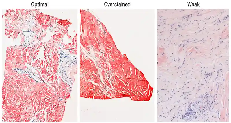
判读时首先检查玻片上组织对照(图 1)。理想情况下,应显示应有淀粉样物质处着色良好,并有阴性成分(例如未着色的血管)。
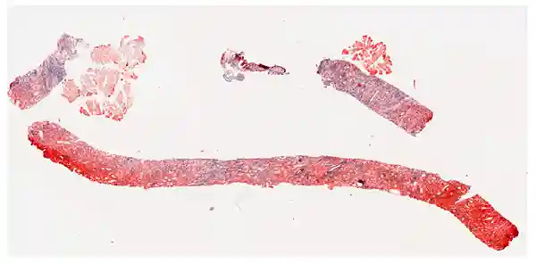
「并非 Congo red 上发红的一切都是淀粉样阳性,」Miller 医生谈到一周前的病例时说(图 2)。「这不是免疫组化,也不是 trichrome。Congo red 非常容易过度着色和非特异着色,所以不要只在 2× 下看一眼就说 Congo red 阳性,哪怕它看起来够红。」
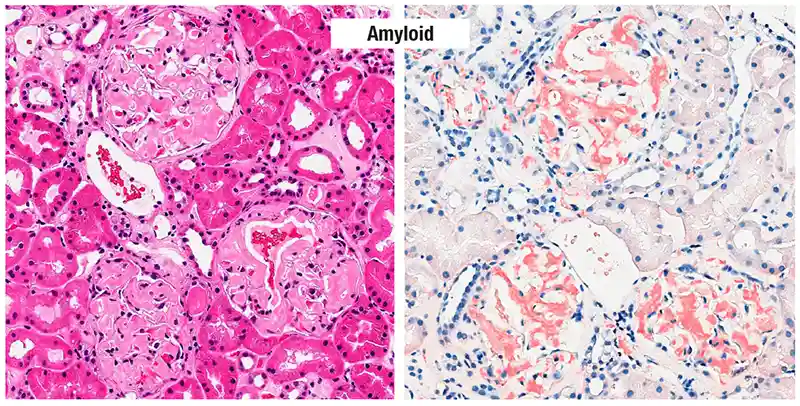
他总是从 H&E 开始。「对我来说,要叫淀粉样,它必须在 H&E 上看起来像淀粉样」——虽不充分。「我不能单凭 H&E 诊断淀粉样,但必须从这里开始。」典型肾肾小球可见粉红玻璃样、平滑或蜡样物质,其中不应嵌有细胞(图 3)。「如果 H&E 看起来可能是淀粉样,且 Congo red 打在可能是淀粉样的所有位置,我就相当怀疑了。几乎可以开口说看起来像淀粉样。」
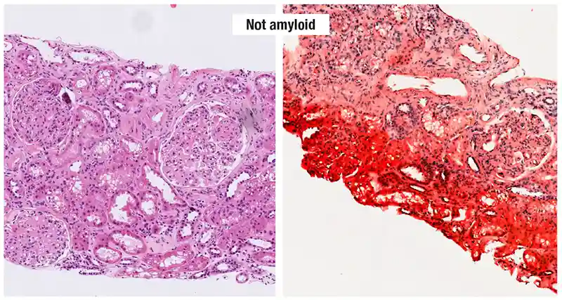
对比之下,糖尿病患者肾活检(图 4)在粉红物质处可见细胞成分与纤维化;高倍下纤维化区可见绳索样胶原或细纤维样质地,而非平滑玻璃样。「真正的淀粉样在 H&E 上不应看到纤维样质地。」若 Congo red 重度过度着色且与 H&E 上可能淀粉样处不匹配,则不可靠。血管壁亦常沉积淀粉样,但需与仅平滑肌/弹力组织的非特异着色区分。
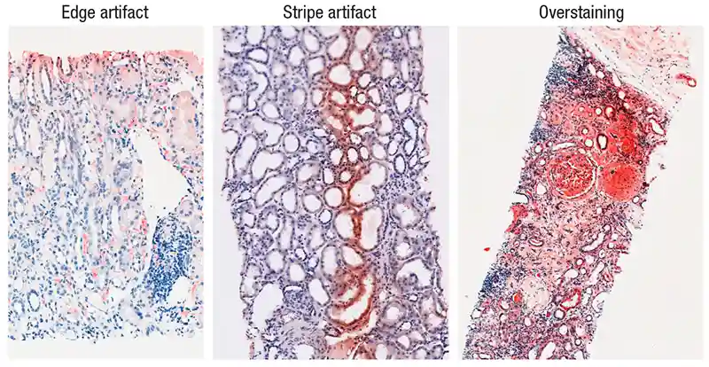
边缘与条带伪影、区域性重度过度着色很常见(图 5)。「至少在我的实验室,过度着色和伪影几乎成了常态,」Miller 医生说。他们用 Dako(Agilent)Artisan 自动染色机做 Congo red,「再也不会回到手工染色。」
非特异刚果红着色(congophilia)是陷阱:软骨样组织、红细胞、骨骼肌及平滑肌、血管弹力组织倾向非特异着色;致密胶原(蓝白双折射)和纤维蛋白也可非特异着 Congo red。「如果在血管看到 Congo red,看起来有点平滑无定形,要小心别只是弹力组织。」
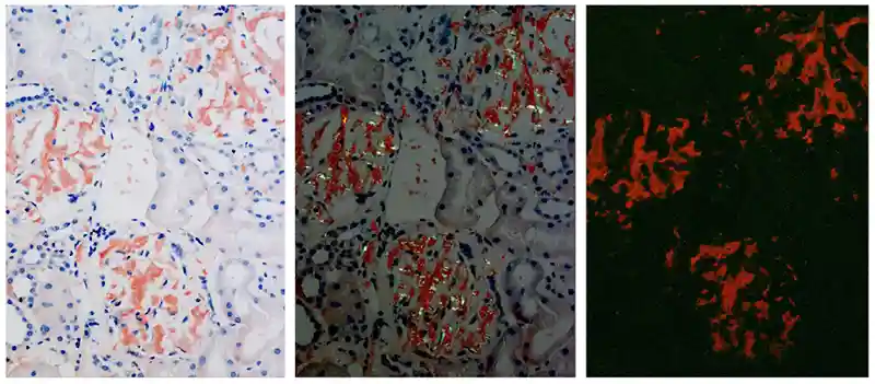
荧光有助于突显 Congo red 阳性(图 6)。「我们在医学院学的是苹果绿色双折射,」Miller 医生说。「我不太确定我见过多少苹果是偏振下那种颜色。对我来说,不太像 Granny Smith,更像 Golden Delicious 那种青苹果绿。」指南使用「特征性双折射」一词——「现在淀粉样命名中更推荐,因为它不止是绿色。」也可见偏橙的色调。「这些应特异,并与非偏振与偏振显微镜下 Congo red 所见相符。」
心脏与脂肪垫活检(Fyfe 医生)
Fyfe 医生说,心内科医生现已可凭扫描自行诊断 ATTR;「淀粉样是老龄病,患者带着 MGUS 和扫描结果来,扫描可能模棱两可。」心活检通常相当直截了当,「但现在有时更棘手。」
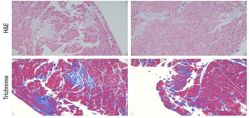
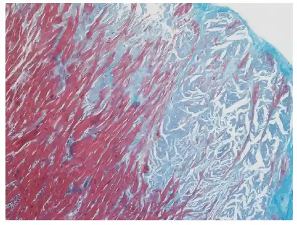
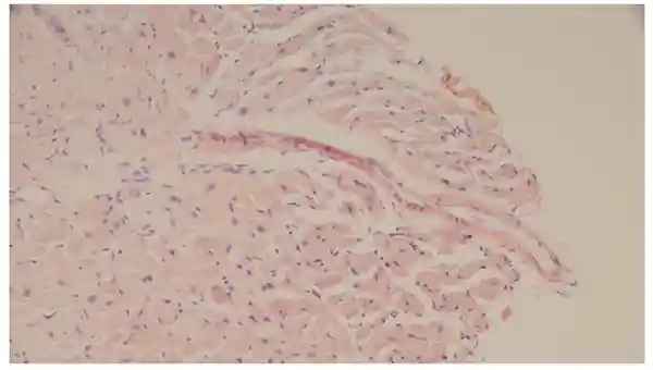
图 7 为一名 MGUS 患者心内膜心肌活检的 H&E(「肯定是淀粉样,或许细微」)与 trichrome。「有淀粉样变的心脏也可以有纤维化」;淀粉样破坏组织后瘢痕替代。trichrome 对淀粉样沉积应略显蓝灰(图 8)。
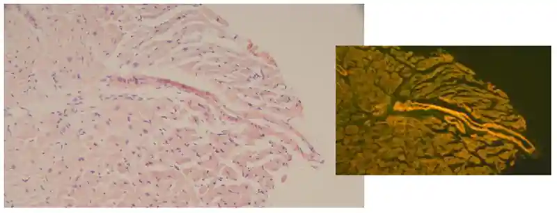
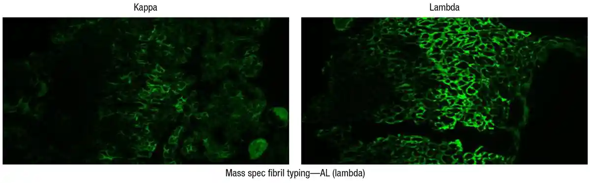
另一例 MGUS 患者 Congo red 在 H&E 上细微(图 9),但 TRITC 荧光显示强血管着色(图 10)。此类病例其机构会冻存组织做轻链免疫荧光。「目前我不只依赖免疫荧光做纤维分型;我也会送质谱。」图 11 为一例 λ 轻链 MGUS:免疫荧光 λ 阳性也有 κ 着色;质谱确认 AL(λ)淀粉样变。
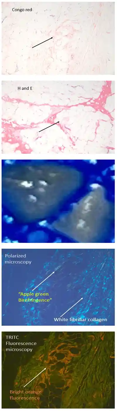
「脂肪垫是另一根刺,」Fyfe 医生说。图 12 中 Congo red 局灶阳性,偏振下可见白色纤维状胶原与双折射,荧光下明亮橙色——对她而言可诊断;患者后续确诊 AL 淀粉样变。
非靶向组织与局限性淀粉样变
指南未覆盖在非靶向组织中筛查淀粉样。「淀粉样变的韦恩图与其他疾病显著重叠,」Miller 医生说,「腕管综合征和腰椎管狭窄恰好是其中两个圆。」指南并不建议对每一例腕管或腰椎管狭窄标本都做 Congo red,但要有意识:若临床医生提出担心,「他们可能说得对,你大概应该做 Congo red。」
指南仅针对系统性淀粉样变;局限性可见于喉、皮肤、肠(损伤区)及罕见泌尿道。「细微之处在于:你可以有局限性淀粉样沉积,而其纤维类型是可以系统性的类型。例如肺的 amyloidoma 可能是 AL λ 型,但并不意味着患者有系统性淀粉样变。」「仅因你在组织活检中诊断了潜在系统性形式,并不意味着他们患有该病的系统形式。」
若患者反复向腹壁脂肪垫注射,某些药物/制剂可错误折叠并以与淀粉样沉积相同的方式沉积。「你可能在脂肪垫活检看到淀粉样,」Miller 医生说,「但若是注射胰岛素多年的糖尿病患者,要非常小心认识到这可能是局限性形式。」被问及 GLP-1 激动剂类注射是否可见类似沉积时,他说「有可能,但大概还需要更多时间演化。」
治疗背景(简述)
AL 治疗包括浆细胞/免疫导向:daratumumab(抗 CD38)、环磷酰胺、地塞米松、belantamab mafodotin(ADC);蛋白酶体抑制剂(硼替佐米);自体干细胞移植;以及结合纤维表位导致巨噬细胞活化与溶解的较新抗淀粉样抗体(CAEL-101、birtamimab、AT-02 等)。
ATTR 的导向治疗截然不同,Fyfe 医生说。「转甲状腺素蛋白在肝脏制造,我们可以尝试用 RNA 沉默抑制其产生。」tafamidis 为 FDA 批准的稳定剂,阻止 TTR 四聚体解离。与轻链类似,亦在探索播种抑制剂与抗体以打散淀粉样纤维。
Amy Carpenter 为 CAP TODAY 高级编辑。
- After the CAP amyloidosis guideline (Arch Pathol Lab Med. 2026;150:179–193), co-chairs shared practical tips for Congo red interpretation and fibril typing at CAP25.
- Fibril type drives therapy; mass spectrometry is the gold standard on paraffin tissue; immunofluorescence often works in renal amyloid, with reflex to MS when equivocal.
- Start with H&E that looks like amyloid; Congo red overstains easily; TRITC/Texas red fluorescence can increase sensitivity.
Amyloidosis has become a treatable disease—if the fibril type is identified accurately and quickly.
That and the variability seen in its workup and diagnosis were the impetus for the recently published CAP amyloidosis guideline (Miller DV, et al. Arch Pathol Lab Med. 2026;150[3]:179–193).
The co-chairs of the guideline expert panel provided, in a CAP25 session, what they described as their own tips and tricks for amyloid detection and fibril typing, reported here.
There are at least 42 amyloid subtypes, said panel co-chair Billie S. Fyfe, MD, professor in the Department of Pathology and Laboratory Medicine at Rutgers Health Robert Wood Johnson Medical School. “And with mass spectrometry, more amyloid types are being identified each year.”
The fibril type dictates the treatment, said co-chair Dylan V. Miller, MD, professor of pathology at the University of Utah School of Medicine and chair of the CAP Immunohistochemistry Committee. “In my mind, that sounds a lot like a predictive marker test,” he said, “like PD-L1 and HER2 and others.”
If left untreated, 25 percent of patients with immunoglobulin light chain (AL) amyloidosis die within six months of diagnosis, and a similar percentage with amyloid transthyretin (ATTR) amyloidosis die within 24 months of diagnosis. All forms of amyloidosis are underdiagnosed (Gertz MA, et al. JAMA. 2020;324[1]:79–89).
Guideline recommendations (“guideposts”)
Recommendation 1. In patients with suspected systemic amyloidosis, pathologists may screen cytology specimens (conventional smears and/or cell blocks) of aspirated abdominal fat for detection of amyloid. Individual laboratories should determine and optimize the best preparation methods and ancillary testing techniques should be validated on cytologic material. If cytologic smears only are prepared in the absence of a cell block, the ability for further testing, including subtyping, is limited.
There are data to support screening of cytology specimens by conventional smears or cell blocks, Dr. Miller said. “If you’re not using a tissue-based method, you should be sure the method you’re using works for detecting amyloid.” If using an air-dried smear, “you should know when you do Congo red on that smear you are able to detect amyloid in your lab.” With a limited sample like an air-dried smear, fibril typing will not be able to be performed. “There may be a need to get more tissue for doing that important part of the diagnostic workup.”
Recommendation 2. When evaluating specimens for the presence of amyloid, pathologists should use the Congo red staining method. Other methods can be used but should be validated against Congo red or electron microscopy and must show equivalency.
Recommendation 3. When assessing Congo red histochemistry, pathologists may add fluorescence microscopy with the tetramethylrhodamine isothiocyanate/Texas red filter to increase sensitivity for amyloid detection. “You don’t need to buy a fluorescence scope to do this,” Dr. Miller said, “but if you have one, this is a helpful thing you can do to improve sensitivity of your Congo red interpretation.”
Recommendation 4. In patients with amyloidosis for whom therapy is being considered, mass spectrometry should be used to identify the fibril protein type in order to optimize diagnostic yield and the use of tissue. In renal amyloidosis, amyloid fibril typing may often be accomplished successfully by immunofluorescence, though reflex to MS-based proteomics should be performed in difficult or equivocal cases.
Good practice statements
- When specimens are received for detection of systemic amyloidosis, pathologists should evaluate for the presence of amyloid using validated method(s), and the validated method(s) used should be identified in the pathology report. For a fat pad or other surrogate biopsy that is negative, “be very clear that that doesn’t exclude the possibility of amyloid in a target organ or elsewhere in the body.”
- If a clinical concern for amyloidosis persists after a negative biopsy from a surrogate site but the potentially affected organ (or organs) was not sampled, then suggesting a biopsy of the potentially affected organ(s) and/or recommending suitable archived specimen(s) to evaluate is appropriate.
- In patients with amyloidosis who are being considered for therapy, pathologists should determine the fibril protein type.
IHC can be effective for fibril identification, but panels are only as good as optimization of each assay; it is a “closed-ended exercise” versus mass spectrometry. Immunoelectron microscopy combines confirming definitive amyloid fibrils with gold-labeled monoclonal antibodies. Mass spectrometry on paraffin tissue is the gold standard, with very high sensitivity and specificity, detecting the amyloid type regardless of fibril peptide—and even multiple overlapping types.
Practical tips for slide reading
Check the on-slide tissue control (Fig. 1). Ideally it should show good staining of what is supposed to be amyloid and have a negative component.
“Not everything that’s red on a Congo red is positive for amyloid,” Dr. Miller said (Fig. 2). “This is not immunohistochemistry, it’s not a trichrome. And Congo red tends to be very prone to overstaining and nonspecific staining, so do not just look at a slide at 2× and say it is Congo red positive even though it’s pretty red.”
He always begins with the H&E. For him to call something amyloid, it has to look like amyloid on the H&E—though that is not sufficient. Prototypical renal glomeruli show pink, glasslike, smooth or waxy material without cells embedded (Fig. 3). “So if the H&E looks like it could be amyloid, and the Congo red is hitting everywhere where it could be amyloid, then I’m pretty suspicious.”
By contrast, in diabetes (Fig. 4), cellularity and fibrosis are visible where there is pink material; fibrotic areas show ropy collagen or fine fibrillar texture rather than smooth glass. “You shouldn’t see any fibrillar material or texture on the H&E in true amyloid.” Heavy overstaining that does not match H&E candidates is unreliable. Amyloid loves vessel walls, but nonspecific staining of smooth muscle/elastic must be excluded.
Edge and stripe artifacts and heavy regional overstaining are common (Fig. 5). “Overstaining and artifacts are almost the rule now, at least in my lab,” Dr. Miller said. They use the Dako (Agilent) Artisan autostainer for Congo red and are “never going back to hand-staining.”
Nonspecific congophilia is a pitfall: chondroid, erythrocytes, skeletal and smooth muscle, and elastic tissue in vessels; dense collagen (blue-white birefringence) and fibrin can also pick up Congo red nonspecifically. “Be careful if you’re seeing Congo red in vessels, and it looks a little smooth and amorphous, that it’s not just elastic tissue.”
Fluorescence can highlight Congo red positivity (Fig. 6). The guideline prefers “characteristic birefringence” rather than only “apple green”; orangish hues can be seen and should match nonpolarized and polarized Congo red findings.
Heart and fat pad biopsies (Dr. Fyfe)
Cardiologists are now diagnosing ATTR on the basis of scans; patients may present with MGUS and an equivocal scan. Heart biopsies are characteristically fairly straightforward but “are sometimes now a bit trickier.”
Fig. 7 shows H&E (“definitely amyloid, maybe subtle”) and trichrome of an endomyocardial biopsy from a patient with MGUS. Hearts with amyloidosis can also have fibrosis; trichrome for amyloid deposits is supposed to look a little bluish-gray (Fig. 8).
Another MGUS case shows subtle Congo red on H&E (Fig. 9) but strong vascular staining on TRITC fluorescence (Fig. 10). Her institution freezes tissue for light-chain immunofluorescence; she also sends mass spectrometry. Fig. 11: lambda MGUS with lambda IF positive and also kappa staining; MS confirmed AL (lambda) amyloidosis.
“Fat pads are another thorn in people’s side,” Dr. Fyfe said. In Fig. 12 Congo red is focally positive, with birefringence and bright orange fluorescence—“So it was diagnostic to me”; the patient went on to AL amyloidosis.
Nontargeted tissues and localized amyloid
The guideline does not suggest staining every carpal tunnel or lumbar stenosis specimen with Congo red, but awareness matters when clinicians raise concern.
The guideline addresses only systemic amyloidosis; localized amyloidosis is seen in larynx, skin, bowel (injury), and rarely urinary tract. A potentially systemic fibril type in a biopsy does not always mean systemic disease (e.g., pulmonary amyloidoma AL lambda). Repeated abdominal fat injections (e.g., long-term insulin) can produce localized amyloid-like deposits. GLP-1 agonist–type injections: “possible but probably needs more time to evolve.”
Therapies (context)
AL therapies include plasma cell– and immune-directed agents (daratumumab, cyclophosphamide, dexamethasone, belantamab mafodotin; proteasome inhibitors; ASCT; newer anti-amyloid antibodies). ATTR therapies differ (RNA silencing of hepatic TTR production; tafamidis stabilizer; seeding inhibitors and antibodies under study).
Amy Carpenter is CAP TODAY senior editor.