- YAP1 融合阳性幕上室管膜瘤:边界清楚的幕上胶质瘤,室管膜形态 + 室管膜免疫表型(GFAP+、Olig2−、EMA+),YAP1 重排(最常见伙伴 MAMLD1);女性 75%,中位年龄 11 个月,5 年 PFS 66%,预后优于 ZFTA 融合型。
- 形态线索:血管周围假菊形团、嗜酸性颗粒小体(PAS+)、钙化;L1-CAM 与 p65 阴性有助于与 ZFTA 融合型区分;按形态可分 CNS WHO 2 或 3 级,但分级与预后关系弱。
- 专题含影像/镜下/免疫组化共 21 幅图、报告示例、6 项鉴别诊断(ZFTA 融合型、血管中心性胶质瘤、MN1 改变星形母细胞瘤、BCOR ITD 等)及 2 道练习题。
- 同日教科书更新另含高级别滤泡细胞起源的非间变性甲状腺癌(细胞学)、BCOR 与 TRPS1 免疫组化、胃双分泌样癌 4 个专题摘要。
收录范围:PathologyOutlines 2026-10-08 博客「Textbook Updates」全文(5 个专题摘要与精选图片),以及其中「YAP1 融合阳性幕上室管膜瘤」专题页全文(全部栏目、21 幅站内图像及图注、报告示例、鉴别诊断、练习题与答案);托管于外站的电镜与 FISH 图仅保留链接。其余 4 个专题仅收博客摘要,正文见各专题链接。
教科书更新 · 2026 年 10 月 8 日
我们已更新以下专题:
CNS 与垂体肿瘤 > 胶质瘤、胶质神经元肿瘤和神经元肿瘤 > 室管膜肿瘤 > YAP1 融合阳性幕上室管膜瘤
作者:Alberto Pietrantoni, M.D., Valeria Barresi, M.D., Ph.D.
专题摘要: YAP1 融合阳性幕上室管膜瘤是一种边界清楚的幕上胶质瘤,呈室管膜形态,并具有涉及 YAP1 基因的特征性融合。常为累及多个脑叶或整个大脑半球的巨大肿块。女性患者更常见(75%);> 60% 的患者年龄小于 4 岁。5 年无进展生存率:66%;预后优于 ZFTA 融合阳性幕上室管膜瘤。治疗:在最大限度保证安全的前提下,尽可能完整地手术切除。
细胞病理学 > 甲状腺 > 高级别滤泡细胞起源的非间变性甲状腺癌
作者:Asma Arshia, M.B.B.S., Heather I-Hsuan Chen-Yost, M.D.
专题摘要: 高级别滤泡细胞起源的非间变性甲状腺癌(HGFCTC)包括低分化甲状腺癌(PDTC)和高级别分化型甲状腺癌(HGDTC),这些甲状腺恶性肿瘤的特征是核分裂活性增高或肿瘤坏死,而不具有间变性细胞学特征。表现为在数月内迅速增大的、质地坚实的巨大甲状腺结节。手术和放射性碘为一线治疗;然而,PDTC 和 HGDTC 常对放射性碘耐受。
染色(免疫组织化学与特殊染色)> BCOR
作者:Nat Pernick, M.D.
专题摘要: BCOR 免疫组织化学(IHC)可检测具有 BCOR 遗传改变的肿瘤中 BCOR 蛋白的过表达,这些改变包括 BCOR 基因融合和 BCOR 内部串联重复。阳性染色:强而弥漫的细胞核反应。对于具有 BCOR 遗传异常的肉瘤以及肾透明细胞肉瘤,该标志物具有很高的敏感性,但由于滑膜肉瘤和其他某些肿瘤类型也可呈阳性,其特异性受到限制。可作为筛查工具,识别可能受益于确证性分子检测的肿瘤。
染色(免疫组织化学与特殊染色)> TRPS1
作者:Di (Andy) Ai, M.D., Ph.D., Qingqing Ding, M.D., Ph.D.
专题摘要: TRPS1(毛发-鼻-指(趾)骨综合征 1 型)是一种 GATA 家族锌指转录因子,参与乳腺上皮细胞生长、乳腺癌发生和细胞存活。呈细胞核染色。用于诊断 ER 阳性乳腺癌(> 95%)、HER2 阳性乳腺癌(> 90%)及三阴性乳腺癌(> 90%,包括化生性与非化生性三阴性乳腺癌)。在免疫组织化学组合中,与广谱细胞角蛋白、ER 和 GATA3 联合用于诊断乳腺来源的转移性癌。
胃 > 癌 > 双分泌样癌
作者:Ihsan Baroudi, M.D., Raul S. Gonzalez, M.D.
专题摘要: 一类罕见的单相性肿瘤,其特征是肿瘤内同时存在神经内分泌与非神经内分泌(外分泌)上皮分化。双向分化可存在于单个肿瘤细胞内,也可表现为形态相似的肿瘤细胞紧密混合。此类肿瘤罕见,大多数资料来自单个病例报告。病例就诊时常已处于晚期,预后似乎不佳。手术切除是主要治疗方式,可考虑辅助化疗或放疗。
精选图片
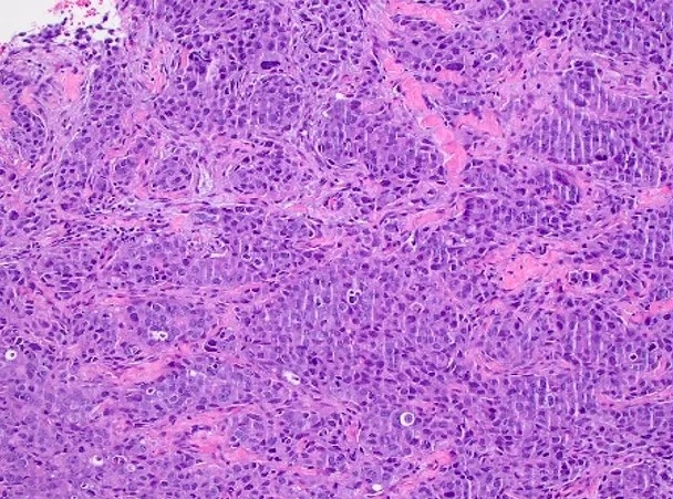
专题全文:YAP1 融合阳性幕上室管膜瘤
作者:Alberto Pietrantoni, M.D., Valeria Barresi, M.D., Ph.D. · 编委:Jared T. Ahrendsen, M.D., Ph.D. · 副主编:Chunyu Cai, M.D., Ph.D. · 作者最近更新:2026 年 10 月 1 日 · 工作人员最近更新:2026 年 10 月 1 日
定义 / 概述
- YAP1 融合阳性幕上室管膜瘤是一种边界清楚的幕上胶质瘤,呈室管膜形态,并具有涉及 YAP1 基因的特征性融合
要点
- 按定义位于幕上
- 具有血管周围假菊形团或室管膜管的胶质肿瘤,具有室管膜免疫表型(GFAP+、Olig2-、EMA+)
- YAP1 基因重排
- 可根据形态学分为 CNS WHO 2 级或 3 级
ICD 编码
- ICD-O: 9396/3 - YAP1 融合阳性幕上室管膜瘤
- ICD-11: 2A00.0Y & XH1511 - 其他特指的脑胶质瘤与室管膜瘤,非特指型
流行病学
- 女性患者(75%)比男性患者更常见(Cancer Cell 2015;27:728)
- 中位年龄:11 个月(范围:1 个月至 51 岁)(Cancer Cell 2015;27:728, Clin Neuroradiol 2024;34:939)
- 超过 60% 的患者年龄小于 4 岁(Cancer Cell 2015;27:728)
- YAP1 融合见于 10 - 11% 的幕上室管膜瘤(Cancer Cell 2015;27:728)
部位
- 按定义累及幕上区域(Brain Pathol 2019;29:205)
- 常为累及多个脑叶或整个大脑半球的巨大肿块(Clin Neuroradiol 2024;34:939)
- YAP1 融合也曾在罕见的后颅窝或脊髓区域室管膜瘤中报道(Acta Neuropathol Commun 2024;12:158, Brain Pathol 2022;32:e13020)
病理生理
- Yes 相关蛋白 1(YAP1)基因重排是主要的驱动改变
- YAP1 是 Hippo 通路的转录共激活因子,调控组织稳态、细胞命运和增殖(Nat Commun 2019;10:3914)
- YAP1 是一种原癌基因,通过与 TEAD 转录因子家族及核因子 I 相互作用发挥功能(Nat Commun 2019;10:3914)
- YAP1 过表达可在动物模型中导致室管膜瘤样肿瘤的形成(Nat Commun 2020;11:2380)
- CDKN2A / CDKN2B 纯合性缺失及拷贝数变异通常不存在(Cancer Cell 2015;27:728)
病因
- 不明
临床特征
- 幕上占位性病变的常见表现(Brain Sci 2021;11:301, Ann Transl Med 2017;5:269)
- 颅内压增高
- 头痛
- 癫痫发作
- 局灶性神经功能缺损
- 行为改变
诊断
- 结合肿瘤部位、组织学和免疫组织化学结果,以及 YAP1 融合的证据(Neuro Oncol 2021;23:1231)
- WHO CNS 2021 规定的必需诊断标准
- 具有室管膜瘤形态学和免疫组织化学特征的幕上肿瘤
- 基因融合涉及 YAP1
- 理想诊断标准
- DNA 甲基化谱符合 YAP1 融合阳性幕上室管膜瘤
- p65(RELA)或 L1-CAM 均无免疫反应性
- PAS 阳性的嗜酸性颗粒小体
- 参考文献:Neuro Oncol 2021;23:1231
影像学描述
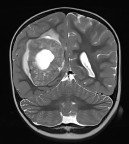
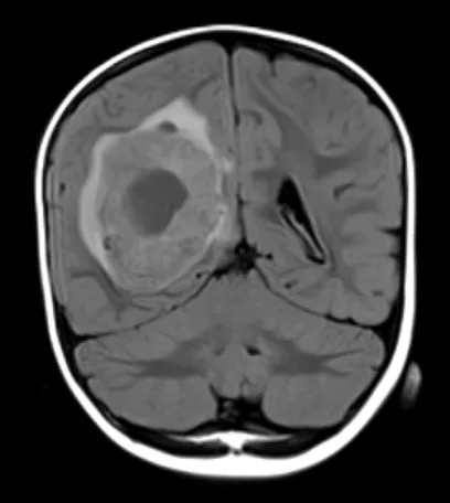
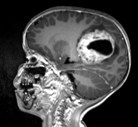
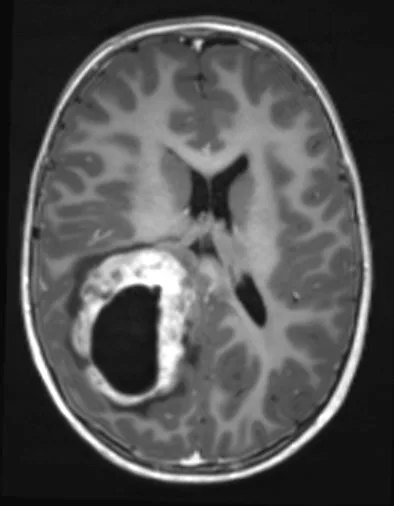
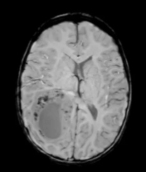
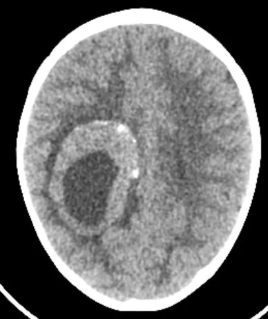
- MRI 表现(Brain Pathol 2019;29:205, Clin Neuroradiol 2024;34:939)
- 囊实性占位性病变
- 位于脑实质内、脑室内或脑室旁
- 实性成分可呈多结节状并伴出血
- 在 T1 和 T2 加权序列上多呈等信号
- 瘤周水肿程度不一
- 可见钙化,CT 显示更清楚
- 不均匀强化
影像学图像
供图:Fabio Martino Doniselli, M.D., Ph.D.
预后因素
- 5 年总生存率:100%(Cancer Cell 2015;27:728)
- 5 年无进展生存率:66%(Cancer Cell 2015;27:728)
- 预后优于 ZFTA 融合阳性幕上室管膜瘤 (Cancer Cell 2015;27:728)
- 大体全切或近全切有利于预后(Neurosurgery 2019;85:41)
- 组织学 WHO 分级(见镜下描述)与预后的相关性较弱或无相关性(J Negat Results Biomed 2011;10:7, Cancer Cell 2015;27:728)
病例报告与病例系列
- 一名有左侧额顶叶肿块的女婴(BMJ Case Rep 2024;17:e261022)
- 15 例诊断时年龄小于 3 岁的儿童患者病例系列(Brain Pathol 2019;29:205)
- 一名 26 岁男性,左额叶肿块(Neuropathology 2021;41:133)
- 一名 35 岁男性,左额叶肿块(Brain Tumor Pathol 2022;39:240)
治疗
- 在最大限度保证安全的前提下,尽可能完整地手术切除(Neuro Oncol 2018;20:445)
- 根据患者年龄及有无肿瘤残留,术后行适形放射治疗(Neuro Oncol 2018;20:445)
- 化疗仍处于研究阶段;当无法进一步手术和放疗时,曾将化疗作为唯一治疗用于儿童及成人复发性室管膜瘤患者(Neuro Oncol 2018;20:445)
大体描述
- 边界清楚的病变
- 可见坏死和钙化区域
冰冻切片描述
- 典型室管膜瘤特征(J Neurosurg Spine 2018;30:133)
- 呈纤维性胶质细胞样外观的细胞增生,细胞形态从单一到中度多形性不等
- 常见血管周围假菊形团
- 可见坏死、微血管增生和核分裂活性
镜下(组织学)描述
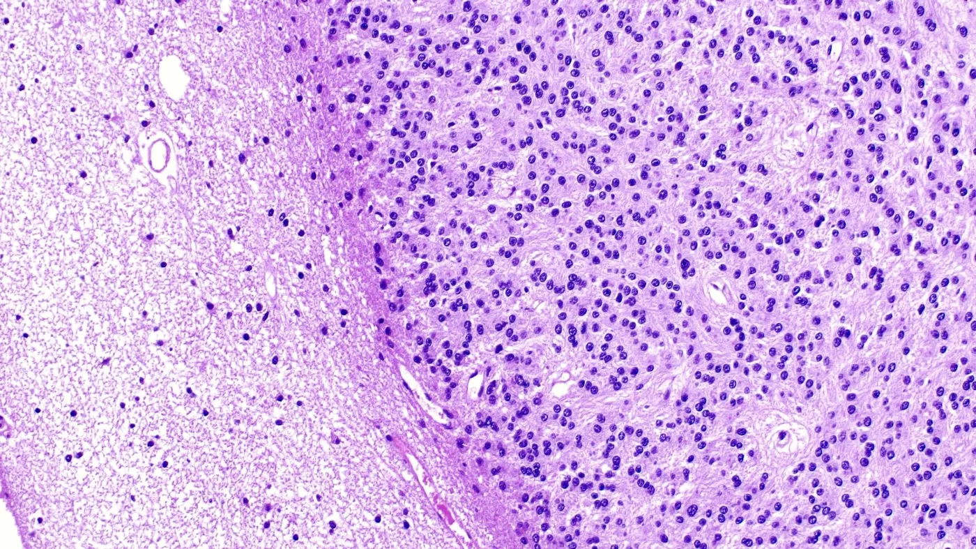
- 与周围脑实质界限清楚,但镜下可见浸润(Brain Pathol 2019;29:205)
- 细胞密度中等或较高(Brain Pathol 2019;29:205)
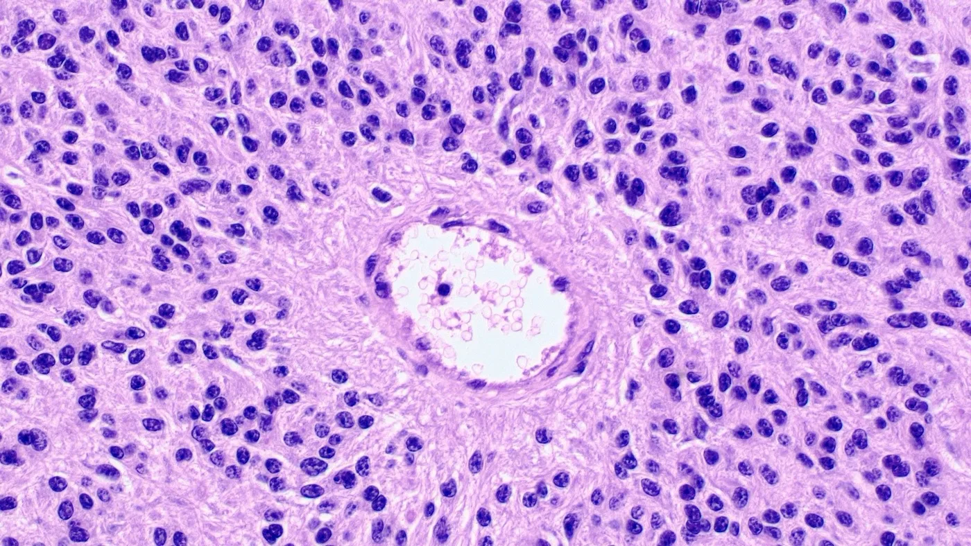
- 血管周围假菊形团是常见表现(Brain Pathol 2019;29:205)
- 真性室管膜菊形团鲜有报道(Brain Pathol 2019;29:205)
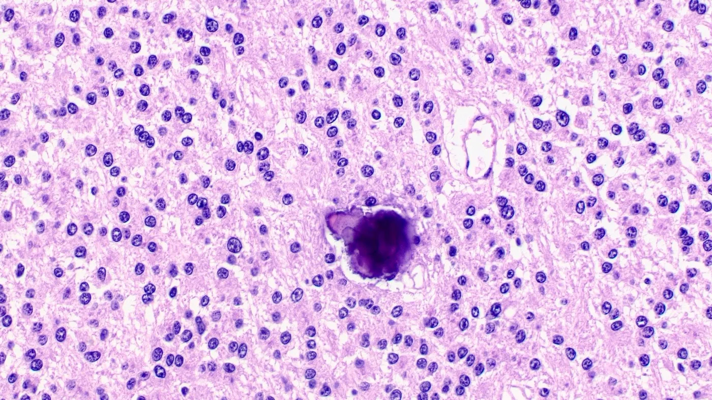
- 细胞核小至中等大小,形态单一,呈圆形或多边形,染色质致密(Brain Pathol 2019;29:205)
- 细胞质嗜酸性,细胞边界多不清楚;局部可见边界清楚、呈颗粒状的嗜酸性细胞质(Brain Pathol 2019;29:205)
- 偶见小簇小型肥胖细胞样细胞(Brain Pathol 2019;29:205)
- 可见嗜酸性颗粒小体和钙化(Brain Pathol 2019;29:205)
- 核分裂指数不一(Brain Pathol 2019;29:205)
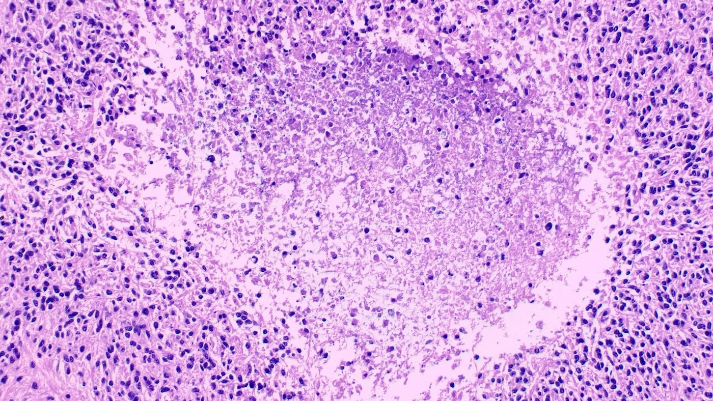
- 可见明显的核多形性、微血管增生和坏死(Brain Pathol 2019;29:205)
- 分级
- CNS WHO 2 级或 3 级(无标准化分级系统)
- 提示 3 级的特征:核分裂活跃、坏死、微血管增生、间变
- 分级与预后的相关性存在争议(J Negat Results Biomed 2011;10:7, Cancer Cell 2015;27:728)
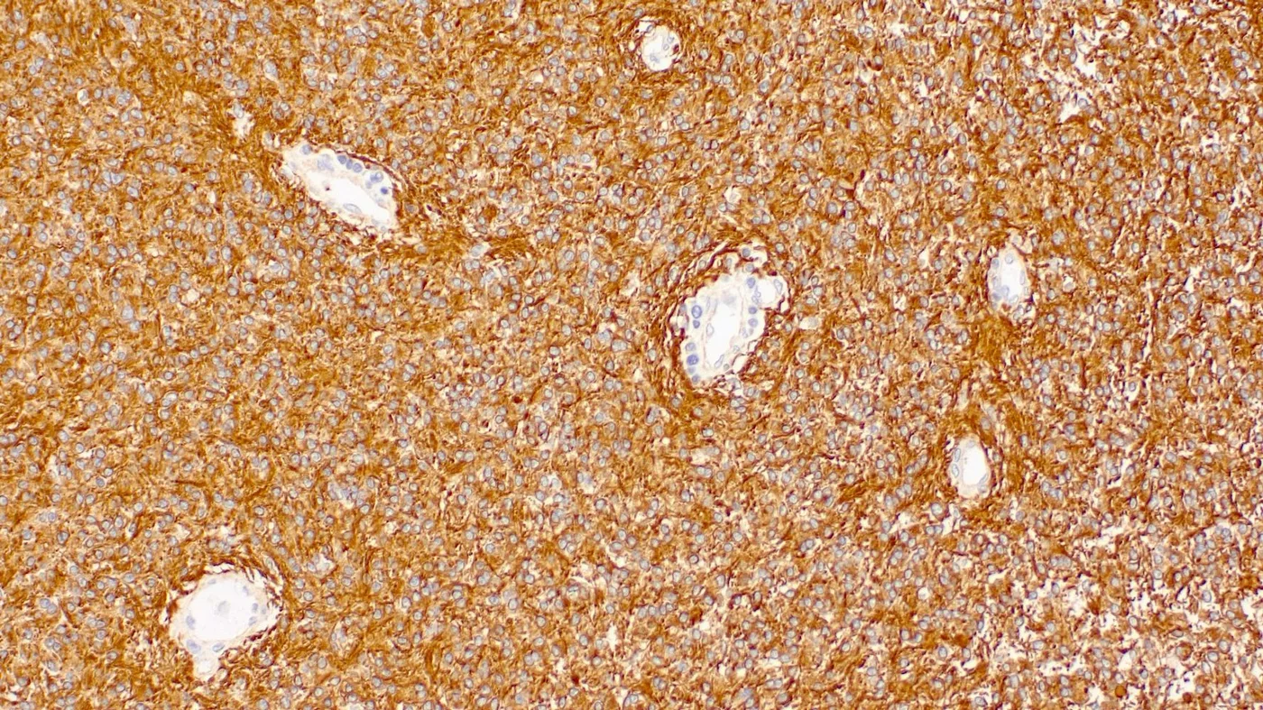
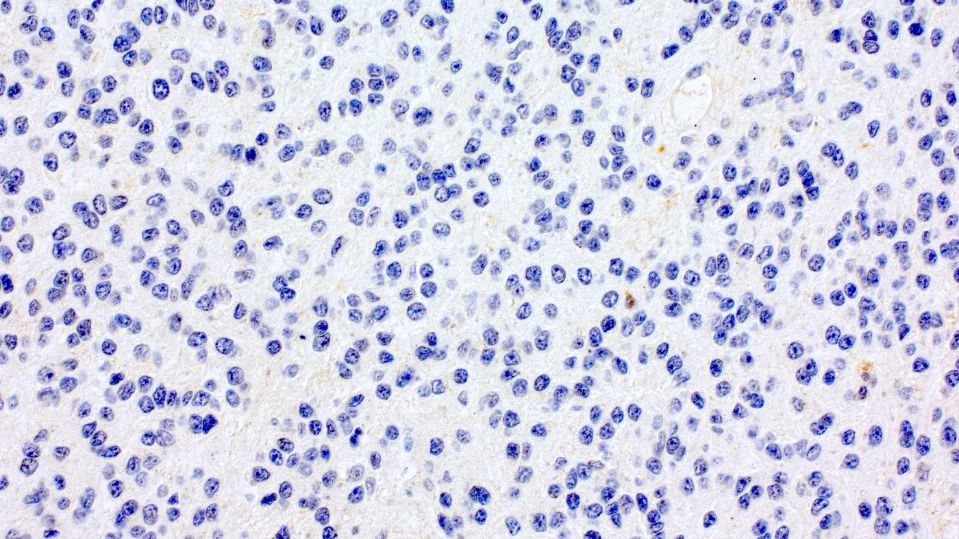
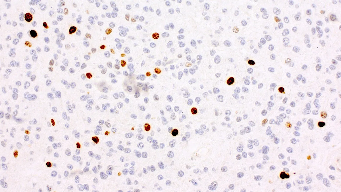
镜下(组织学)图像
供图:Alberto Pietrantoni, M.D.
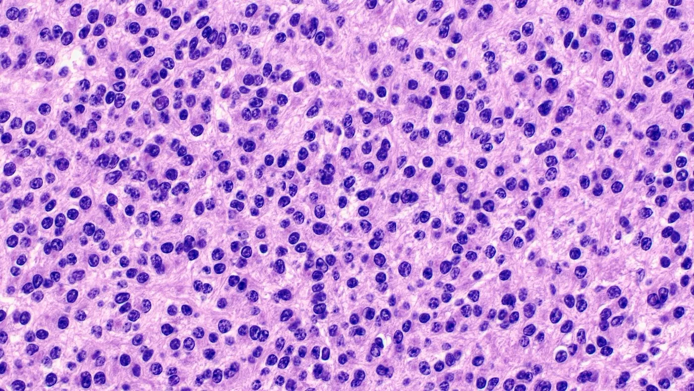
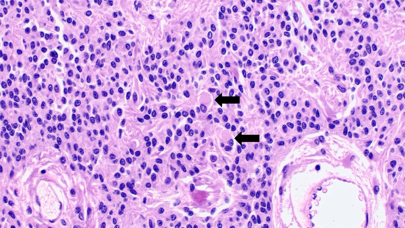
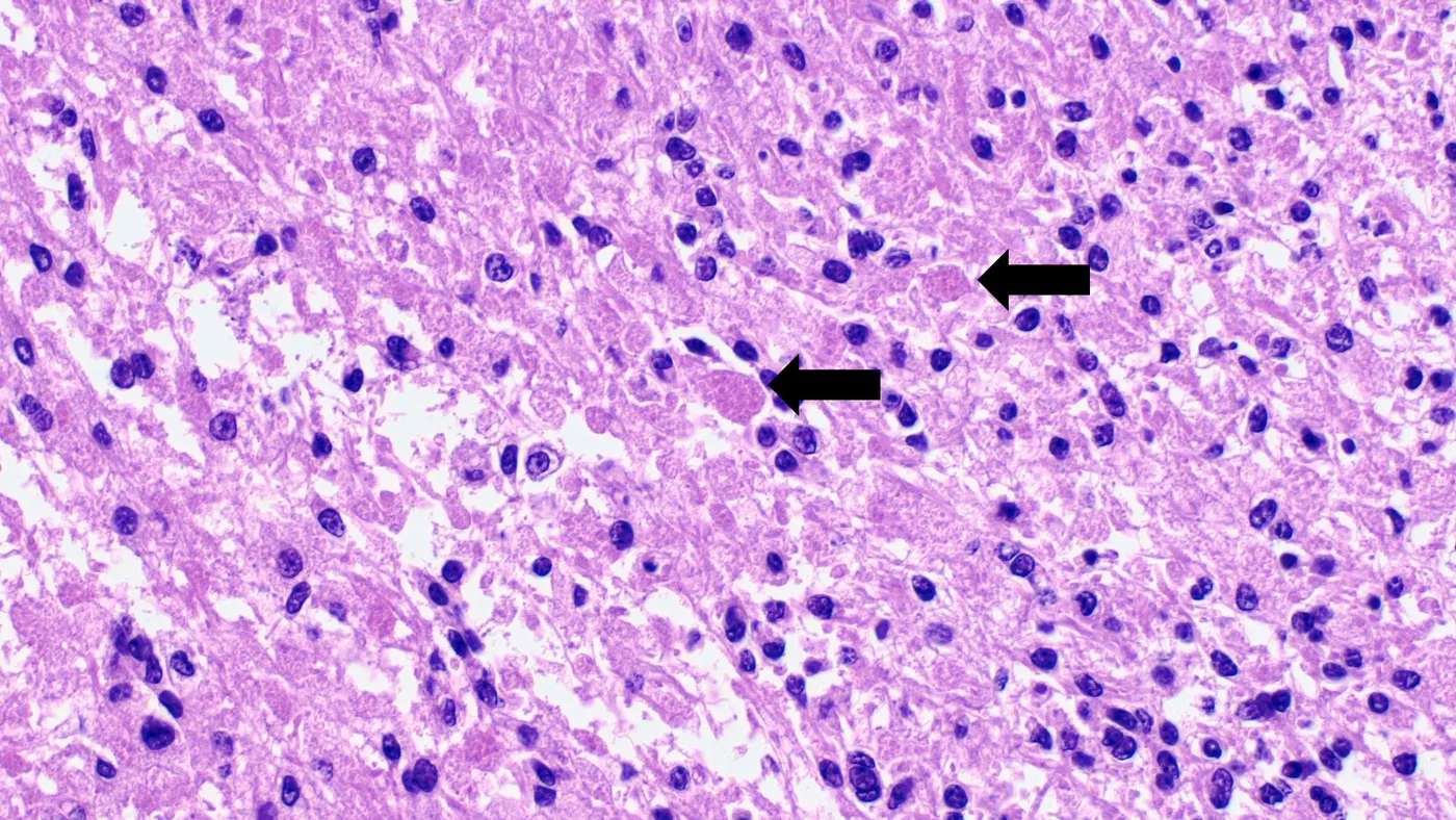
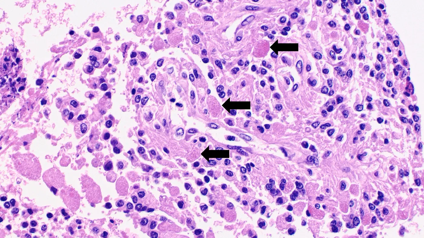
细胞学描述
- 脑脊液细胞学检查通常用于分期
- 术中检查时制作涂片
- 细胞学表现(Cytopathology 2024;35:556)
- 具有粗大细胞质突起的纤维性胶质细胞
- 细胞核呈圆形,形态从单一到中度多形性不等,染色质呈盐和胡椒状
- 术中涂片显示细胞量中等,常见细胞围绕血管聚集(血管周围假菊形团)
- 真性室管膜菊形团少见
- 血管有时发生玻璃样变
阳性染色
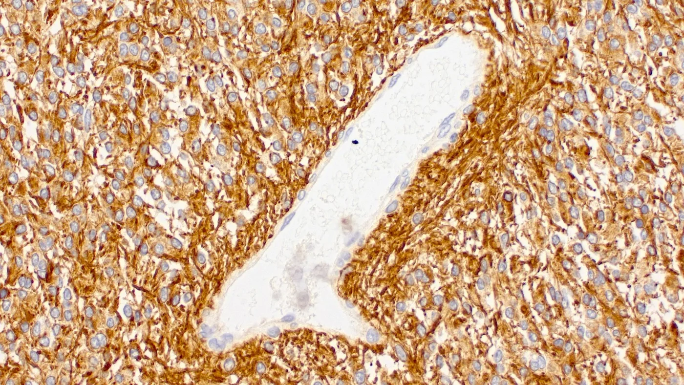
- GFAP:强度不一;血管周围假菊形团内表达更强(Brain Pathol 2019;29:205)
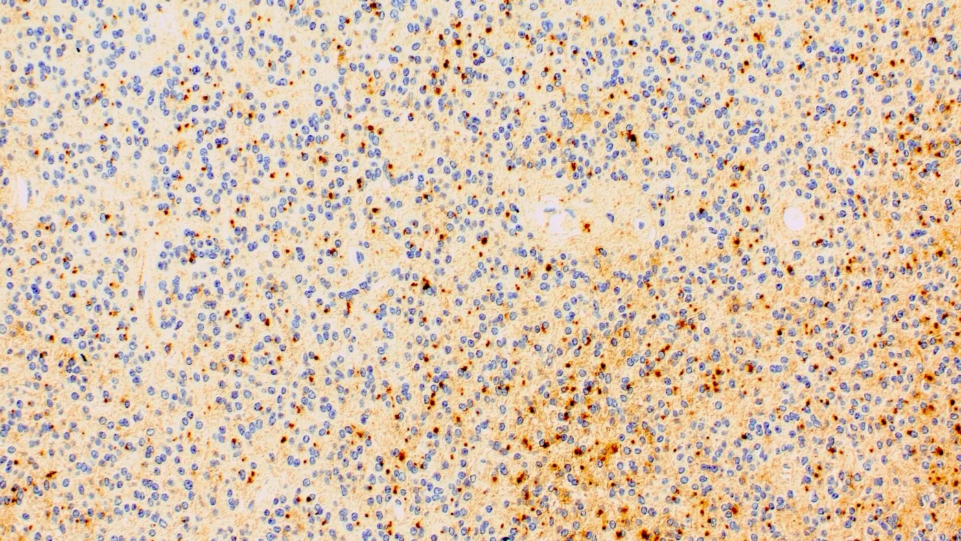
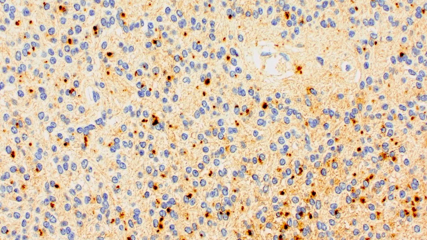
- EMA:细胞质核旁点状或环状阳性(Brain Pathol 2019;29:205)
- MAP2:表达程度不一(Brain Pathol 2019;29:205)
- S100, CD56, 平足蛋白(podoplanin,D2-40):阳性,但无特异性(Clin Neuropathol 2009;28:373)
- 突触素, 细胞角蛋白:散在细胞可呈阳性;少数病例中表达更广泛(Appl Immunohistochem Mol Morphol 2000;8:25)
- PAS:嗜酸性颗粒小体阳性(Brain Pathol 2019;29:205)
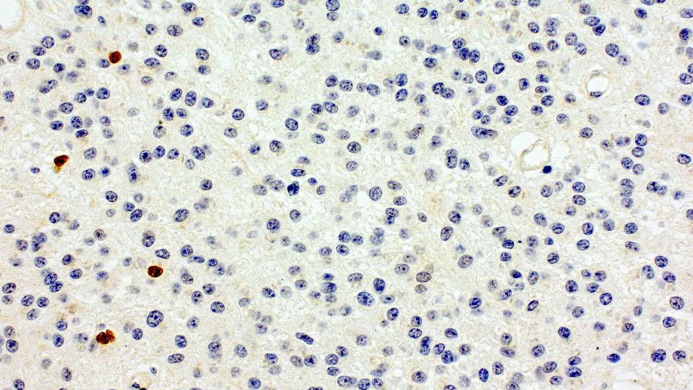
- Ki67 标记指数:不一(Brain Pathol 2019;29:205)
阴性染色
- Olig2:通常阴性;罕见的孤立细胞可呈阳性(Brain Pathol 2019;29:205)
- L1-CAM, p65 (Brain Pathol 2019;29:205)
- 神经丝 (Brain Pathol 2019;29:205)
电镜描述
- 超微结构检查发现发育不全的纤毛可证实室管膜分化(Brain Pathol 2019;29:205)
电镜图像
图像托管于其他服务器:
分子 / 细胞遗传学描述
- YAP1 重排,MAMLD1 为最常见的融合伙伴(Brain Pathol 2019;29:205, Cancer Cell 2015;27:728)
- 其他较少见的融合伙伴基因:MAML2, FAM118B (Cancer Cell 2015;27:728, Brain Tumor Pathol 2022;39:240)
- 已有病例报道存在 ZFTA::YAP1 融合,这不符合现行分类标准(Acta Neuropathol Commun 2026;14:183)
- 基因组相对稳定(拷贝数变异发生频率低)(Cancer Cell 2015;27:728)
- YAP1 融合阳性幕上室管膜瘤具有其特有的 DNA 甲基化谱(Cancer Cell 2015;27:728)
- 曾报道一例幕上室管膜瘤,具有 TEAD::NCOA2 融合而无 YAP1 重排,其 DNA 甲基化谱与 YAP1 融合阳性幕上室管膜瘤相同 (Acta Neuropathol 2025;149:14)
分子 / 细胞遗传学图像
图像托管于其他服务器:
病理报告示例
- 脑,颞叶,切除标本:
- YAP1 融合阳性幕上室管膜瘤,CNS WHO 3 级(见备注)
- 备注:胶质细胞呈边界清楚的增生,细胞核小而圆,可见血管周围假菊形团及散在嗜酸性颗粒小体。核分裂指数为 5 个核分裂象/mm2。可见坏死和微血管增生。肿瘤细胞表达 GFAP 和 EMA。Olig2 和 L1-CAM 阴性。热点区域 Ki67 增殖指数为 20%。FISH 分析检出 YAP1 基因重排。未发现 ZFTA 基因重排。
鉴别诊断
- ZFTA 融合阳性幕上室管膜瘤:
- 通常无嗜酸性颗粒小体
- L1-CAM+
- p65+,如果 ZFTA 与 RELA 融合
- 无 YAP1 重排
- ZFTA 重排
- 极罕见的幕上室管膜瘤病例具有 ZFTA::YAP1 融合(Acta Neuropathol Commun 2026;14:183)
- 血管中心性胶质瘤:
- 无增强后强化
- 弥漫性生长,可见合体样细胞和梭形细胞
- 无 YAP1 重排
- MYB 重排(最常见为 MYB::QKI 融合)
- MN1 改变的星形母细胞瘤:
- 星形母细胞性假菊形团,血管及细胞周围玻璃样变
- Olig2 至少局灶阳性
- 无 YAP1 重排
- MN1 重排
- 伴 BCOR 内部串联重复的 CNS 肿瘤:
- 形成菊形团的胶质神经元肿瘤:
- 伴多层菊形团的胚胎性肿瘤:
- 菊形团呈多层结构
- 胶质标志物多为阴性;神经毡样基质中的神经元标志物阳性
- LIN28A+
- 无 YAP1 重排
- 发生改变的基因为 C19MC 或 DICER1
其他参考文献
练习题 #1
上图所示为一名儿童患者的脑室内、边界清楚的幕上脑肿瘤中反复出现的特征。未观察到明显的核分裂活性、坏死或微血管增生。肿瘤与周围脑实质界限清楚。肿瘤 GFAP 和 EMA 阳性,Olig2 和 L1-CAM 阴性。此类肿瘤最可能发生以下哪一种基因的改变?
- CDKN2A / CDKN2B
- EWSR1
- STAT6
- YAP1
- ZFTA
练习题答案 #1
D.YAP1。图像显示形态大多单一的细胞围绕血管排列,并有血管周围无核区。这一特征结合所述免疫表型及临床资料,可能提示幕上室管膜瘤,此类肿瘤可出现 YAP1 或 ZFTA 基因重排。选项 E 错误,因为本例 L1-CAM 阴性,因此 ZFTA 重排的可能性较小。因此,YAP1 融合阳性幕上室管膜瘤是所给选项中最可能的诊断。选项 A 错误,因为 CDKN2A / CDKN2B 在室管膜瘤中可发生缺失,但在 YAP1 融合阳性室管膜瘤中通常保留。选项 B 和 C 错误,因为 EWSR1 和 STAT6 的改变见于间叶性肿瘤(分别如尤文肉瘤和孤立性纤维性肿瘤),这些肿瘤似乎与本病例不符。
参考文献:YAP1 融合阳性幕上室管膜瘤
练习题 #2
儿童幕上室管膜瘤应检测以下哪一种分子改变?
- BRAF 突变
- EGFR 扩增
- FGFR1 突变
- IDH 突变
- YAP1 融合
练习题答案 #2
E.YAP1 融合。YAP1 融合已在幕上室管膜瘤中报道,并据此定义了此类肿瘤的一个亚型。选项 A、B、C 和 D 错误,因为其他选项是其他 CNS 肿瘤的特征性分子改变:BRAF 和 FGFR1 突变见于多种低级别胶质瘤和胶质神经元肿瘤,EGFR 扩增见于高级别胶质瘤,包括胶质母细胞瘤,IDH 野生型;而 IDH1 / IDH2 突变见于 IDH 突变型星形细胞瘤和少突胶质细胞瘤。
参考文献:YAP1 融合阳性幕上室管膜瘤
- Supratentorial ependymoma, YAP1 fusion positive: circumscribed supratentorial glioma with ependymal morphology and immunophenotype (GFAP+, Olig2−, EMA+) and YAP1 rearrangement (most often with MAMLD1); 75% female, median age 11 months, 5-year PFS 66%, better prognosis than the ZFTA-fused type.
- Clues: perivascular pseudorosettes, PAS+ eosinophilic granular bodies, calcifications; L1-CAM and p65 negativity help separate it from ZFTA-fused ependymoma; CNS WHO grade 2 or 3 on morphology, with weak prognostic correlation.
- The topic includes 21 radiology/histology/IHC images, a sample report, six differentials and two practice questions.
- The same Textbook Updates post also summarises four other topics: high grade follicular cell derived nonanaplastic thyroid carcinoma (cytology), BCOR and TRPS1 IHC, and gastric amphicrine-like carcinoma.
Scope: the full PathologyOutlines Textbook Updates blog post of 8 October 2026 (five topic summaries and the featured image) and the complete topic page “Supratentorial ependymoma, YAP1 fusion positive” (all sections, 21 on-site images with legends, sample report, differential diagnosis, practice questions and answers). Externally hosted EM and FISH images are linked only. The other four topics are covered by their blog summaries.
Textbook Updates · 8 October 2026
We have posted updates of the following topics:
CNS & pituitary tumors > Gliomas, glioneuronal tumors and neuronal tumors > Ependymal tumors > Supratentorial ependymoma, YAP1 fusion positive
by Alberto Pietrantoni, M.D., Valeria Barresi, M.D., Ph.D.
Topic summary: Supratentorial ependymoma, YAP1 fusion positive is a circumscribed supratentorial glioma showing an ependymal morphology and a distinctive fusion involving the YAP1 gene. Frequently large mass involving multiple lobes or the entire hemisphere. More frequent in female patients (75%); > 60% of patients are younger than 4 years. 5 year progression free survival: 66%; better prognosis than supratentorial ependymoma, ZFTA fusion positive. Treatment: safest maximal surgical resection.
Cytopathology > Thyroid > High grade follicular cell derived nonanaplastic thyroid carcinoma
by Asma Arshia, M.B.B.S., Heather I-Hsuan Chen-Yost, M.D.
Topic summary: High grade follicular cell derived nonanaplastic thyroid carcinoma (HGFCTC) encompasses poorly differentiated thyroid carcinoma (PDTC) and high grade differentiated thyroid carcinoma (HGDTC), which are malignant thyroid neoplasms characterized by elevated mitotic activity or tumor necrosis without anaplastic cytologic features. Presents with a large, firm thyroid nodule that has grown rapidly over a period of months. Surgery and radioactive iodine are first line for treatment; however, PDTC and HGDTC are often radioactive iodine resistant.
Stains (IHC & special) > BCOR
by Nat Pernick, M.D.
Topic summary: BCOR immunohistochemistry (IHC) detects overexpression of the BCOR protein in tumors harboring BCOR genetic alterations, including BCOR gene fusions and BCOR internal tandem duplications. Positive staining: strong, diffuse nuclear reactivity. Highly sensitive marker for sarcomas with BCOR genetic abnormalities and clear cell sarcoma of the kidney, although specificity is limited by positivity in synovial sarcomas and some other tumor types. Screening tool to identify tumors that may benefit from confirmatory molecular testing.
Stains (IHC & special) > TRPS1
by Di (Andy) Ai, M.D., Ph.D., Qingqing Ding, M.D., Ph.D.
Topic summary: TRPS1 (trichorhinophalangeal syndrome type 1) is a GATA family zinc finger transcription factor involved in mammary epithelial cell growth, breast cancer development and cell survival. Nuclear staining. Used to diagnose ER positive breast cancer (> 95%), HER2 positive breast cancer (> 90%) and triple negative breast cancer (> 90%, including metaplastic and nonmetaplastic triple negative breast cancer). In immunohistochemical panel along with pancytokeratin, ER and GATA3 to diagnose metastatic carcinoma of breast origin.
Stomach > Carcinoma > Amphicrine-like carcinoma
by Ihsan Baroudi, M.D., Raul S. Gonzalez, M.D.
Topic summary: Category of rare monophasic neoplasms characterized by the coexistence of neuroendocrine and nonneuroendocrine (exocrine) epithelial differentiation within the tumor. Dual differentiation may be present either within individual tumor cells or as a tight, intimate admixture of morphologically similar tumor cells. Rare, with most data coming from single case reports. Cases often present at advanced stage and prognosis appears to be poor. Surgical resection is the main treatment, with consideration of adjuvant chemotherapy or radiotherapy.
Featured Image
Topic full text: Supratentorial ependymoma, YAP1 fusion positive
Authors: Alberto Pietrantoni, M.D., Valeria Barresi, M.D., Ph.D. · Editorial Board Member: Jared T. Ahrendsen, M.D., Ph.D. · Deputy Editor-in-Chief: Chunyu Cai, M.D., Ph.D. · Last author update: 1 October 2026 · Last staff update: 1 October 2026
Definition / general
- Supratentorial ependymoma, YAP1 fusion positive is a circumscribed supratentorial glioma showing an ependymal morphology and a distinctive fusion involving the YAP1 gene
Essential features
- Supratentorial by definition
- Glial neoplasm with perivascular pseudorosettes or ependymal canals, with an ependymal immunophenotype (GFAP+, Olig2-, EMA+)
- Rearrangement of the YAP1 gene
- It can be graded as WHO CNS grade 2 or 3 on morphological grounds
ICD coding
- ICD-O: 9396/3 - supratentorial ependymoma, YAP1 fusion positive
- ICD-11: 2A00.0Y & XH1511 - other specified gliomas of brain & ependymoma, NOS
Epidemiology
- More frequent in female patients (75%) than male patients (Cancer Cell 2015;27:728)
- Median age: 11 months (range: 1 month - 51 years) (Cancer Cell 2015;27:728, Clin Neuroradiol 2024;34:939)
- Over 60% of patients are younger than 4 years (Cancer Cell 2015;27:728)
- YAP1 fusion found in 10 - 11% of supratentorial ependymomas (Cancer Cell 2015;27:728)
Sites
- By definition it affects the supratentorial compartment (Brain Pathol 2019;29:205)
- Frequently large mass involving multiple lobes or the entire hemisphere (Clin Neuroradiol 2024;34:939)
- YAP1 fusion has also been reported in rare ependymomas of the posterior fossa or the spinal region (Acta Neuropathol Commun 2024;12:158, Brain Pathol 2022;32:e13020)
Pathophysiology
- Rearrangement of the yes associated protein 1 (YAP1) gene is the main driver alteration
- YAP1 is a Hippo pathway transcriptional coactivator that regulates tissue homeostasis, cell fate and proliferation (Nat Commun 2019;10:3914)
- YAP1 is a proto-oncogene and exerts its function through interaction with the family of TEAD transcription factors and nuclear factor I (Nat Commun 2019;10:3914)
- YAP1 overexpression leads to formation of ependymoma-like tumors in animal models (Nat Commun 2020;11:2380)
- CDKN2A / CDKN2B homozygous deletion and copy number variations are usually absent (Cancer Cell 2015;27:728)
Etiology
- Unknown
Clinical features
- Usual features of supratentorial mass forming lesions (Brain Sci 2021;11:301, Ann Transl Med 2017;5:269)
- Increased intracranial pressure
- Headache
- Seizures
- Focal neurological deficits
- Behavioral changes
Diagnosis
- Combination of location, histological and immunohistochemical findings and evidence of a YAP1 fusion (Neuro Oncol 2021;23:1231)
- Essential diagnostic criteria according to WHO CNS 2021
- Supratentorial tumor with morphological and immunohistochemical features of ependymoma
- Gene fusion involving YAP1
- Desirable diagnostic criteria
- DNA methylation profile aligned with supratentorial ependymoma, YAP1 fusion positive
- No immunoreactivity for p65 (RELA) or L1-CAM
- PAS positive eosinophilic granular bodies
- Reference: Neuro Oncol 2021;23:1231
Radiology description
- MRI findings (Brain Pathol 2019;29:205, Clin Neuroradiol 2024;34:939)
- Cystic and solid mass forming lesion
- Intraparenchymal, intraventricular or paraventricular
- Solid component can display multinodular pattern and hemorrhage
- Mostly isointense on T1 and T2 weighted sequences
- Variable peritumoral edema
- Calcifications can be present, better highlighted with CT
- Heterogeneous contrast enhancement
Radiology images
Contributed by Fabio Martino Doniselli, M.D., Ph.D.
Prognostic factors
- 5 year overall survival: 100% (Cancer Cell 2015;27:728)
- 5 year progression free survival: 66% (Cancer Cell 2015;27:728)
- Better prognosis than supratentorial ependymoma, ZFTA fusion positive (Cancer Cell 2015;27:728)
- Gross total or near total resection favorably affects prognosis (Neurosurgery 2019;85:41)
- Histological WHO grade (see Microscopic description) shows weak or no correlation with prognosis (J Negat Results Biomed 2011;10:7, Cancer Cell 2015;27:728)
Case reports & case series
- Infant girl with a left frontoparietal mass (BMJ Case Rep 2024;17:e261022)
- Case series of 15 pediatric patients younger than 3 years at diagnosis (Brain Pathol 2019;29:205)
- 26 year old man with a left frontal mass (Neuropathology 2021;41:133)
- 35 year old man with a left frontal mass (Brain Tumor Pathol 2022;39:240)
Treatment
- Safest maximal surgical resection (Neuro Oncol 2018;20:445)
- Postsurgical conformal radiation therapy depending on patient's age and presence or absence of tumor residue (Neuro Oncol 2018;20:445)
- Chemotherapy is still investigational and has been administered as exclusive therapy in children and in adults with recurring ependymoma when further surgery and radiation are no longer feasible (Neuro Oncol 2018;20:445)
Gross description
- Circumscribed lesion
- Necrotic and calcified areas can be present
Frozen section description
- Classic ependymoma features (J Neurosurg Spine 2018;30:133)
- Proliferation of fibrillary glial looking monomorphic to moderately pleomorphic cells
- Perivascular pseudorosettes are frequently seen
- Necrosis, microvascular proliferation and mitotic activity can be present
Microscopic (histologic) description
- Sharp demarcation from surrounding parenchyma, although microscopic infiltration can be seen (Brain Pathol 2019;29:205)
- Moderate or high cellular density (Brain Pathol 2019;29:205)
- Perivascular pseudorosettes are a common finding (Brain Pathol 2019;29:205)
- True ependymal rosettes are rarely reported (Brain Pathol 2019;29:205)
- Monomorphic small to medium size nuclei, with round or polygonal shape and dense chromatin (Brain Pathol 2019;29:205)
- Eosinophilic cytoplasm with mostly indistinct borders; well demarcated, granular eosinophilic cytoplasm may be seen focally (Brain Pathol 2019;29:205)
- Small clusters of small gemistocyte-like cells are occasionally observed (Brain Pathol 2019;29:205)
- Eosinophilic granular bodies and calcifications can be seen (Brain Pathol 2019;29:205)
- Variable mitotic index (Brain Pathol 2019;29:205)
- Marked nuclear pleomorphism, microvascular proliferation and necrosis can be present (Brain Pathol 2019;29:205)
- Grading
- CNS WHO grade 2 or 3 (no standardized grading system)
- Features suggesting grade 3: brisk mitotic activity, necrosis, microvascular proliferation, anaplasia
- Controversial prognostic relevance of grading (J Negat Results Biomed 2011;10:7, Cancer Cell 2015;27:728)
Microscopic (histologic) images
Contributed by Alberto Pietrantoni, M.D.
Cytology description
- Cytology of cerebrospinal fluid usually performed for staging purposes
- Smears performed during intraoperative examination
- Cytology findings (Cytopathology 2024;35:556)
- Fibrillary glial cells with plump cytoplasmic processes
- Round, monomorphic to moderately pleomorphic nuclei, with salt and pepper chromatin
- Intraoperative smears show moderate cellularity, frequently with aggregates of cells around blood vessels (perivascular pseudorosettes)
- True ependymal rosettes are rarely seen
- Vessels are sometimes hyalinized
Positive stains
- GFAP: variable intensity; stronger expression within perivascular pseudorosettes (Brain Pathol 2019;29:205)
- EMA: cytoplasmic paranuclear dot-like or ring-like positivity (Brain Pathol 2019;29:205)
- MAP2: variable expression (Brain Pathol 2019;29:205)
- S100, CD56, podoplanin (D2-40): positive, although not specific (Clin Neuropathol 2009;28:373)
- Synaptophysin, cytokeratins: may be positive in scattered cells; more extensive expression in rare cases (Appl Immunohistochem Mol Morphol 2000;8:25)
- PAS: positive in eosinophilic granular bodies (Brain Pathol 2019;29:205)
- Ki67 labeling index: variable (Brain Pathol 2019;29:205)
Negative stains
- Olig2: generally negative; rare isolated cells may be positive (Brain Pathol 2019;29:205)
- L1-CAM, p65 (Brain Pathol 2019;29:205)
- Neurofilament (Brain Pathol 2019;29:205)
Electron microscopy description
- Ependymal differentiation can be confirmed by the presence of abortive cilia on ultrastructural examination (Brain Pathol 2019;29:205)
Electron microscopy images
Images hosted on other servers:
Molecular / cytogenetics description
- YAP1 rearrangement with MAMLD1 being the most common partner (Brain Pathol 2019;29:205, Cancer Cell 2015;27:728)
- Other less common fusion partner genes: MAML2, FAM118B (Cancer Cell 2015;27:728, Brain Tumor Pathol 2022;39:240)
- Case with ZFTA::YAP1 fusion has been reported, which defies current classification criteria (Acta Neuropathol Commun 2026;14:183)
- Relatively stable genome (low frequency of copy number variations) (Cancer Cell 2015;27:728)
- Supratentorial ependymoma, YAP1 fusion positive has its own DNA methylation profile (Cancer Cell 2015;27:728)
- Case of supratentorial ependymoma with a TEAD::NCOA2 fusion and no YAP1 rearrangement was reported to have the same DNA methylation profile as supratentorial ependymoma, YAP1 fusion positive (Acta Neuropathol 2025;149:14)
Molecular / cytogenetics images
Images hosted on other servers:
Sample pathology report
- Brain, temporal lobe, excision:
- Supratentorial ependymoma, YAP1 fusion positive, CNS WHO grade 3 (see comment)
- Comment: Circumscribed proliferation of glial cells, with small round nuclei, perivascular pseudorosettes and scattered eosinophilic granular bodies. The mitotic index is 5 mitoses/mm2. Necrosis and microvascular proliferation are present. Tumor cells express GFAP and EMA. Olig2 and L1-CAM are negative. Ki67 proliferation index is 20% in hotspot areas. FISH analysis identified a rearrangement of the YAP1 gene. No rearrangement of the ZFTA gene was found.
Differential diagnosis
- Supratentorial ependymoma, ZFTA fusion positive:
- Usually no eosinophilic granular bodies
- L1-CAM+
- p65+ if ZFTA is fused with RELA
- No YAP1 rearrangement
- ZFTA rearrangement
- Very rare cases of supratentorial ependymomas with ZFTA::YAP1 fusion (Acta Neuropathol Commun 2026;14:183)
- Angiocentric glioma:
- No contrast enhancement
- Diffuse growth, syncytial and spindle cells
- No YAP1 rearrangement
- MYB rearrangement (MYB::QKI fusion most frequently)
- Astroblastoma, MN1 altered:
- Astroblastic pseudorosettes, vascular and pericellular hyalinization
- Olig2 at least focally positive
- No YAP1 rearrangement
- MN1 rearrangement
- CNS tumor with BCOR internal tandem duplication:
- Rosette forming glioneuronal tumor:
- Embryonal tumor with multilayered rosettes:
- Rosettes are multilayered
- Mostly negative for glial markers; neuronal markers positive in neuropil-like matrix
- LIN28A+
- No YAP1 rearrangement
- Alterations of C19MC or DICER1
Additional references
Practice question #1
The image shown above is a recurring feature of an intraventricular, circumscribed, supratentorial brain tumor in a pediatric patient. No significant mitotic activity, necrosis or microvascular proliferation is observed. The tumor has a sharp demarcation from surrounding brain parenchyma. The tumor is positive for GFAP and EMA and negative for Olig2 and L1-CAM. Which of the following genes is most likely to be altered in such a tumor?
- CDKN2A / CDKN2B
- EWSR1
- STAT6
- YAP1
- ZFTA
Practice answer #1
D.YAP1. The image shows a perivascular disposition of mostly monomorphic cells, with a perivascular anucleate zone. This feature, alongside the reported immunophenotype and clinical information, may indicate a supratentorial ependymoma, which can exhibit rearrangement of the YAP1 or ZFTA genes. Answer E is incorrect because in this case, ZFTA rearrangement is less likely given the negativity for L1-CAM. Therefore, supratentorial ependymoma, YAP1 fusion positive is the most likely diagnosis out of the given options. Answer A is incorrect because CDKN2A / CDKN2B can be deleted in ependymomas but they are usually preserved in YAP1 fusion positive ependymomas. Answers B and C are incorrect because EWSR1 and STAT6 are altered in mesenchymal tumors (like Ewing sarcoma and solitary fibrous tumor, respectively), which do not seem to be compatible with the case.
Reference: Supratentorial ependymoma, YAP1 fusion positive
Practice question #2
Which of the following is a molecular alteration that should be searched for in pediatric supratentorial ependymomas?
- BRAF mutation
- EGFR amplification
- FGFR1 mutation
- IDH mutation
- YAP1 fusion
Practice answer #2
E.YAP1 fusion. YAP1 fusion is reported in supratentorial ependymomas and it defines a subtype of these tumors. Answers A, B, C and D are incorrect because the other options are molecular features that characterize other CNS neoplasms: BRAF and FGFR1 mutations for various low grade gliomas and glioneuronal tumors, EGFR amplification for high grade gliomas including glioblastoma, IDH wild type and IDH1 / IDH2 mutation for IDH mutant astrocytoma and oligodendroglioma.