- 单一癌症中心 2020–2024 年 7 例转移性乳腺癌女性(瑞博西利 6 例、哌柏西利 1 例),用药期间肝酶升高并行肝活检,RUCAM ≥6 且排除其他病因;至首次肝酶升高平均 60 天。
- 组织学:重度急性肝炎 5 例(71.4%)、轻度小叶性肝炎 1 例、伴早期纤维化的亚急性肝炎 1 例;所有急性活检均有中央静脉周围(3 区)融合性坏死,3 例伴桥接坏死;汇管区炎症见于全部活检,淋巴细胞性胆管炎 5 例(71.4%)。
- 3 例 ANA 阳性,但均无自身免疫性肝炎组织学特征;停药后全部生化恢复(平均 63 天),6 例换用另一种 CDK4/6 抑制剂未复发。
- 附 5 幅图(转氨酶时间曲线及 HE、Masson 三色与 CK7 染色组织学)与 3 个表(临床特征、实验室指标、组织病理特征),含逐例描述。
收录范围:Histopathology 开放获取全文(CC BY 4.0)摘要、引言、材料与方法、结果、讨论,5 幅图及 3 个表;缩略语表、作者贡献、基金、利益冲突、数据可用性声明、补充材料(表 S1–S2)与参考文献未收录,上标数字为原文参考文献序号。
摘要
目的
肝毒性是 CDK4/6 抑制剂一种日益受到认识且可能限制剂量的不良反应,但其组织病理学谱系尚未得到完整阐明。我们旨在描述经组织学明确特征的 CDK4/6 抑制剂所致肝损伤的临床病理特征。
方法与结果
我们回顾性筛选了单一综合性癌症中心在 2020–2024 年期间接受 CDK4/6 抑制剂治疗时出现肝酶升高并接受肝活检的转移性乳腺癌患者。纳入要求为 RUCAM 评分 ≥6,并排除其他病因。7 例女性患者符合标准(瑞博西利(ribociclib),n = 6;哌柏西利(palbociclib),n = 1)。至首次肝酶升高的平均间隔为 60 天(范围 20–114 天),平均 RUCAM 评分为 8 分。3 例患者抗核抗体(ANA)阳性,扩展自身免疫检测组合均为阴性。肝活检显示,5 例患者(71.4%)为重度急性肝炎,1 例(14.3%)为轻度小叶性肝炎,1 例(14.3%)为伴早期纤维化的亚急性肝炎。所有急性肝炎活检均显示中央静脉周围(3 区)融合性坏死,其中 3 份活检还存在桥接坏死。所有活检均见汇管区炎症,5 份(71.4%)活检见淋巴细胞性胆管炎。ANA 阳性患者均未显示自身免疫性肝炎的组织学特征。所有患者停药后生化指标均恢复正常(自峰值起平均 63 天;范围 30–129 天),其中 6 例成功换用另一种药物。
结论
CDK4/6 抑制剂所致肝损伤主要表现为伴 3 区坏死和淋巴细胞性胆管炎的急性肝细胞性损伤。尽管组织学异常严重,但停药后可消退,且药物之间有限的交叉反应性支持采用个体化治疗策略。
引言
细胞周期蛋白依赖性激酶 4 和 6(CDK4/6)抑制剂联合内分泌治疗已成为激素受体阳性、人表皮生长因子受体 2(HER2)阴性乳腺癌的标准治疗。1 瑞博西利、哌柏西利和阿贝西利(abemaciclib)是经 FDA 批准、具有口服生物利用度的小分子药物,通过结合 CDK4 和/或 CDK6 的 ATP 裂隙发挥作用。2 将其加入内分泌治疗可显著延长无进展生存期(PFS)。尽管这些药物的疗效相似,但肝毒性已成为 CDK4/6 抑制剂的剂量限制性不良反应,在各项临床试验中的发生率为 14%–25%。3, 4 2017 年 3 月,瑞博西利获 FDA 批准与来曲唑联合用于激素受体阳性、HER2 阴性(转移性或局部晚期)乳腺癌的一线治疗。1 在实际临床中,药物选择受患者个体因素影响,包括绝经状态、既往治疗史、合并症和耐受性情况。2, 5, 6
瑞博西利是处方使用最广泛的 CDK4/6 抑制剂之一,具有明确的生存获益,但其不良事件负担较重;在 MONALEESA 试验中,几乎所有接受治疗的患者均发生至少一种治疗期间出现的不良事件,发生 III–IV 级事件的患者比例高达 84%。6 瑞博西利常见的毒性反应包括发热、乏力、腹泻和血小板减少,而严重反应包括 QTc 延长和重度中性粒细胞减少。1 当临床优先考虑尽量降低肝毒性风险时,通常更倾向于选择哌柏西利,因为其肝脏安全性始终更佳。5, 7 阿贝西利的肝脏风险处于中间水平。3, 5 真实世界病例系列以及有关 IV 级肝毒性和暴发性肝炎的个案报告,也反映了瑞博西利所致的肝毒性。8-10
尽管 CDK4/6 抑制剂所致肝损伤的临床重要性日益增加,但其组织病理学特征仍缺乏充分描述,给病理医师带来了诊断挑战。
本研究报告了 7 例特征资料明确的晚期乳腺癌患者的详细组织病理学和生化结果。这些患者在接受瑞博西利(n = 6)和哌柏西利(n = 1)治疗后出现临床显著的肝损伤,并已排除其他肝损伤原因,从而确认了 CDK4/6 抑制剂所致的药物性肝损伤(DILI)。
材料与方法
研究设计
在获得本机构伦理委员会必要的批准后,我们回顾性审查了 2020 年 1 月至 2024 年 12 月期间在单一综合性癌症中心接受 CDK4/6 抑制剂治疗、出现肝酶升高并接受肝活检的乳腺癌患者。位于其他机构的共同作者(R.S.、S.M.)以远程方式独立进行了组织病理学复核;其他中心均未提供额外病例。
收集了临床和实验室数据,包括人口学特征、乳腺癌亚型、所用 CDK4/6 抑制剂的具体种类及给药方案、从开始治疗至肝酶升高的时间、生化参数以及基础肝病。所有肝活检均接受详细的组织病理学评估,以明确肝损伤模式并评估是否合并其他肝脏病变。在首次记录到异常之前,肝功能检测的中位监测间隔为 15 天(范围 1–30 天),此后的中位监测间隔为 7 天。所有患者在首次异常之前的相邻两次检测间隔均未超过 30 天(表 S1)。
患者选择
符合以下标准的患者被纳入研究:(1)经组织学明确特征的 DILI,且 Roussel Uclaf 因果关系评估法(RUCAM)评分 ≥6(2022 年修订版电子 RUCAM)11;(2)排除其他病因,包括病毒性肝炎(HCV RNA、HBsAg、HBV DNA 均阴性)、自身免疫性肝炎(AIH)(扩展自身免疫性肝病检测组合阴性,抗 LKM <1:40,IgG ≤16 g/L)、胆道梗阻(磁共振胰胆管成像[MRCP]正常)以及肝转移性疾病;(3)无其他可能导致肝损伤的原因,包括 30 天内同时使用具有肝毒性的药物、大量饮酒(超过 20 g/天)和既存慢性肝病。
至首次肝酶升高的时间定义为从开始使用 CDK4/6 抑制剂至首次 ALT(丙氨酸氨基转移酶)或 AST(天冬氨酸氨基转移酶)数值超过实验室参考范围的间隔。具有临床意义的肝毒性,即暂停用药并开展进一步评估的阈值,定义为 ALT 或 AST 数值超过正常值上限(ULN)的 3 倍,对应《不良事件通用术语标准》(CTCAE)5.0 版的 ≥2 级毒性。12 所有患者在开始治疗前均有记录证实基线肝脏生化指标正常,并通过治疗前的代谢危险因素评估、饮酒史和影像学检查排除了慢性肝病,包括代谢功能障碍相关脂肪性肝病(MASLD)、酒精相关性肝病和代谢功能障碍与酒精相关性肝病(MetALD)。当无创检查后肝酶升高的原因仍不明确,或停药后异常仍持续存在时,进行活检。
数据收集
从电子病历中提取所有符合条件患者的临床参数。原发乳腺肿瘤特征包括肿瘤组织学类型和分级,以及激素受体(雌激素受体[ER]和孕激素受体[PgR])与 HER2 状态。评估的实验室参数包括肝功能检测(LFTs;ALT、AST、碱性磷酸酶[ALP]及总胆红素)、病毒血清学指标(乙型肝炎表面抗原、丙型肝炎抗体、HBV DNA、HCV RNA)以及自身免疫标志物(抗核抗体[ANA]、抗肝肾微粒体抗体[ALKMA]、抗平滑肌抗体[ASMA]、抗线粒体抗体[AMA-M2]及 IgG 水平)。
记录每例患者肝酶(ALT、AST)开始升高的时间及肝酶峰值。峰值定义为随访期间任一时间点记录到的单次最高数值,并根据完整的连续监测数据集加以核实(表 S1)。生化指标恢复正常所需时间从转氨酶达到峰值当天起,计算至 ALT 和 AST 均首次恢复至参考范围内的当天。R 值按 R =(ALT/ULN)÷(ALP/ULN)计算,肝损伤分为肝细胞型(R ≥ 5)、胆汁淤积型(R ≤ 2)或混合型(R = 2–5)。13
组织病理学评估
所有肝活检均由 3 名病理医师(R.M.、R.S.、S.M.)独立复核,每位医师均不知晓临床病史。每例均检查苏木精-伊红(HE)、Masson 三色及细胞角蛋白 7(CK7)染色切片,并将每份活检归为肝炎型、胆汁淤积型或混合型。采用改良 Ishak 系统对汇管区炎症(分为极轻度、轻度、中度或重度)、界面活动度(0–4)、小叶坏死性炎症活动度(1–4)及纤维化(0–6)进行分级;14 同时报告相应的 METAVIR 纤维化分期(0–4)。15 对于不伴界板破坏的汇管区周围炎症外溢(Ishak 界面评分 1 分),与明确的界面性肝炎(评分 ≥2 分)分开记录,因为前者常伴随急性小叶损伤。对炎症浸润进行半定量评估,分为以淋巴细胞为主、富含浆细胞或富含嗜酸性粒细胞。记录胆管损伤,包括淋巴细胞性胆管炎和上皮退行性改变;胆管减少定义为在 HE 和 CK7 染色中,超过 50% 的汇管区缺失小叶间胆管。记录每份活检中的汇管区数量,作为标本充分性的衡量指标。记录融合性坏死、中央静脉周围坏死和桥接坏死,并在 HE 染色上估算融合性坏死累及的实质比例,分为无或 <5%、5%–10%、10%–30% 或 >30%。如存在脂肪变性、肉芽肿、胆汁淤积及胆红素淤积(毛细胆管内或肝细胞内胆栓),则予以记录。
结果
临床发现
9 例接受 CDK4/6 抑制剂治疗的晚期乳腺癌女性患者因肝功能检测异常接受了肝活检;其中 2 例因同时使用具有潜在肝毒性的补充替代药物而被排除。其余 7 例(瑞博西利,n = 6;哌柏西利,n = 1)符合 CDK4/6 抑制剂所致 DILI 的纳入标准,RUCAM 评分均 ≥6(平均 8 分;范围 7–9 分)。年龄范围为 36–72 岁(平均 50.4 岁;中位数 48 岁),所有患者均为激素受体阳性癌(表 1)。瑞博西利的给药剂量为每日一次 600 mg,哌柏西利为每日一次 125 mg,两者均采用用药 3 周、停药 1 周的方案;发现肝毒性时所有患者均使用标准剂量,此前均未减量(表 S2)。
| 病例 | 年龄(岁) | 肿瘤类型 | 生物标志物状态 | 转移部位 | 初始治疗 | CDK4/6 抑制剂及剂量 | DILI 处理 | 换用另一种 CDK4/6 抑制剂 |
|---|---|---|---|---|---|---|---|---|
| 1 | 38 | IDC (G3) | ER+、PgR+、HER2 阴性 | 肺、纵隔、骨、淋巴结 | 他莫昔芬 + 瑞博西利 | 瑞博西利 600 mg/天, 21/7 | 瑞博西利停药;加用泼尼松龙 | 哌柏西利 |
| 2 | 43 | IDC (G3) | ER+、PgR+、HER2 过表达(IHC 3+) | 骨、胸膜、胰腺 | 他莫昔芬 + 氟维司群 + 瑞博西利 | 瑞博西利 600 mg/天, 21/7 | 瑞博西利停药;加用泼尼松龙 | 阿贝西利 |
| 3 | 36 | IDC (G2) | ER+、PgR+、HER2 阴性 | 淋巴结、皮肤 | 他莫昔芬 + 瑞博西利 | 瑞博西利 600 mg/天, 21/7 | 瑞博西利停药;加用泼尼松龙 | 阿贝西利 + 阿那曲唑 |
| 4 | 48 | 混合性 IDC–ILC(G2) | ER+、PgR+、HER2 阴性 | 肝 | 亮丙瑞林 + 瑞博西利 + 阿那曲唑 | 瑞博西利 600 mg/天, 21/7 | 瑞博西利停药;加用泼尼松龙 | 哌柏西利 |
| 5 | 68 | IDC (G3) | ER+、PgR+、HER2 阴性 | 肝、脑、骨 | 来曲唑 + 瑞博西利 | 瑞博西利 600 mg/天, 21/7 | 瑞博西利停药 | 阿贝西利 |
| 6 | 72 | ILC (G2) | ER+、PgR+、HER2 阴性 | 淋巴结、骨 | 哌柏西利 | 哌柏西利 125 mg/天, 21/7 | 哌柏西利停药 | 无(失访) |
| 7 | 48 | IDC (G2) | ER+、PgR+、HER2 阴性 | 淋巴结、骨 | 尼拉帕利 + 贝伐珠单抗 + 替西罗莫司 + 氟维司群 + 瑞博西利 | 瑞博西利 600 mg/天, 21/7 | 瑞博西利停药 | 阿贝西利 |
注:21/7 = 用药 3 周,停药 1 周。每例患者均在下一次计划监测随访时发现 ≥2 级转氨酶升高后停用 CDK4/6 抑制剂;同时全程继续内分泌治疗。发现肝毒性时,所有患者均接受说明书规定的标准剂量,此前均未减量。
缩略语:DILI,药物性肝损伤;ER,雌激素受体;G,分级;HER2,人表皮生长因子受体 2;IDC,非特殊型浸润性导管癌;IHC,免疫组织化学;ILC,浸润性小叶癌;LN,淋巴结;PgR,孕激素受体。
ALT 峰值范围为 243–1588 U/L(平均 674 U/L),AST 峰值范围为 106–1059 U/L(平均 480 U/L)(表 2,图 1)。2 例患者总胆红素超过参考范围,其中患者 3 明显升高(9.6 mg/dL),患者 5 轻度升高(2.8 mg/dL)。ALP 正常或仅轻度升高(平均 190 U/L)。在 ALP 超过参考范围的患者中(患者 4–7),ALP 在转氨酶达到峰值的同时或其后 1–2 周内达到峰值,符合以肝细胞损伤为主、伴继发性胆道受累的损伤。3 例患者的 ANA 滴度 ≥1:160,这些患者均无其他自身免疫性肝病标志物;1 例患者仅有低滴度抗 dsDNA 阳性,而无临床系统性红斑狼疮(SLE)。至首次肝酶升高的平均间隔为 60 天(中位数 45 天;范围 20–114 天),至升高峰值的平均间隔为 81 天(中位数 60 天;范围 20–188 天)。4 例患者(1、5、6 和 7)均在约 2 个月内达到上述两个时间点;其余患者出现升高较晚,患者 3 为第 68 天,患者 2 为第 100 天,患者 4 为第 114 天,分别于第 104、120 和 188 天达到峰值。R 值分析显示,5/7(71.4%)为肝细胞型(R ≥ 5),2/7(28.6%)为混合型,数值范围为 2.6–33.7。
| 病例 | ALT 峰值(7–56 U/L) | AST 峰值(15–46 U/L) | ALP 峰值(38–126 U/L) | 总胆红素(Bil (T))(0.2–1.3 mg/dL) | 至首次肝酶升高的天数 | 至肝酶升高峰值的天数 | 至肝酶恢复正常的天数 | R 值 | RUCAM 评分 | ANA | 其他 |
|---|---|---|---|---|---|---|---|---|---|---|---|
| 1 | 1588 | 1059 | 106 | 0.6 | 38 | 43 | 30 | 33.7 | 8 | 阴性(1:40) | 无可用资料 |
| 2 | 243 | 106 | 95 | 0.4 | 100 | 120 | 54 | 5.8 | 7 | 阳性(>1:2560) | 无可用资料 |
| 3 | 800 | 571 | 123 | 9.6 | 68 | 104 | 72 | 14.6 | 8 | 阴性(1:40) | 无可用资料 |
| 4 | 612 | 504 | 272 | 1.1 | 114 | 188 | 129 | 5.1 | 7 | 阴性(1:40) | 无可用资料 |
| 5 | 367 | 388 | 216 | 2.8 | 35 | 35 | 61 | 3.8 | 9 | 阳性(1:160) | 无可用资料 |
| 6 | 783 | 492 | 230 | 1.0 | 45 | 60 | 63 | 7.7 | 9 | 阴性(1:40) | 无可用资料 |
| 7 | 328 | 237 | 286 | 1.1 | 20 | 20 | 34 | 2.6 | 8 | 阳性(1:160) | dsDNA (+) |
注:括号中的数值表示实验室参考(正常)范围。峰值表示随访期间任一时间点记录到的单次最高数值,并根据完整的连续监测数据集加以核实(表 S1)。“至峰值天数”指至转氨酶(ALT/AST)升高峰值的时间。“至恢复正常天数”从转氨酶达到峰值当天起,计算至 ALT 和 AST 均首次恢复至参考范围内的当天。R 值 =(ALT/ULN)÷(ALP/ULN);肝细胞型 R ≥ 5,混合型 R 2–5,胆汁淤积型 R ≤ 2。
缩略语:ALP,碱性磷酸酶;ALT,丙氨酸转氨酶;ANA,抗核抗体;AST,天冬氨酸转氨酶;Bil(T),总胆红素;dsDNA,双链 DNA;NA,无可用资料;RUCAM,Roussel Uclaf 因果关系评估法。
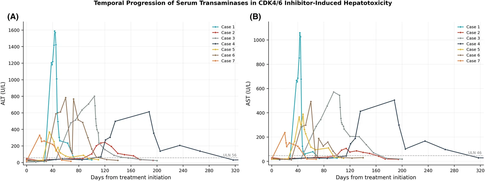
所有患者均在下一次计划随访时发现 ≥2 级转氨酶升高后停用 CDK4/6 抑制剂,同时全程继续内分泌治疗。3 例患者因转氨酶持续升高而接受糖皮质激素治疗,另 1 例(患者 3)接受经验性试验治疗。停药后至开始使用激素的中位时间为 32 天(范围 5–45 天),采用固定剂量口服泼尼松龙 50–100 mg/天(患者 1 先接受静脉注射甲泼尼龙;表 S2)。所有患者肝酶均在峰值后 30–129 天恢复正常(平均 63 天;中位数 61 天)。6 例患者(85.7%)换用另一种 CDK4/6 抑制剂,其中 4 例换为阿贝西利,2 例换为哌柏西利,在中位 18.5 个月(范围 5–34 个月)的随访期间未复发。患者 6 未换药,并在 9 个月后失访。
组织学发现
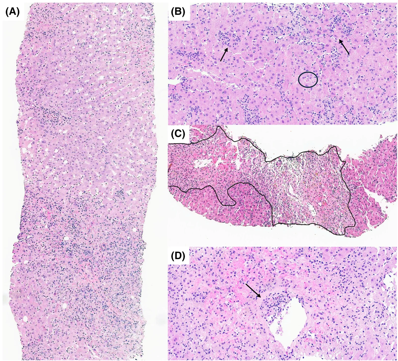
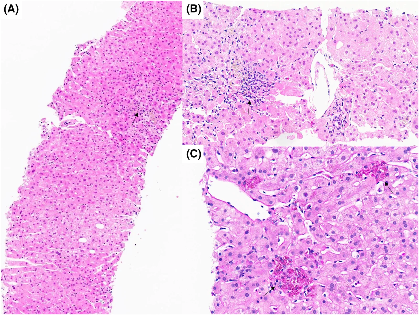
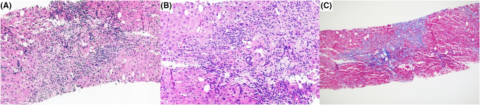
所有病例的活检标本均充分,平均含 17 个汇管区(范围 12–27 个);结果汇总于表 3。主要模式为急性肝炎,共 5 例患者(71.4%),轻度小叶性肝炎和伴早期纤维化的亚急性肝炎各 1 例(14.3%)。5 例急性病例均见中央静脉周围(3 区)融合性坏死,其中 3 例还见桥接坏死(图 2)。患者 1 的融合性坏死累及超过 30% 的实质,患者 3、5 和 6 为 10%–30%,患者 7 为 5%–10%;患者 2 和 4 未见融合性坏死。坏死区域均伴有淋巴单个核细胞浸润,证实损伤具有坏死性炎症性质。轻度小叶性肝炎患者可见偶发的小叶炎症及消退期特征,包括中央静脉周围蜡样质巨噬细胞(图 3)。
| 病例 | 活检时 ALT/AST/ALP(U/L) | 活检间隔(天) | 汇管区数量(n) | 汇管区炎症 | 炎症浸润 | 淋巴细胞性胆管炎 | 小叶炎症 | 融合性坏死 | 桥接坏死 | 纤维化(Ishak/METAVIR) | 损伤模式 |
|---|---|---|---|---|---|---|---|---|---|---|---|
| 1 | 1588/1059/106 | 5 | 15 | 轻度 | 以淋巴细胞为主,伴浆细胞、嗜酸性粒细胞 | 有(轻度) | 中度 | >30% | 有 | 0/F0 | 重度急性肝炎 |
| 2 | 243/77/95 | 20 | 16 | 极轻度 | 以淋巴细胞为主,伴浆细胞 | 无 | 轻度 | 无 | 无 | 0/F0 | 轻度小叶性肝炎 |
| 3 | 358/289/119 | 42 | 14 | 轻度 | 以淋巴细胞为主,伴浆细胞、嗜酸性粒细胞 | 有(轻度) | 中度 | 10%–30% | 有 | 0/F0 | 重度急性肝炎 |
| 4 | 206/167/170 | 121 | 12 | 中度 | 富含浆细胞,伴淋巴细胞、嗜酸性粒细胞 | 有 | 中度 | 无 | 无 | 1/F1 | 亚急性肝炎,早期纤维化 |
| 5 | 181/388/105 | 13 | 27 | 中度 | 以淋巴细胞为主,伴浆细胞、嗜酸性粒细胞 | 有 | 中度 | 10%–30% | 有 | 0/F0 | 重度急性肝炎 |
| 6 | 515/126/193 | 34 | 22 | 轻度 | 以淋巴细胞为主,伴浆细胞、嗜酸性粒细胞 | 有 | 中度 | 10%–30% | 无 | 0/F0 | 重度急性肝炎 |
| 7 | 204/108/276 | 22 | 12 | 轻度 | 以淋巴细胞为主,伴浆细胞、嗜酸性粒细胞 | 无 | 中度 | 5%–10% | 无 | 0/F0 | 重度急性肝炎 |
注:参考范围:ALT 7–56 U/L,AST 15–46 U/L,ALP 38–126 U/L。活检间隔 = 从首次肝功能检测异常至活检的天数。所有融合性坏死均位于中央静脉周围(3 区)。仅患者 4 存在明确的界面性肝炎(Ishak 界面评分 ≥2 分);患者 5 存在炎症向汇管区周围外溢(评分 1 分),但无真正的界面性肝炎。所有活检均未见明显脂肪变性、肉芽肿或胆汁淤积型(胆道为主)模式。除患者 3 可见局灶性小叶中央区毛细胆管内胆栓外,其余所有活检均未见毛细胆管内胆红素淤积。
缩略语:ALT,丙氨酸转氨酶;ALP,碱性磷酸酶;AST,天冬氨酸转氨酶;F,METAVIR 纤维化分期;LFT,肝功能检测;Ishak,改良 Ishak 纤维化分期。
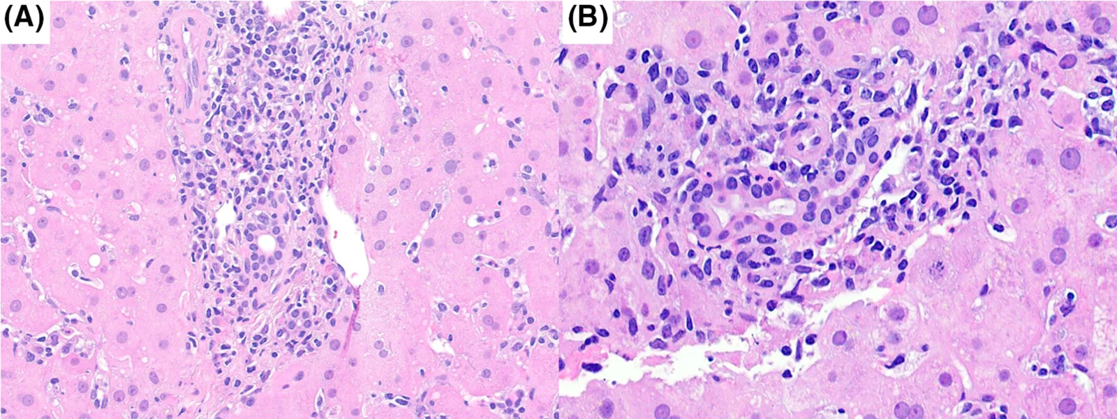
2 份活检(28.6%)的汇管区炎症为中度,4 份(57.1%)为轻度,1 份(14.3%)为极轻度。5 份(71.4%)活检存在淋巴细胞性胆管炎及胆管损伤(图 4),伴轻至中度细胆管反应,两者均经 CK7 染色证实;所有活检均未见胆管减少。5 份活检(71.4%)可见炎症向汇管区周围外溢,但仅 1 份(患者 4)存在明确的界面性肝炎(Ishak 评分 ≥2 分),该例同时可见汇管区周围纤维化(Ishak 1 期;METAVIR F1)(图 5)。其他活检均未见明显纤维化。未见明显脂肪变性或肉芽肿,也未见胆汁淤积型(胆道为主)模式。6 份活检未见毛细胆管内胆红素淤积;患者 3 的小叶中央区(3 区)可见少量胆栓;该患者的总胆红素也最高(9.6 mg/dL)。
个别病例描述
有 2 例患者单独描述。患者 6(唯一接受哌柏西利治疗的患者)的肝损伤在形态学上与瑞博西利所致者无法区分:呈肝细胞型(R = 7.7),伴 3 区融合性坏死及淋巴细胞性胆管炎,但无桥接坏死或纤维化。她仅通过停药即完全恢复。
患者 4 的时间进程及形态学表现与其他患者不同。她的基线代谢评估正常,无 MASLD 特征,饮酒量可忽略不计。连续肝功能检测至第 85 天均正常,第 114 天首次出现轻度升高(ALT 69 U/L),第 121 天明显升高;两次检测之间的间隔均未超过 30 天。活检显示中度汇管区淋巴浆细胞性炎症,伴明确的界面活动、胆管损伤及汇管区周围纤维化,无融合性坏死或桥接坏死。鉴于基线正常且无既存肝病,我们认为此处使用“慢性肝炎”不合适,并将这一模式重新命名为伴早期纤维化的亚急性肝炎。值得注意的是,该活检于首次异常后 121 天、峰值后 47 天获取,此时已开始糖皮质激素治疗,肝功能检测结果正在改善;取样较晚和既往免疫抑制治疗均可能促成其纤维化而非急性坏死的表现。
临床病理相关性
在呈急性肝炎模式的患者中,转氨酶升高幅度与坏死范围大致平行,最高 ALT 峰值(患者 1,1588 U/L)对应最广泛的融合性坏死(>30%)。然而,这种对应关系并不完全一致:患者 5 的 ALT 峰值为 367 U/L 时存在桥接坏死,而患者 6 的 ALT 峰值为 783 U/L 时却无桥接坏死;患者 4 肝酶明显升高,但无融合性坏死。
糖皮质激素的使用与坏死范围并不一致:4 例接受激素治疗的患者中仅 2 例存在融合性坏死或桥接坏死,而 3 例仅通过停药即恢复的患者均存在融合性坏死。决定因素是生化指标的变化趋势,即停药后转氨酶未下降,而非任何组织学特征。广泛坏死本身并不意味着必须使用免疫抑制治疗,相对轻微的活检表现也不能排除其必要性。ANA 阳性同样与组织学严重程度无关:最高滴度(患者 2,>1:2560)伴随最轻微的活检改变,而最广泛的坏死发生于一例 ANA 阴性患者。淋巴细胞性胆管炎主要发生于 R 值属肝细胞型的患者,与单纯 ALP 升高并不一致,提示胆道损伤是以肝细胞损伤为主的过程中的继发性组成部分。
讨论
CDK4/6 抑制剂改变了激素受体阳性、HER2 阴性乳腺癌的治疗格局。3 肝毒性是此类药物一项重要的剂量限制性不良事件。1, 3, 4 FDA 不良事件报告系统(FAERS)的药物警戒数据显示,基于报告比值比(ROR),瑞博西利(ROR = 2.60;95% CI:2.48–2.72)和阿贝西利(ROR = 2.37;95% CI:2.18–2.58)均存在显著的 DILI 不成比例信号,而哌柏西利未见此信号(ROR = 0.70;95% CI:0.67–0.73)。16 在近期一篇综述中,Wong 等得出结论,瑞博西利的肝毒性风险最高,哌柏西利最低,阿贝西利处于中间水平。5
瑞博西利相关肝毒性可能涉及胆盐输出泵(BSEP)受抑制,以及广泛的细胞色素 P450 3A4(CYP3A4)介导的代谢,从而产生反应性中间产物。17 药物–基因相互作用的网络分析已将 STAT3、HSP90AA1 和 EP300 等关键基因确定为 CDK4/6 抑制剂肝毒性的潜在介导因素。16
尽管哌柏西利被认为是 CDK4/6 抑制剂中肝脏安全性最佳的药物,但仍有罕见的重度肝毒性病例报告。在 PALOMA-3 试验中,哌柏西利组有 1 例患者在肝转移进展的背景下发生致死性肝衰竭,个案报告也描述了归因于哌柏西利的重度急性肝炎。18-21
尽管对 CDK4/6 抑制剂相关肝毒性的临床和药代动力学认识不断增加,但全面的组织病理学特征描述仍然有限。迄今仅发表了少数描述瑞博西利相关肝损伤的报告。6, 8, 9, 22 据我们所知,这一包含 7 例患者的病例系列是首项针对经组织学明确特征的 CDK4/6 抑制剂所致肝损伤开展的系统性临床病理研究。
我们的组织病理学分析显示,重度急性肝细胞性损伤呈现出高度一致的模式,多数活检(71.4%)以明显的小叶炎症及中央静脉周围(3 区)融合性坏死为特征,其中 3 例还可见桥接坏死。这些发现与既往有关瑞博西利相关急性肝炎的描述高度一致。8, 9, 22 呈现这一急性肝炎模式的患者均较早出现肝毒性(开始治疗后 20–68 天),生化恢复也更快(峰值后 30–72 天)。相比之下,唯一呈伴早期纤维化的亚急性模式的患者(患者 4)无既存肝病,肝功能检测指标升高的出现明显延迟(114 天),肝酶于第 188 天达到峰值(ALT 612 U/L;AST 504 U/L),且在停药及给予激素后恢复间隔最长(129 天)。
本病例系列中一项值得注意的发现是胆管损伤的发生率较高(71.4%),以淋巴细胞性胆管炎为特征,这与不断出现的提示胆管上皮细胞在 DILI 中易受损伤的证据相符。23 由于细胞色素 P450 活性较低且谷胱甘肽储备减少,胆管上皮细胞的解毒能力有限,可能使其易于受到毒性损伤。23 伴随的细胆管反应可能代表对上皮损伤的代偿性再生反应。23
3 例患者 ANA 滴度升高,提示药物诱导的自身免疫性肝炎(DI-AIH)的可能,近期瑞博西利病例报告中也有此类描述。24, 25 然而,ANA 阳性患者的活检均未显示急性损伤背景下明确的界面性肝炎、富含浆细胞的浸润或真正 AIH 的其他特征。相反,唯一显示明确界面活动并伴富含浆细胞浸润的活检(患者 4),来自一例 ANA 阴性、血清 IgG 正常的患者。综合来看,这些观察提示,在缺乏相应组织学证据时,不应过度解读单独的 ANA 阳性;而提示自身免疫性的组织学特征也可能在 ANA 阴性时出现。
本队列观察到的组织病理学特征与免疫检查点抑制剂(ICI)所致肝毒性有一些相似之处。26, 27 两者均表现为以小叶模式为主的肝细胞损伤,但严重程度和分布存在重要差别。本队列的小叶炎症明显更重,常伴 3 区融合性坏死,数例还存在桥接坏死;而 Zen 等和 Cohen 等描述的 ICI 病例系列通常表现为散在的小叶坏死灶,而无广泛的 3 区融合性坏死。26, 27 ICI 相关肝毒性中的胆道病变比以往认识到的更常见,尤其见于第二代检查点抑制剂。28, 29 本研究 71.4% 的活检存在淋巴细胞性胆管炎,这一表现连同显著的细胆管反应,可能反映了胆管上皮细胞对 BSEP 抑制和反应性代谢产物的特殊易感性。每例患者的胆管损伤均伴随以肝细胞损伤为主的损伤。重要的是,这种胆管损伤是以肝细胞损伤为主的过程中的继发性组成部分;我们未在任何病例中观察到类似原发性胆汁性胆管炎的胆管病型(胆道为主)损伤模式。ICI 相关损伤中描述的脂肪性肝炎和单纯性胆汁淤积也均未出现。26, 27
CDK4/6 抑制剂与 ICI 肝毒性具有共同的形态学和免疫学特征,提示两者可能存在重叠的免疫失调通路。然而,临床过程似乎有所不同:约三分之一至二分之一的 ICI 所致肝损伤患者无需糖皮质激素治疗即可自行消退,而本病例系列中 57% 的患者需要糖皮质激素。25, 26 这一发现与法国 REFHEPS 多中心研究一致,该研究中 41% 的 CDK4/6 抑制剂相关 DILI 病例(主要由瑞博西利所致)需要使用糖皮质激素进行免疫抑制治疗。4 与系统综述和药物警戒分析一致,我们的观察支持对重度瑞博西利所致肝损伤患者进行密切监测并尽早考虑糖皮质激素治疗,尤其是在停药后转氨酶升高未改善时。3, 4, 16
我们发现,85.7% 的患者成功换用另一种 CDK4/6 抑制剂,在中位 18.5 个月的随访期间未再次出现肝毒性,这与近期报告和综述一致,后者描述了在瑞博西利所致 III–IV 级肝毒性后换用哌柏西利或阿贝西利的安全性和可行性。5, 30, 31 这些观察提示 CDK4/6 抑制剂之间的交叉反应性有限,并支持肝毒性可能为具体药物特异性而非整个药物类别共有的观点。30 然而,这一推论仍需审慎:这 3 种药物具有若干共同的药理学特性,包括 CYP3A4 介导的代谢及胆汁排泄途径,因此药物类别层面的机制仍可能影响个别患者的易感性。
本研究提供的详细组织病理学特征描述,可帮助病理医师识别 CDK4/6 抑制剂相关 DILI 的特征性损伤模式,从而提高临床复杂病例的诊断信心。31 进行基线肝功能评估和治疗期间的定期监测仍属审慎之举,是否停药应依据肝功能检测异常的严重程度及变化趋势。尽管预测 CDK4/6 抑制剂所致肝损伤患者能否从糖皮质激素治疗中获益的确切标准仍需前瞻性研究确定,但我们及 REFHEPS 队列的观察提示,对停药后转氨酶未改善的患者,可能有必要尽早考虑免疫抑制治疗;在本病例系列中,促使开始治疗的是这一生化变化趋势,而非活检中的坏死范围。
本研究的局限性包括回顾性设计、队列规模较小,以及监测间隔和活检时机不一致。尤其是患者 4 的活检在临床病程较晚且已开始糖皮质激素治疗后获取,因此该活检的纤维化表现,既可能反映疾病演变阶段及既往免疫抑制治疗的影响,也可能反映一种本质上不同的损伤模式;因此,应谨慎解读这一单例。需要开展具有更大规模、标准化队列的前瞻性研究,以验证这些发现并确定 CDK4/6 抑制剂相关肝毒性的预测性生物标志物。
总之,CDK4/6 抑制剂所致肝损伤是一项重要的不良事件,其特征性模式以急性肝细胞性损伤为主,主要累及 3 区,部分病例可较严重。尽管生化和组织学损伤程度较重,多数患者在停药及有指征时接受糖皮质激素治疗后可完全恢复。换用另一种 CDK4/6 抑制剂后肝毒性复发率较低,凸显了药物之间有限的交叉反应性,并支持通过个体化治疗调整继续使用此类药物。明确 CDK4/6 抑制剂肝毒性的组织病理学谱系,可增强诊断信心,并指导接受这些药物治疗的转移性乳腺癌患者的临床管理。
- Seven women with metastatic breast cancer at a single cancer centre (2020–2024; ribociclib 6, palbociclib 1) developed liver enzyme elevation on a CDK4/6 inhibitor and underwent liver biopsy; RUCAM ≥6 with alternative causes excluded; mean 60 days to first enzyme elevation.
- Histology: severe acute hepatitis in 5 (71.4%), mild lobular hepatitis in 1 and subacute hepatitis with early fibrosis in 1; all acute biopsies had perivenular (zone 3) confluent necrosis, 3 with bridging necrosis; portal inflammation in all and lymphocytic cholangitis in 5 (71.4%).
- Three were ANA-positive without histological features of autoimmune hepatitis; all normalised after withdrawal (mean 63 days) and six were switched to another CDK4/6 inhibitor without recurrence.
- Five figures (transaminase time courses and H&E, Masson trichrome and CK7 histology) and three tables (clinical, laboratory and histopathological features), with individual case descriptions.
Scope: open-access full text (CC BY 4.0) from Histopathology — abstract, introduction, material and methods, results and discussion, with 5 figures and 3 tables; the abbreviations list, author contributions, funding, conflict of interest, data availability statement, supporting information (Tables S1–S2) and references are not included. Superscript numbers are the original reference numbers.
Abstract
Aims
Hepatotoxicity is an increasingly recognised and potentially dose-limiting adverse effect of CDK4/6 inhibitors, but their histopathological spectrum remains incompletely characterised. We aimed to describe the clinicopathological features of histologically characterised CDK4/6 inhibitor-induced liver injury.
Methods and results
We retrospectively identified patients with metastatic breast cancer who developed liver enzyme elevation and underwent liver biopsy while receiving a CDK4/6 inhibitor at a single comprehensive cancer centre (2020–2024). Inclusion required a RUCAM score ≥6 and exclusion of alternative aetiologies. Seven female patients met the criteria (ribociclib, n = 6; palbociclib, n = 1). The mean interval to first enzyme elevation was 60 days (range 20–114) and the mean RUCAM score was 8. Antinuclear antibody (ANA) positivity was noted in three patients, with negative extended autoimmune panels. Liver biopsy demonstrated severe acute hepatitis in five patients (71.4%), mild lobular hepatitis in one (14.3%) and subacute hepatitis with early fibrosis in one (14.3%). All acute biopsies showed perivenular (zone 3) confluent necrosis, with additional bridging necrosis in three biopsies. Portal inflammation was identified in all biopsies and lymphocytic cholangitis in five (71.4%) biopsies. No ANA-positive patient showed histological features of autoimmune hepatitis. All patients achieved biochemical normalisation after drug withdrawal (mean 63 days from peak; range 30–129), and six were successfully switched to an alternative agent.
Conclusions
CDK4/6 inhibitor-induced liver injury predominantly manifests as acute hepatocellular injury with zone 3 necrosis and lymphocytic cholangitis. Despite severe histological abnormalities, it resolves with drug withdrawal, and limited cross-reactivity between agents supports individualised therapeutic strategies.
Introduction
Cyclin-dependent kinase 4 and 6 (CDK4/6) inhibitors combined with endocrine therapy have become the standard treatment for hormone receptor-positive, human epidermal growth factor receptor 2 (HER2) negative breast cancer.1 Ribociclib, palbociclib and abemaciclib are FDA-approved orally bioavailable small molecules that act by binding to the ATP cleft of CDK4 and/or CDK6.2 They significantly increase progression-free survival (PFS) when added to endocrine therapy. Although these drugs have shown similar efficacy, hepatotoxicity emerges as a dose-limiting adverse effect of CDK4/6 inhibitors, occurring in 14%–25% of patients across clinical trials.3, 4 Ribociclib received FDA approval in March 2017 in combination with letrozole for the first-line treatment of hormone receptor-positive, HER2-negative (metastatic or locally advanced) breast carcinoma.1 The choice between the drugs, in practice, is influenced by patient-specific factors including menopausal status, prior treatment history, comorbidities and tolerability profile.2, 5, 6
Ribociclib is among the most widely prescribed CDK4/6 inhibitors and confers a clear survival benefit, but its adverse-event burden is substantial; in the MONALEESA trials, nearly all treated patients experienced at least one treatment-emergent adverse event, and grade III–IV events occurred in up to 84% of patients.6 Common toxicities of ribociclib include fever, fatigue, diarrhoea and thrombocytopaenia, while severe reactions include QTc prolongation and severe neutropaenia.1 Palbociclib is often preferred when minimising hepatotoxicity risk is a clinical priority, given its consistently more favourable hepatic safety profile.5, 7 Abemaciclib occupies an intermediate position with respect to hepatic risk.3, 5 Hepatotoxicity caused by ribociclib is echoed in real-world series and in isolated case reports of grade IV hepatotoxicity and fulminant hepatitis.8-10
Despite its growing clinical relevance, histopathological characterisation of CDK4/6 inhibitor-induced liver injury remains inadequately described, posing diagnostic challenges for pathologists.
In this study, we present detailed histopathological and biochemical findings from seven well-characterised patients with advanced-stage breast carcinoma, who developed clinically apparent liver injury following treatment with ribociclib (n = 6) and palbociclib (n = 1), and in whom other causes of liver injury were excluded, confirming drug-induced liver injury (DILI) due to CDK4/6 inhibitors.
Material and methods
Study Design
After obtaining requisite approval from the Institute Ethics Committee, a retrospective review of breast cancer patients receiving CDK4/6 inhibitor therapy who had elevated liver enzymes and underwent liver biopsy at a single comprehensive cancer centre from January 2020 through December 2024 was performed. Histopathological review was performed independently and remotely by co-authors based at other institutions (R.S., S.M.); no additional cases were contributed by any other centre.
Clinical and laboratory data were collected, including demographic characteristics, breast cancer subtype, specific CDK4/6 inhibitor used and dosing schedule, duration from therapy initiation to liver enzyme elevation, biochemical parameters and underlying liver disease. All liver biopsies underwent detailed histopathological evaluation to characterise the liver injury pattern and assess for concurrent hepatic pathology. Liver function tests were monitored at a median interval of 15 days (range 1–30) prior to the first recorded abnormality, and at a median interval of 7 days thereafter. No interval between successive measurements before the first abnormality exceeded 30 days in any patient (Table S1).
Patient Selection
Patients were included if they met these criteria: (1) Histologically characterised DILI with Roussel Uclaf Causality Assessment Method (RUCAM) score ≥6 (revised electronic RUCAM, 2022)11; (2) Exclusion of alternative aetiologies, including viral hepatitis (negative HCV RNA, HBsAg, HBV DNA), autoimmune hepatitis (AIH) (negative extended autoimmune liver disease panel, anti-LKM <1:40, IgG ≤16 g/L), biliary obstruction (normal magnetic resonance cholangiopancreatography [MRCP]) and hepatic metastatic disease; (3) Absence of competing causes of liver injury, including concurrent use of hepatotoxic medications within 30 days, significant alcohol consumption (more than 20 g/day) and pre-existing chronic liver disease.
Time to first liver enzyme elevation was defined as the interval from initiation of the CDK4/6 inhibitor to the first ALT (alanine aminotransferase) or AST (aspartate aminotransferase) value above the laboratory reference range. Clinically significant hepatotoxicity, the threshold at which the drug was withheld and further evaluation undertaken, was defined as an ALT or AST value greater than three times the upper limit of normal (ULN), corresponding to grade ≥2 toxicity by the Common Terminology Criteria for Adverse Events (CTCAE) version 5.0.12 All patients had documented normal baseline liver biochemistry before starting therapy, and chronic liver disease, including metabolic dysfunction-associated steatotic liver disease (MASLD), alcohol-related liver disease and MetALD, was excluded on pretreatment metabolic risk-factor assessment, alcohol history and imaging. Biopsy was performed when the cause of the enzyme elevation remained unclear after non-invasive investigation, or when the abnormality persisted despite drug withdrawal.
Data Collection
Clinical parameters for all eligible patients were extracted from the electronic medical records. Primary breast tumour characteristics included histological tumour type and grade, as well as hormone receptor (oestrogen receptor [ER] and progesterone receptor [PgR]) and HER2 status. Laboratory parameters evaluated consisted of liver function tests (LFTs; ALT, AST, alkaline phosphatase [ALP], and total bilirubin), viral serologies (hepatitis B surface antigen, hepatitis C antibody, HBV DNA, HCV RNA), and autoimmune markers (antinuclear antibody [ANA], anti-liver–kidney microsomal antibody [ALKMA], anti-smooth muscle antibodies [ASMA], anti-mitochondrial antibodies [AMA-M2], and IgG levels).
The time to onset of liver enzyme elevation (ALT, AST) as well as peak enzyme values were recorded for each patient. Peak values were defined as the single highest recorded value at any timepoint during follow-up, verified against the complete serial monitoring dataset (Table S1). Time to biochemical normalisation was measured from the day of peak transaminase elevation to the first day on which both ALT and AST returned within the reference range. The R value was calculated as R = (ALT/ULN) ÷ (ALP/ULN), and liver injury classified as hepatocellular (R ≥ 5), cholestatic (R ≤ 2) or mixed (R = 2–5).13
Histopathological Evaluation
All liver biopsies were independently reviewed by three pathologists (R.M., R.S., S.M.), each blinded to the clinical history. Haematoxylin and eosin (H&E), Masson's trichrome and cytokeratin 7 (CK7) stained sections were examined in every case, and each biopsy was classified as hepatitic, cholestatic or mixed. Portal inflammation (graded as minimal, mild, moderate or severe), interface activity (0–4), lobular necroinflammatory activity (1–4) and fibrosis (0–6) were graded using the modified Ishak system;14 the equivalent METAVIR fibrosis stage (0–4) is also reported.15 Periportal inflammatory spill-over without destruction of the limiting plate (Ishak interface score 1) was recorded separately from definite interface hepatitis (score ≥2), as the former commonly accompanies acute lobular injury. The inflammatory infiltrate was assessed semi-quantitatively as predominantly lymphocytic, plasma cell-rich or eosinophil-rich. Bile duct injury, including lymphocytic cholangitis and epithelial degenerative change, was documented, and ductopenia defined as loss of interlobular bile ducts in more than 50% of portal tracts on H&E and CK7. The number of portal tracts was recorded in each biopsy as a measure of adequacy. Confluent, perivenular and bridging necrosis were recorded, with the proportion of parenchyma involved by confluent necrosis estimated on H&E as none or <5%, 5%–10%, 10%–30%, or >30%. Steatosis, granulomas, cholestasis and bilirubinostasis (canalicular or hepatocellular bile plugs) were noted when present.
Results
Clinical Findings
Nine female patients with advanced breast cancer receiving CDK4/6 inhibitor therapy underwent liver biopsy for abnormal liver tests; two were excluded because of concurrent complementary-alternative medication use with potential hepatotoxicity. The remaining seven (ribociclib, n = 6; palbociclib, n = 1) met inclusion criteria for CDK4/6 inhibitor-induced DILI, with RUCAM scores ≥6 (mean 8; range 7–9). Ages ranged from 36 to 72 years (mean 50.4; median 48) and all had hormone receptor-positive carcinoma (Table 1). Ribociclib was given at 600 mg and palbociclib at 125 mg once daily, both on a 3-weeks-on/1-week-off schedule; all patients were on standard doses when hepatotoxicity was identified and none had undergone a prior dose reduction (Table S2).
| Case | Age (y) | Tumour type | Biomarker status | Metastatic site | Primary treatment | CDK4/6 inhibitor and dose | DILI management | Switch to alternative CDK4/6 inhibitor |
|---|---|---|---|---|---|---|---|---|
| 1 | 38 | IDC (G3) | ER+, PgR+, HER2-negative | Lung, mediastinum, bone, LN | Tamoxifen + Ribociclib | Ribociclib 600 mg/day, 21/7 | Ribociclib withdrawal; prednisolone added | Palbociclib |
| 2 | 43 | IDC (G3) | ER+, PgR+, HER2 overexpressed (3+ by IHC) | Bone, pleura, pancreas | Tamoxifen + Fulvestrant + Ribociclib | Ribociclib 600 mg/day, 21/7 | Ribociclib withdrawal; prednisolone added | Abemaciclib |
| 3 | 36 | IDC (G2) | ER+, PgR+, HER2-negative | LN, skin | Tamoxifen + Ribociclib | Ribociclib 600 mg/day, 21/7 | Ribociclib withdrawal; prednisolone added | Abemaciclib + Anastrozole |
| 4 | 48 | Mixed IDC–ILC (G2) | ER+, PgR+, HER2-negative | Liver | Leuprorelin + Ribociclib + Anastrozole | Ribociclib 600 mg/day, 21/7 | Ribociclib withdrawal; prednisolone added | Palbociclib |
| 5 | 68 | IDC (G3) | ER+, PgR+, HER2-negative | Liver, brain, bone | Letrozole + Ribociclib | Ribociclib 600 mg/day, 21/7 | Ribociclib withdrawal | Abemaciclib |
| 6 | 72 | ILC (G2) | ER+, PgR+, HER2-negative | LN, bone | Palbociclib | Palbociclib 125 mg/day, 21/7 | Palbociclib withdrawal | None (lost to follow-up) |
| 7 | 48 | IDC (G2) | ER+, PgR+, HER2-negative | LN, bone | Niraparib + Bevacizumab + Temsirolimus + Fulvestrant + Ribociclib | Ribociclib 600 mg/day, 21/7 | Ribociclib withdrawal | Abemaciclib |
Note: 21/7 = 3 weeks on, 1 week off. In every patient, the CDK4/6 inhibitor was withdrawn on identification of grade ≥2 transaminase elevation at the next scheduled monitoring visit; concurrent endocrine therapy was continued throughout. All patients were receiving standard label doses at the time hepatotoxicity was identified and none had undergone a prior dose reduction.
Abbreviations: DILI, drug-induced liver injury; ER, oestrogen receptor; G, grade; HER2, human epidermal growth factor receptor 2; IDC, invasive ductal carcinoma of no special type; IHC, immunohistochemistry; ILC, invasive lobular carcinoma; LN, lymph node; PgR, progesterone receptor.
Peak ALT ranged from 243 to 1588 U/L (mean 674) and peak AST from 106 to 1059 U/L (mean 480) (Table 2, Figure 1). Total bilirubin exceeded the reference range in two patients, markedly in Patient 3 (9.6 mg/dL) and mildly in Patient 5 (2.8 mg/dL). ALP was normal or only mildly elevated (mean 190 U/L). Where ALP exceeded the reference range (Patients 4–7), it peaked concomitantly with or within 1–2 weeks after the transaminase peak, consistent with a hepatocellular-predominant injury with secondary biliary involvement. ANA titres ≥1:160 were found in three patients, none of whom had other markers of autoimmune liver disease; one patient had isolated low-titre anti-dsDNA without clinical SLE. The mean interval to first enzyme elevation was 60 days (median 45; range 20–114) and to peak elevation 81 days (median 60; range 20–188). Four patients (1, 5, 6 and 7) reached both within approximately 2 months; onset was later in the others; Day 68 in Patient 3, Day 100 in Patient 2 and Day 114 in Patient 4, with peaks at Days 104, 120 and 188. R value analysis indicated a hepatocellular pattern (R ≥ 5) in 5/7 (71.4%) and a mixed pattern in 2/7 (28.6%), ranging from 2.6 to 33.7.
| Case | Peak ALT (7–56 U/L) | Peak AST (15–46 U/L) | Peak ALP (38–126 U/L) | Bil (T) (0.2–1.3 mg/dL) | Days to first enzyme elevation | Days to peak enzyme elevation | Days to normalisation of enzymes | R value | RUCAM score | ANA | Others |
|---|---|---|---|---|---|---|---|---|---|---|---|
| 1 | 1588 | 1059 | 106 | 0.6 | 38 | 43 | 30 | 33.7 | 8 | Negative (1:40) | NA |
| 2 | 243 | 106 | 95 | 0.4 | 100 | 120 | 54 | 5.8 | 7 | Positive (>1:2560) | NA |
| 3 | 800 | 571 | 123 | 9.6 | 68 | 104 | 72 | 14.6 | 8 | Negative (1:40) | NA |
| 4 | 612 | 504 | 272 | 1.1 | 114 | 188 | 129 | 5.1 | 7 | Negative (1:40) | NA |
| 5 | 367 | 388 | 216 | 2.8 | 35 | 35 | 61 | 3.8 | 9 | Positive (1:160) | NA |
| 6 | 783 | 492 | 230 | 1.0 | 45 | 60 | 63 | 7.7 | 9 | Negative (1:40) | NA |
| 7 | 328 | 237 | 286 | 1.1 | 20 | 20 | 34 | 2.6 | 8 | Positive (1:160) | dsDNA (+) |
Note: Values in parentheses denote the laboratory reference (normal) range. Peak values represent the single highest recorded value at any timepoint during follow-up, verified against the complete serial monitoring dataset (Table S1). ‘Days to peak’ refers to peak transaminase (ALT/AST) elevation. ‘Days to normalisation’ is measured from the day of peak transaminase elevation to the first day on which both ALT and AST returned within the reference range. R value = (ALT/ULN) ÷ (ALP/ULN); hepatocellular R ≥ 5, mixed R 2–5, cholestatic R ≤ 2.
Abbreviations: ALP, alkaline phosphatase; ALT, alanine transaminase; ANA, antinuclear antibody; AST, aspartate transaminase; Bil(T), total bilirubin; dsDNA, double-stranded DNA; NA, not available; RUCAM, Roussel Uclaf Causality Assessment Method.
The CDK4/6 inhibitor was discontinued in all patients on identification of grade ≥2 transaminase elevation at the next scheduled visit while concurrent endocrine therapy was continued throughout. Corticosteroids were given in three patients for persistent transaminase elevation and in one (Patient 3) as an empirical therapeutic trial. Steroids began a median of 32 days after withdrawal (range 5–45), as fixed-dose oral prednisolone 50–100 mg/day (Patient 1 received intravenous methylprednisolone first; Table S2). Enzymes normalised in all patients, 30–129 days after peak (mean 63; median 61). Six patients (85.7%) were switched to an alternative CDK4/6 inhibitor, four to abemaciclib and two to palbociclib, without recurrence over a median follow-up of 18.5 months (range 5–34). Patient 6 was not switched and was lost to follow-up after 9 months.
Histological Findings
Biopsies were adequate in all cases, with a mean of 17 portal tracts (range 12–27); findings are summarised in Table 3. The predominant pattern was acute hepatitis in five patients (71.4%), with mild lobular hepatitis and subacute hepatitis with early fibrosis in one each (14.3%). All five acute cases showed confluent perivenular (zone 3) necrosis and three also showed bridging necrosis (Figure 2). Confluent necrosis involved more than 30% of the parenchyma in Patient 1, 10%–30% in Patients 3, 5 and 6, and 5%–10% in Patient 7; none was identified in Patients 2 or 4. The necrotic zones were consistently accompanied by a lymphomononuclear infiltrate, confirming the necroinflammatory nature of the injury. The patient with mild lobular hepatitis showed occasional lobular inflammation and resolving features, including perivenular ceroid-laden macrophages (Figure 3).
| Case | ALT/AST/ALP at biopsy (U/L) | Biopsy interval (days) | No. of portal tracts (n) | Portal inflammation | Inflammatory infiltrate | Lymphocytic cholangitis | Lobular inflammation | Confluent necrosis | Bridging necrosis | Fibrosis (Ishak/METAVIR) | Pattern of injury |
|---|---|---|---|---|---|---|---|---|---|---|---|
| 1 | 1588/1059/106 | 5 | 15 | Mild | Lymphocyte predominant, plasma cells, eosinophils | Present (mild) | Moderate | >30% | Present | 0/F0 | Severe acute hepatitis |
| 2 | 243/77/95 | 20 | 16 | Minimal | Lymphocyte predominant, plasma cells | Absent | Mild | None | Absent | 0/F0 | Mild lobular hepatitis |
| 3 | 358/289/119 | 42 | 14 | Mild | Lymphocyte predominant, plasma cells, eosinophils | Present (mild) | Moderate | 10%–30% | Present | 0/F0 | Severe acute hepatitis |
| 4 | 206/167/170 | 121 | 12 | Moderate | Plasma cell rich, lymphocytes, eosinophils | Present | Moderate | None | Absent | 1/F1 | Subacute hepatitis, early fibrosis |
| 5 | 181/388/105 | 13 | 27 | Moderate | Lymphocyte predominant, plasma cells, eosinophils | Present | Moderate | 10%–30% | Present | 0/F0 | Severe acute hepatitis |
| 6 | 515/126/193 | 34 | 22 | Mild | Lymphocyte predominant, plasma cells, eosinophils | Present | Moderate | 10%–30% | Absent | 0/F0 | Severe acute hepatitis |
| 7 | 204/108/276 | 22 | 12 | Mild | Lymphocyte predominant, plasma cells, eosinophils | Absent | Moderate | 5%–10% | Absent | 0/F0 | Severe acute hepatitis |
Note: Reference ranges: ALT 7–56 U/L, AST 15–46 U/L, ALP 38–126 U/L. Biopsy interval = days from first LFT abnormality to biopsy. All confluent necrosis was perivenular (zone 3). Definite interface hepatitis (Ishak interface score ≥2) was present only in Patient 4; Patient 5 showed periportal spill-over (score 1) without true interface hepatitis. No biopsy showed significant steatosis, granulomas, or a cholestatic (biliary-dominant) pattern. Canalicular bilirubinostasis was absent in all biopsies except Patient 3, which showed focal centrizonal canalicular bile plugs.
Abbreviations: ALT, alanine transaminase; ALP, alkaline phosphatase; AST, aspartate transaminase; F, METAVIR fibrosis stage; LFT, liver function test; Ishak, modified Ishak fibrosis stage.
Portal inflammation was moderate in two biopsies (28.6%), mild in four (57.1%) and minimal in one (14.3%). Lymphocytic cholangitis and bile duct injury were present in five (71.4%) (Figure 4) with mild to moderate ductular reaction, both corroborated by CK7; no biopsy showed ductopenia. Periportal spill-over of inflammation was seen in five biopsies (71.4%), but definite interface hepatitis (Ishak score ≥2) in only one (Patient 4), which also showed periportal fibrosis (Ishak stage 1; METAVIR F1) (Figure 5). No other biopsy showed significant fibrosis. There was no significant steatosis or granulomas, and no cholestatic (biliary-dominant) pattern. Canalicular bilirubinostasis was absent in six biopsies; in Patient 3, a few bile plugs were present in centrizonal (zone 3) areas; this patient also had the highest total bilirubin (9.6 mg/dL).
Individual Case Descriptions
Two patients are described separately. Hepatic injury in Patient 6 (the only patient treated with palbociclib) was morphologically indistinguishable from that seen with ribociclib: a hepatocellular pattern (R = 7.7) with confluent zone 3 necrosis and lymphocytic cholangitis, but no bridging necrosis or fibrosis. She recovered fully on drug withdrawal alone.
The time course and morphology differed from the rest in Patient 4. Her baseline metabolic evaluation was normal, with no features of MASLD and negligible alcohol intake. Serial liver tests were normal to Day 85, with a first mild elevation at Day 114 (ALT 69 U/L) and a marked rise at Day 121; no gap between measurements exceeded 30 days. Biopsy showed moderate portal lymphoplasmacytic inflammation with definite interface activity, bile duct injury and periportal fibrosis, without confluent or bridging necrosis. Given the normal baseline and absence of pre-existing liver disease, we consider ‘chronic hepatitis’ inappropriate here and have re-designated this pattern as subacute hepatitis with early fibrosis. Notably, this biopsy was obtained 121 days after the first abnormality and 47 days after the peak, by which time corticosteroids had been started and liver tests were improving; both the late sampling and prior immunosuppression may have contributed to its fibrosing rather than acutely necrotic appearance.
Clinicopathological Correlations
Among patients with an acute hepatitic pattern, the magnitude of transaminase elevation broadly paralleled the extent of necrosis, the highest peak ALT (Patient 1, 1588 U/L) corresponding to the most extensive confluent necrosis (>30%). Yet, the correspondence was not exact: bridging necrosis was present in Patient 5 at a peak ALT of 367 U/L but absent in Patient 6 at 783 U/L, and Patient 4 showed marked enzyme elevation without confluent necrosis.
Corticosteroid use did not track with the extent of necrosis: only two of the four steroid-treated patients showed confluent or bridging necrosis, whereas all three who recovered on withdrawal alone did so despite confluent necrosis being present. The determinant was the biochemical trajectory, failure of transaminases to fall after withdrawal, rather than any histological feature. Extensive necrosis did not by itself mandate immunosuppression, nor did a relatively bland biopsy preclude it. ANA positivity likewise showed no relationship to histological severity: the highest titre (Patient 2, >1:2560) accompanied the mildest biopsy, while the most extensive necrosis occurred in an ANA-negative patient. Lymphocytic cholangitis occurred predominantly in patients with a hepatocellular R value and did not track with isolated ALP elevation, suggesting that the biliary injury is a secondary component of a predominantly hepatocellular process.
Discussion
CDK4/6 inhibitors have transformed the treatment landscape for hormone receptor-positive, HER2-negative breast cancer.3 Hepatotoxicity is an important dose-limiting adverse event of this class.1, 3, 4 Pharmacovigilance data from the FDA Adverse Event Reporting System (FAERS) demonstrate significant disproportionality signals for DILI, based on the reporting odds ratio (ROR), for ribociclib (ROR = 2.60; 95% CI: 2.48–2.72) and abemaciclib (ROR = 2.37; 95% CI: 2.18–2.58), whereas no signal is observed for palbociclib (ROR = 0.70; 95% CI: 0.67–0.73).16 In a recent review, Wong et al. concluded that ribociclib carries the highest risk of hepatotoxicity, palbociclib the lowest and abemaciclib an intermediate risk profile.5
Ribociclib-associated hepatotoxicity may involve inhibition of the bile salt export pump (BSEP) and extensive cytochrome P450 3A4 (CYP3A4)-mediated metabolism, leading to the generation of reactive intermediates.17 Network analysis of drug–gene interactions has identified key genes such as STAT3, HSP90AA1 and EP300 as potential mediators of CDK4/6 inhibitor hepatotoxicity.16
Although palbociclib is considered to have the most favourable hepatic safety profile among CDK4/6 inhibitors, severe cases of hepatotoxicity have been rarely reported. In the PALOMA-3 trial, one patient in the palbociclib arm developed fatal hepatic failure in the setting of progressive liver metastases, and isolated reports describe severe acute hepatitis attributed to palbociclib.18-21
Despite growing clinical and pharmacokinetic insights into CDK4/6 inhibitor-associated hepatotoxicity, comprehensive histopathological characterisation remains limited. Only a few reports describing ribociclib-related liver injury have been published to date.6, 8, 9, 22 To the best of our knowledge, this series of seven patients represents the first systematic clinicopathological study of histologically characterised CDK4/6 inhibitor-induced liver injury.
Our histopathological analysis demonstrates a strikingly consistent pattern of severe acute hepatocellular injury, marked by prominent lobular inflammation and confluent perivenular (zone 3) necrosis in most biopsies (71.4%), with three cases also showing bridging necrosis. These findings closely parallel previously reported descriptions of ribociclib-associated acute hepatitis.8, 9, 22 Patients exhibiting this acute hepatitic pattern uniformly showed early onset of hepatotoxicity (20–68 days after therapy initiation) and more rapid biochemical recovery (30–72 days after peak). In contrast, the single patient with a subacute pattern with early fibrosis (Patient 4) had no pre-existing liver disease and experienced a markedly delayed onset of LFT elevation (114 days), peak enzyme levels at 188 days (ALT 612 U/L; AST 504 U/L), and the longest recovery interval (129 days) following drug withdrawal and steroid administration.
A notable finding in our series was the high incidence of bile duct injury (71.4%), characterised by lymphocytic cholangitis, which is supported by emerging evidence implicating cholangiocyte vulnerability in DILI.23 Cholangiocytes possess limited detoxification capacity owing to lower cytochrome P450 activity and reduced glutathione reserves, which may predispose them to toxic injury.23 The accompanying ductular reaction likely represents a compensatory regenerative response to epithelial damage.23
Elevated ANA titres in three patients raised the possibility of drug-induced autoimmune hepatitis (DI-AIH), as described in recent ribociclib case reports.24, 25 However, none of the biopsies from the ANA-positive patients demonstrated definite interface hepatitis in the context of acute injury, plasma cell-rich infiltrates or other features of true AIH. Conversely, the only biopsy showing definite interface activity with a plasma cell-rich infiltrate (Patient 4) came from an ANA-negative patient with a normal serum IgG. Taken together, these observations indicate that ANA positivity alone should not be over-interpreted without corroborative histological findings, and that histological features suggestive of autoimmunity may occur in its absence.
The histopathological features observed in our cohort share some similarities with immune checkpoint inhibitor (ICI)-induced hepatotoxicity.26, 27 Both demonstrate a predominantly lobular pattern of hepatocellular damage, but important distinctions emerge in severity and distribution. In our cohort lobular inflammation was markedly more severe, frequently accompanied by confluent zone 3 necrosis and, in several cases, bridging necrosis, whereas the ICI series described by Zen et al. and Cohen et al. typically exhibited scattered foci of lobular necrosis without extensive zone 3 confluent necrosis.26, 27 Biliary lesions in ICI-related hepatotoxicity are more common than was once appreciated, particularly with second-generation checkpoint inhibitors.28, 29 Lymphocytic cholangitis was present in 71.4% of our biopsies, which, together with the conspicuous ductular reaction, may reflect a particular vulnerability of cholangiocytes to BSEP inhibition and reactive metabolites. In every case, the bile duct damage accompanied a predominantly hepatocellular injury. Importantly, this bile duct injury occurred as a secondary component of a predominantly hepatocellular process; we did not observe a cholangiopathic (biliary-dominant) pattern of injury analogous to primary biliary cholangitis in any case. Steatohepatitis and bland cholestasis, described in ICI-associated injury, were likewise absent.26, 27
The shared morphological and immunological features between CDK4/6 inhibitor and ICI hepatotoxicity suggest overlapping pathways of immune dysregulation. However, the clinical course appears to differ: ICI-induced liver injury resolves spontaneously in approximately one-third to one-half of patients without corticosteroid therapy, whereas 57% of patients in our series required corticosteroids.25, 26 This finding parallels the French REFHEPS multicenter study, in which 41% of CDK4/6 inhibitor-associated DILI cases (predominantly ribociclib) required immunosuppression with corticosteroids.4 Consistent with systematic reviews and pharmacovigilance analyses, our observations support the need for close monitoring and early consideration of corticosteroid therapy in patients with severe ribociclib-induced liver injury, particularly when transaminase elevation fails to improve after withdrawal of the drug.3, 4, 16
Our finding that 85.7% of patients were successfully switched to an alternative CDK4/6 inhibitor without recurrence of hepatotoxicity, over a median follow-up of 18.5 months, aligns with recent reports and reviews describing the safety and feasibility of switching to palbociclib or abemaciclib following ribociclib-induced grade III–IV hepatotoxicity.5, 30, 31 These observations suggest limited cross-reactivity among CDK4/6 inhibitors and support the concept that hepatotoxicity may be agent-specific rather than class-wide.30 This inference should nonetheless be tempered: the three agents share several pharmacological properties, including CYP3A4-mediated metabolism and biliary elimination pathways, so class-level mechanisms may still contribute to susceptibility in individual patients.
The detailed histopathological characterisation provided by our study may aid pathologists in recognising the distinctive injury patterns associated with CDK4/6 inhibitor-related DILI, thereby improving diagnostic confidence in clinically complex cases.31 Baseline liver function evaluation and scheduled monitoring during treatment remain prudent, with drug discontinuation guided by the severity and trajectory of liver test abnormalities. While the precise criteria predicting benefit from corticosteroid therapy in CDK4/6 inhibitor-induced liver injury remain to be established in prospective studies, our observations and those from the REFHEPS cohort suggest that early consideration of immunosuppression may be warranted in patients whose transaminases fail to improve after drug withdrawal; in our series this biochemical trajectory, rather than the extent of necrosis on biopsy, was what prompted treatment.
Limitations of the study include the retrospective design, small cohort size and variability in monitoring intervals and biopsy timing. In particular, the biopsy in Patient 4 was obtained late in the clinical course and after corticosteroid therapy had begun, so the fibrosing appearance of that biopsy may reflect the stage of evolution and prior immunosuppression as much as an intrinsically distinct pattern of injury; this single case should therefore be interpreted with caution. Prospective studies with larger, standardised cohorts are needed to validate these findings and identify predictive biomarkers of CDK4/6 inhibitor-related hepatotoxicity.
To conclude, CDK4/6 inhibitor-induced liver injury represents an important adverse event with a characteristic pattern of predominantly acute hepatocellular injury, predominantly involving zone 3, which may be severe in some cases. Despite the intensity of biochemical and histological injury, most patients experience full recovery with drug discontinuation and, when indicated, corticosteroid therapy. The low incidence of recurrent hepatotoxicity on switching to an alternative CDK4/6 inhibitor underscores the limited cross-reactivity among agents and supports continued use of this drug class through individualised therapeutic adjustments. Characterising the histopathological spectrum of CDK4/6 inhibitor hepatotoxicity can enhance diagnostic confidence and guide clinical management in patients receiving these therapies for metastatic breast cancer.