- 硬脑膜 MALT 淋巴瘤:发生于硬脑膜的结外低级别黏膜相关淋巴组织 B 细胞淋巴瘤,常伴单型性浆细胞分化;成人,中位年龄 50~59 岁,男:女 = 1:5–7;边缘区淋巴瘤是硬脑膜最常见的淋巴瘤,且比脑实质 MALT 淋巴瘤多见。
- MRI 可呈单发或多发斑块样硬脑膜增厚、酷似脑膜瘤;WHO 第 5 版必要诊断标准为硬脑膜起源、边缘区样特征及 B 细胞标记伴滤泡树突细胞网(CD21 / CD23 / CD35+)。镜下可见滤泡植入、陷入的脑膜上皮,偶见轻链型淀粉样变性;浆细胞常 IgG4+。
- 分子:TNFAIP3 失活多见于浆细胞分化病例(含 3 例 IgG4+),NOTCH2 激活突变多见于单核细胞样形态并常伴 TBL1XR1 失活;IGH::MALT1 仅 1 例报道。
- 专题含 12 幅镜下与免疫组化图、病理报告示例、鉴别诊断(淋巴浆细胞淋巴瘤、CLL / SLL、套细胞淋巴瘤、滤泡性淋巴瘤、浆细胞肿瘤、脑膜瘤、IgG4 相关疾病、黑色素瘤、SFT、尤因肉瘤等)及 2 道练习题;同日更新另含脱屑性间质性肺炎、寻常痤疮、着色芽生菌病、NUTM1 重排肉瘤 4 个专题摘要。
收录范围:PathologyOutlines 2026-10-12 博客「Textbook Updates」全文(5 个专题摘要与精选图片),以及其中「硬脑膜 MALT 淋巴瘤」专题页全文(全部栏目、12 幅站内图像及图注、病理报告示例、鉴别诊断、练习题与答案)。其余 4 个专题仅收博客摘要,正文见各专题链接。
教科书更新 · 2026 年 10 月 12 日
我们已发布以下专题的更新:
中枢神经系统与垂体肿瘤 > 淋巴瘤与组织细胞肿瘤 > 硬脑膜 MALT 淋巴瘤
作者:Giovanni Martino, M.D., Flavia Adotti, M.D., Antonio d’Amati, M.D., Martina Mandarano, M.D., Ph.D., Marco Gessi, M.D.
专题摘要:发生于硬脑膜的结外低级别黏膜相关淋巴组织(MALT)B 细胞淋巴瘤;可伴浆细胞分化。已有报道发现TNFAIP3, NOTCH2和TBL1XR1基因突变及其他染色体异常。与其他结外边缘区淋巴瘤不同,迄今尚未阐明其发病机制。表现为压迫症状;最常见的是头痛、癫痫发作和局灶性神经功能缺损。临床病程惰性;多数患者可从局部治疗策略中获益。
肺 > 吸烟相关间质性肺疾病 > 肺泡巨噬细胞肺炎(脱屑性间质性肺炎)
作者:Akira Yoshikawa, M.D., Andrey Bychkov, M.D., Ph.D., Sakda Sathirareuangchai, M.D.
专题摘要:一种罕见的特发性间质性肺炎,其特征为肺泡内巨噬细胞聚集,胞质内含细颗粒状棕色色素。多数病例与吸烟有关;也可与结缔组织病、药物、吸入性暴露、误吸或遗传性疾病有关。诊断依据临床特征、影像学和组织病理学。最常见的症状为呼吸困难,其次为干咳。预后通常良好,对治疗反应良好。
皮肤非肿瘤性疾病 > 皮肤附属器其他疾病 > 寻常痤疮
作者:Andrew Kulbiski, M.D., Jayden Ray, D.O., Sepideh Nikki Asadbeigi, M.D.
专题摘要:寻常痤疮是一种常见的炎症性皮肤病,主要累及面部和躯干。临床皮损可从无炎症的粉刺到炎症性丘疹、脓疱、结节和囊肿。其发病机制涉及多种因素,包括皮脂分泌增加、毛囊过度角化、痤疮皮肤杆菌过度增殖及由此引发的炎症性免疫反应。在西方国家,寻常痤疮在青少年中患病率很高。一线治疗为外用过氧化苯甲酰、外用维 A 酸类药物和外用抗生素。
皮肤非肿瘤性疾病 > 感染性疾病 > 着色芽生菌病
作者:Peyker Temiz, M.D.
专题摘要:着色芽生菌病是一种少见的真菌感染,散发于热带和亚热带地区,由多种深色色素性真菌(暗色真菌)引起。可见肉芽肿性组织反应伴中性粒细胞脓肿。由于致病的多种真菌存在于土壤、木材和腐败植物材料中,因此主要累及农村劳动者。以中年男性最常见。感染通常出现于暴露部位的皮肤,尤其是小腿和手臂。感染进展缓慢,可持续多年,且治疗困难。
软组织 > 分化不确定 > NUTM1 重排肉瘤
作者:Divya Bansal, M.B.B.S., M.D., Sunil Pasricha, M.B.B.S., M.D.
专题摘要: NUTM1重排肉瘤是一组新近认识的侵袭性软组织肉瘤,以涉及 15q14 染色体区域上的NUTM1(NUT 中线癌家族成员 1)基因的染色体重排为定义性特征,与更为人熟知的 NUT 癌不同。这是一种极其罕见的肿瘤类型。表现为生长迅速、通常位于深部、常伴疼痛或可触及的肿块。最常见于躯体软组织、胸部区域、胃肠道及体腔。50% 的成人病例就诊时已出现转移;总生存期为 13.4 个月。
精选图片
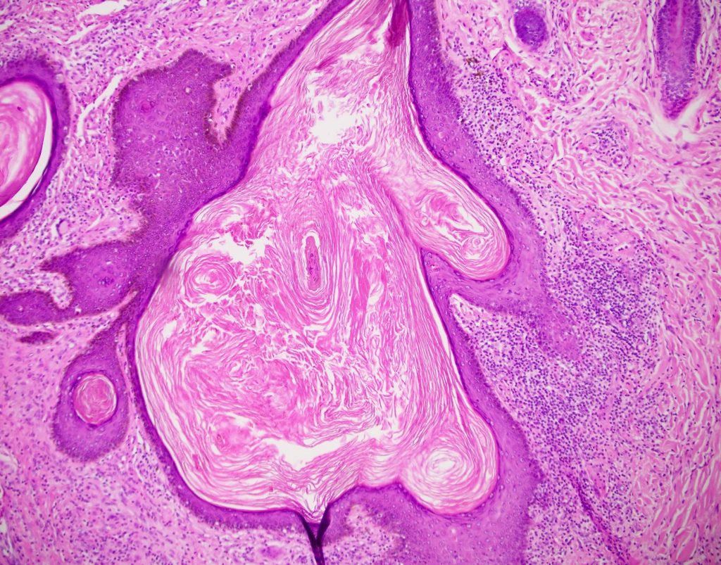
专题全文:硬脑膜 MALT 淋巴瘤
作者:Giovanni Martino, M.D., Flavia Adotti, M.D., Antonio d'Amati, M.D., Martina Mandarano, M.D., Ph.D., Marco Gessi, M.D. · 编委:Jared T. Ahrendsen, M.D., Ph.D. · 工作人员最近更新:2026 年 10 月 6 日(更新进行中)
定义 / 概述
- 发生于硬脑膜的结外低级别黏膜相关淋巴组织(MALT)B 细胞淋巴瘤
- 可伴浆细胞分化
要点
- 发生于硬脑膜、由小至中等大小细胞构成的边缘区 B 细胞淋巴瘤
- 具有 B 细胞表型(CD20、CD79a),但不具有其他淋巴瘤亚型的特异性表型
- 通常伴浆细胞分化,浆细胞成分呈单型性
- 临床病程惰性;多数患者可从局部治疗策略中获益
术语
- 黏膜相关淋巴组织结外边缘区淋巴瘤
- MLD(硬脑膜 MALT 淋巴瘤)
ICD 编码
- ICD-O: 9699/3 - 硬脑膜 MALT 淋巴瘤
- ICD-10: C88.4 - 黏膜相关淋巴组织结外边缘区 B 细胞淋巴瘤(MALT 淋巴瘤)
- ICD-11: 2A85.3 - 结外边缘区 B 细胞淋巴瘤,原发部位不包括胃或皮肤
流行病学
- 罕见疾病;发病率不明(Acta Neurol Belg 2023;123:1189)
- 见于成人,中位年龄在 50~59 岁之间
- 男:女 = 1:5~7(WHO)
- 边缘区淋巴瘤(MZL)是硬脑膜最常见的淋巴瘤
- 较脑实质内 MALT 淋巴瘤更常见(Clin Lymphoma Myeloma Leuk 2018;18:34, Cancer Med 2020;9:663)
部位
- 位于硬脑膜
- 以颅内硬脑膜最常见
- 颅外硬膜(脊髓)较少见(Mod Pathol 2011;24:355, Am J Clin Pathol 2020;154:124)
- 据报道,约 23% 的患者有多灶性病变;全身性复发少见(Leuk Lymphoma 2017;58:882)
病理生理
- 与其他结外边缘区淋巴瘤不同,迄今尚未阐明该部位淋巴瘤的发病机制
- 已有报道发现TNFAIP3, NOTCH2和TBL1XR1基因突变以及其他染色体异常(见分子 / 细胞遗传学描述)(Oncotarget 2016;7:43052)
病因
- 尚未完全明确
- 尚未证实与慢性炎症有关(与胃等其他部位的 MALT 淋巴瘤不同)
- 已有在多发性硬化、鹦鹉热衣原体感染及丙型肝炎背景下发生本病的个案报道(Leuk Res 2012;36:e155, Hum Pathol 2011;42:738, World J Oncol 2020;11:122)
临床特征
- 压迫症状;最常见的是
- 头痛
- 癫痫发作
- 局灶性神经功能缺损
- 参考文献:Hum Pathol 2025;156:105683, Cancer 2020;126:2811
诊断
- 诊断需要组织学检查(J Clin Oncol 2005;23:5718)
- WHO 第 5 版诊断标准
- 必需:位于硬脑膜,具有边缘区样特征和 B 细胞标记,并伴滤泡树突细胞(FDC)网(CD21 / CD23 / CD35+)
- 推荐:若免疫表型尚未证实克隆性,则行 FISH 分析和基因重排克隆性检测
实验室检查
- 当有软脑膜浸润时,脑脊液中可见肿瘤细胞(Neurology 2006;66:1763)
影像学描述
- MRI 表现(Neurology 2006;66:1763)
- 硬脑膜单发或多发斑块状增厚
- 可类似脑膜瘤
- 脑外病变可见钆增强
- 已有报道可见下方脑实质血管源性水肿、骨质增生或骨侵蚀
预后因素
- 预后良好,可达完全缓解(优于脑实质 MZL)(Am J Clin Pathol 2020;154:124)
- 3 年无进展生存率:89%(Leuk Lymphoma 2017;58:882)
病例报告
- 42 岁女性,既往有腮腺 MZL 病史,4 年后出现颅内复发(Acta Neurol Belg 2023;123:1189)
- 49 岁女性,原发性硬脑膜 MZL,表现类似脑膜瘤(Surg Neurol Int 2024;15:113)
- 52 岁女性,原发性硬脑膜 MZL,最初误诊为脑膜瘤,病变进展并行开颅手术后确诊(Acta Neurol Belg 2023;123:1189)
- 63 岁女性,硬脑膜 MZL 伴大量 κ 轻链型淀粉样变性(J Neurosurg 2002;96:368)
- 66 岁女性,同时出现位于 2 个不同区域的 2 处颅内 MZL(Front Oncol 2023;12:927086)
- 83 岁女性,硬脑膜 MZL 伴淋巴脑膜上皮病变(Int J Surg Pathol 2023;31:879)
治疗
- 通常优先选择局部治疗策略而非全身治疗(J Clin Oncol 2005;23:5718)
- 手术切除
- 辅助放疗或化疗,可采用不同的组合(Clin Lymphoma Myeloma Leuk 2018;18:34, Cancer 2020;126:2811)
大体描述
冰冻切片描述
- 术中冰冻切片可能有助于排除侵袭性更强的肿瘤或脑膜瘤
- 通常可见小而成熟的淋巴细胞,伴或不伴浆细胞分化
- 据报道核分裂象少见或不见
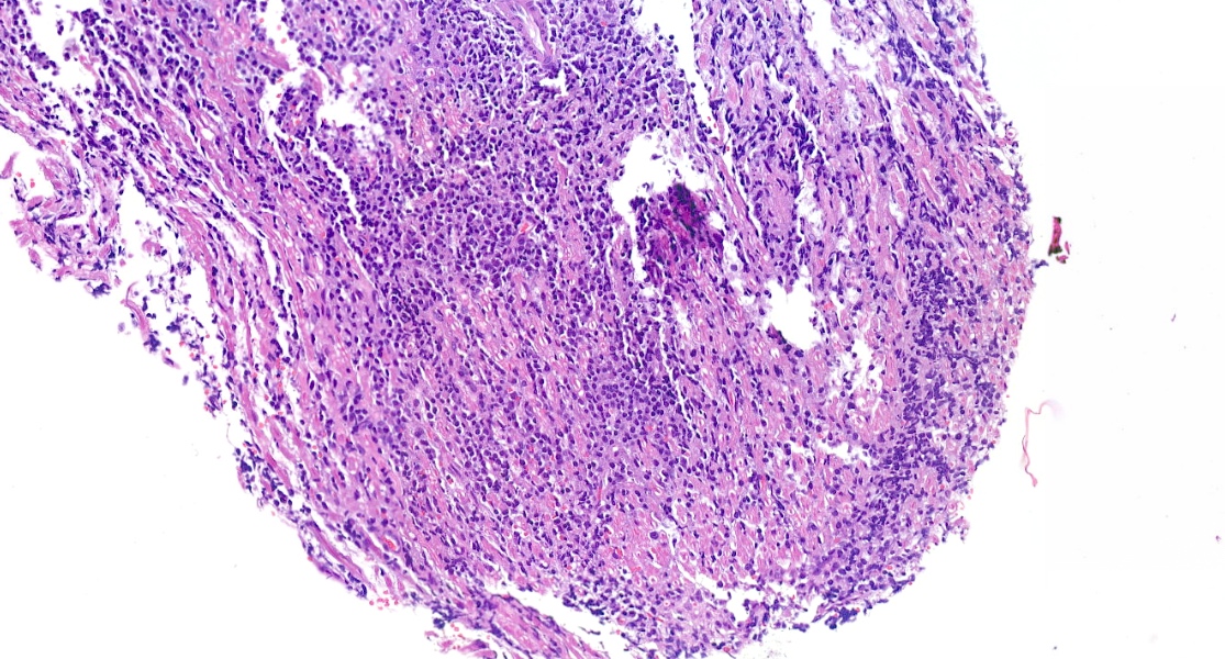
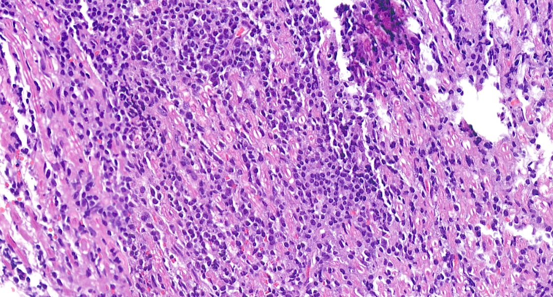
镜下(组织学)描述
- 小淋巴细胞增生并形成肿块(Am J Clin Pathol 2020;154:124)
- 形态(J Clin Oncol 2005;23:5718, Leuk Lymphoma 2017;58:882)
- 明显的浆细胞分化
- 呈单核细胞样外观(胞质透明)
- 生长方式
- 模糊的结节状
- 弥漫性
- 可有不同程度的反应性生发中心
- 可见滤泡植入(滤泡殖民化)
- 少量大细胞(< 5%)
- 浆细胞成分(与脑实质 MZL 相比,其数量通常较少)(Am J Clin Pathol 2020;154:124)
- 陷入的脑膜上皮
- 少见 Virchow-Robin 间隙和脑实质浸润
- 偶见轻链型淀粉样变性(J Clin Oncol 2005;23:5718)
镜下(组织学)图像
供图:Giovanni Martino, M.D. 和 Martina Mandarano, M.D., Ph.D.
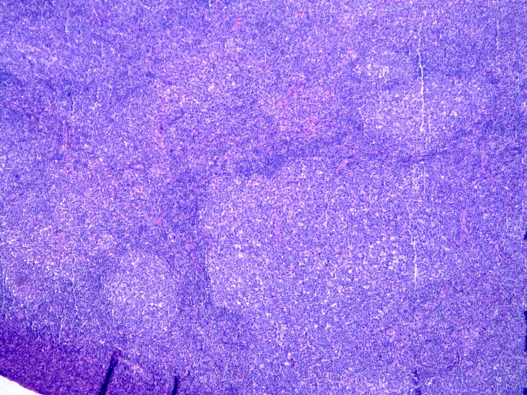
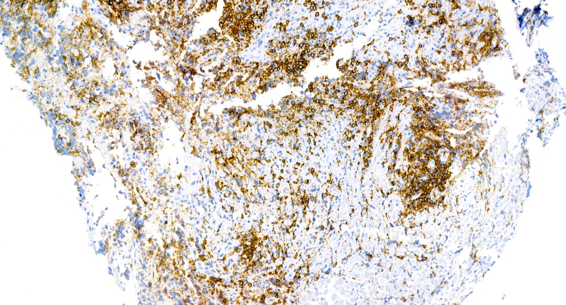
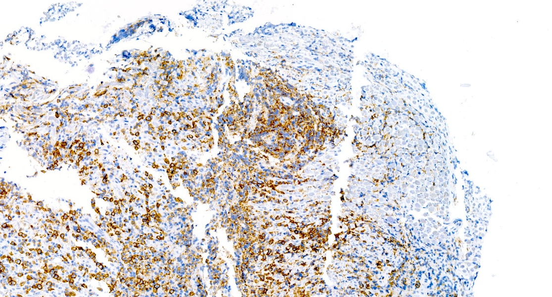
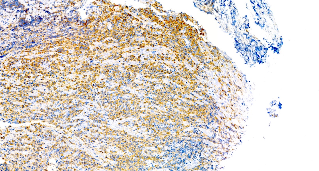
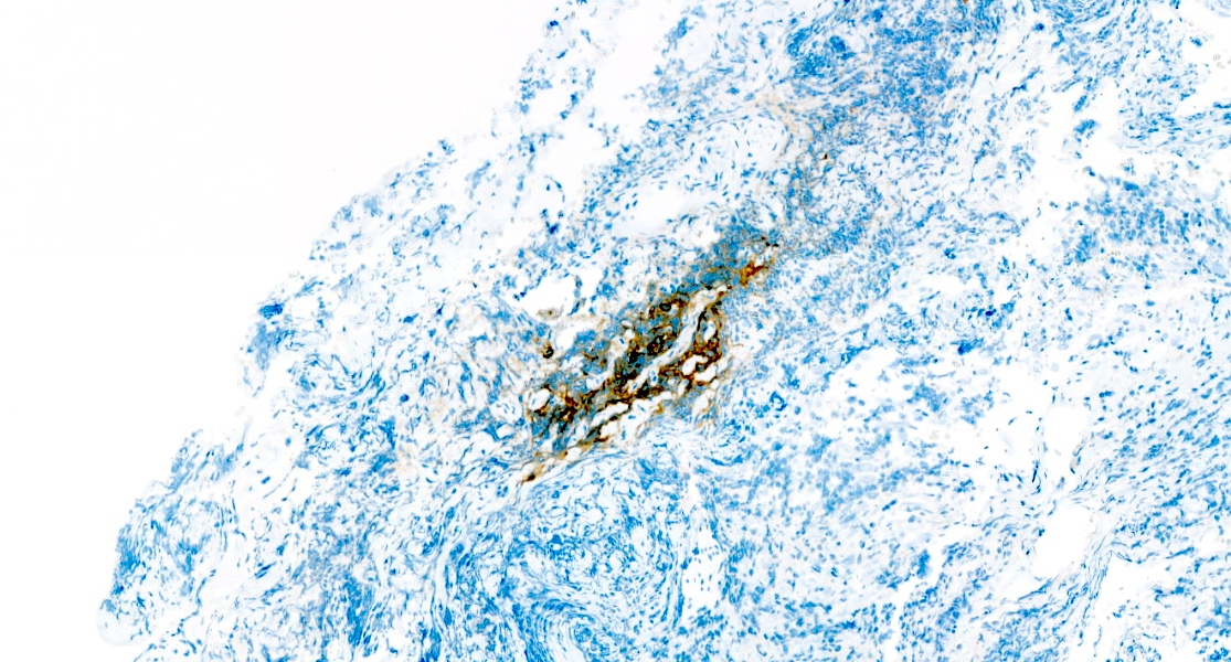
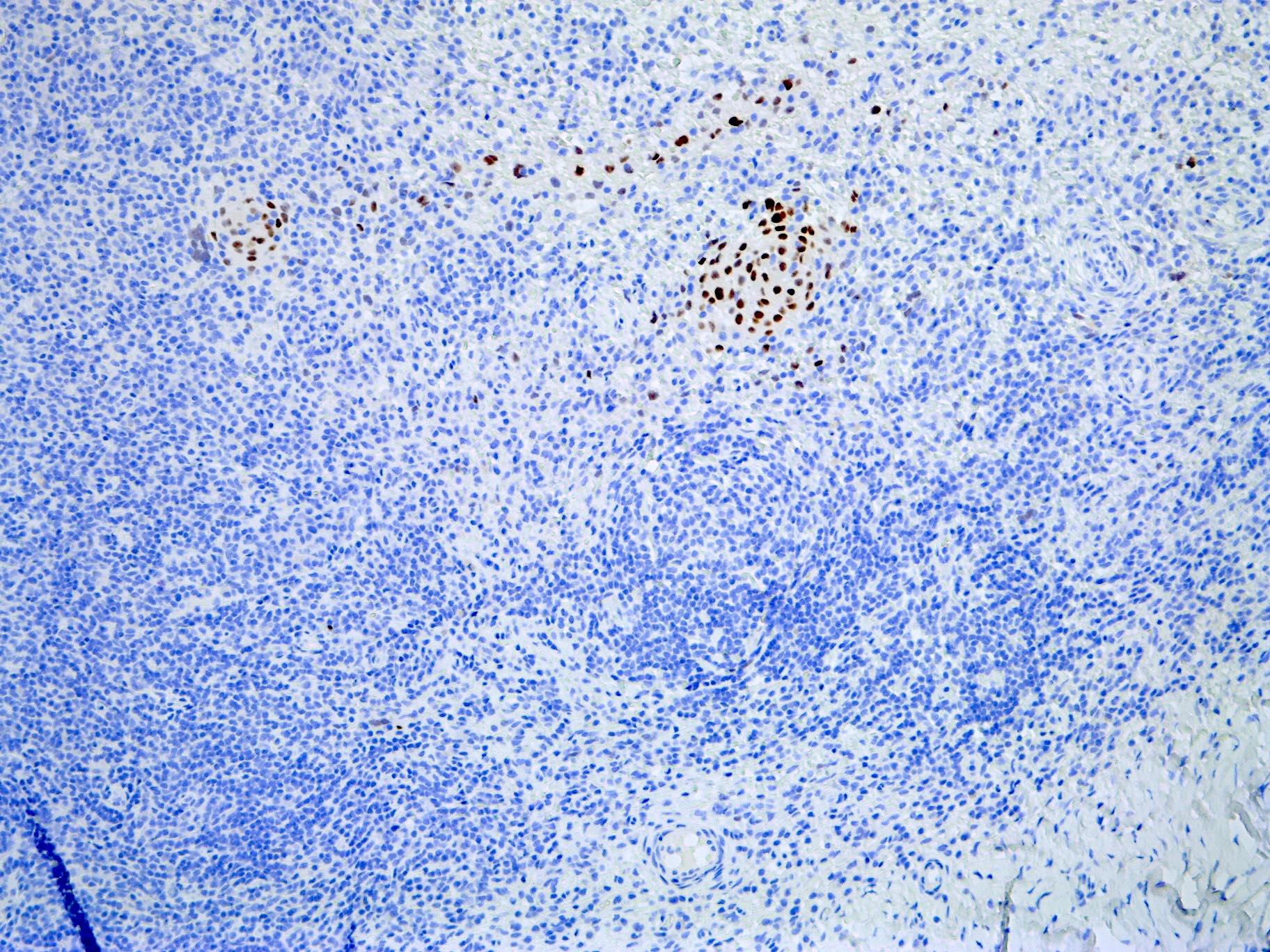
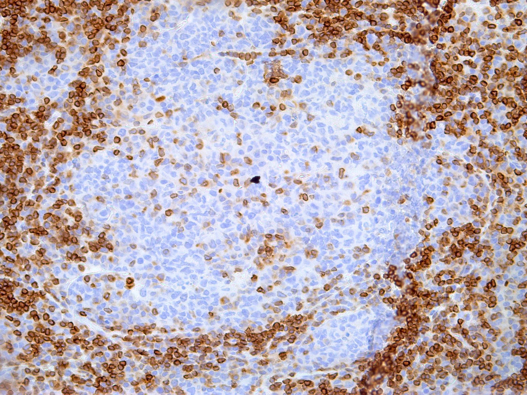
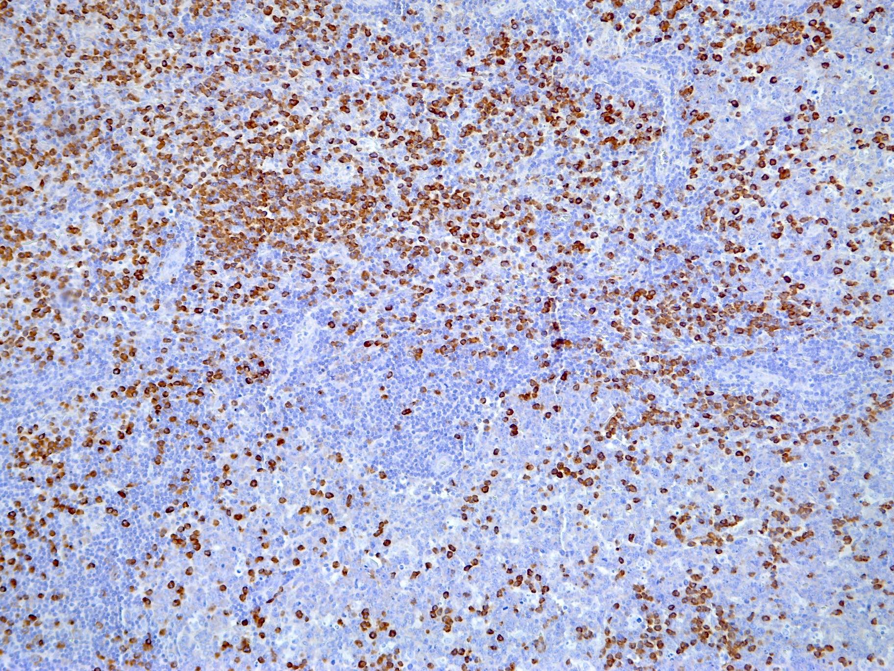
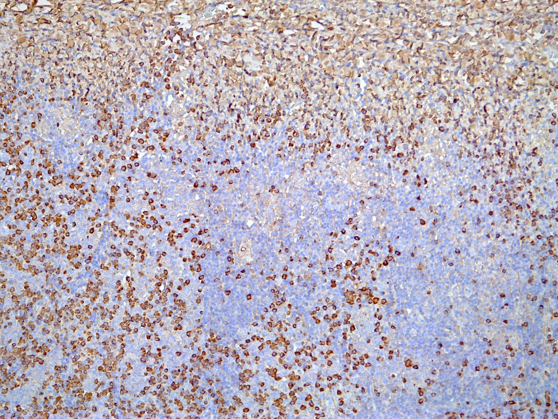
细胞学描述
- 组织涂片中可见数量不等的成熟淋巴细胞、淋巴浆细胞和浆细胞;部分病例报道可见 Dutcher 小体等核内包涵体,可作为诊断线索
- 多数已报道病例无脑脊液受累或受累极为有限;因此,脑脊液细胞学检查对本淋巴瘤的价值可能有限(Am J Clin Pathol 2020;154:124)
阳性染色
- B 细胞谱系标记:CD20, CD79a (J Clin Oncol 2005;23:5718)
- MUM1 / IRF4(表达不一)
- BCL2
- κ 和 λ 轻链:用于显示可能存在的轻链限制
- 增殖指数低(Ki67)(Clin Lymphoma Myeloma Leuk 2018;18:34)
- 与原发性皮肤 MZL(PCMZL)相似,浆细胞常为IgG4+(不伴 IgG4 相关疾病)(Mod Pathol 2011;24:355)
- CD21, CD23和CD35用于显示可能存在的破坏的滤泡树突细胞网(滤泡植入〔滤泡殖民化〕)
- Congo red(刚果红)可显示淀粉样物质沉积(苹果绿双折光)(J Neurosurg 2002;96:368)
- 参考文献:Cancer 2020;126:2811, Hum Pathol 2025;156:105683
阴性染色
- CD5, CD23, cyclin D1, CD10, BCL6 (J Clin Oncol 2005;23:5718)
- EBER (J Clin Oncol 2005;23:5718)
- 细胞角蛋白
- PR, SSTR2A
- STAT6, S100, MelanA, PRAME, SOX10
分子 / 细胞遗传学描述
- IG基因克隆性检测用于确认该增生的克隆性
- 总体上,其基因组特征与其他 MZL 亚型相似,但 3 号和 18 号染色体受累较少(Oncotarget 2016;7:43052)
- 特征性表现为相互排斥的遗传学改变与不同的形态表型相关
- 单等位基因和双等位基因的TNFAIP3失活更常见于表现出浆细胞分化的病例,包括 3 例 IgG4+ 病例(Oncotarget 2016;7:43052)
- 激活性NOTCH2突变主要见于具有单核细胞样外观的病例报道
- NOTCH2突变病例通常也存在失活性TBL1XR1突变
- 其他反复出现的突变涉及KLHL6和MLL2
- 染色体异常,例如 1p36.32 区域缺失(涵盖TNFRSF14, PRDM16, RPL22和CAMTA1基因)以及 6p25.3 区域增益(涵盖IRF4基因)
- IGH::MALT1融合 [t(14;18)(q32;q21)] 已在 1 例病例中报道(J Hematop 2008;1:131)
病理报告示例
- 中枢神经系统(如额叶)硬脑膜病变,活检 / 手术切除:
- 该部位见伴浆细胞分化的成熟 B 细胞肿瘤,符合黏膜相关淋巴组织结外边缘区淋巴瘤(见附注)
- 附注:建议进行分期检查(影像学检查和骨髓活检),以排除全身性病变并确认原发于硬脑膜。
鉴别诊断
- 与其他小至中等大小 B 细胞淋巴瘤鉴别,例如(J Neurosurg 2002;96:368, J Clin Oncol 2005;23:5718):
- 浆细胞肿瘤,在浆细胞分化极为显著的病例中需与之鉴别:
- 鉴别诊断可能十分困难,尽管孤立性骨外浆细胞肿瘤非常罕见
- FISH 分析可能有助于区分这些不同疾病(Semin Diagn Pathol 2025;42:150944)
- 脑膜瘤:
- 在某些病例中,增强、骨侵蚀以及无钙化等影像学特征可能有助于淋巴瘤的诊断(Neuroradiology 2021;63:1215)
- 明确诊断必须进行组织学检查
- IgG4 相关疾病的形成占位性病变的变型,当存在明显浆细胞成分时需与之鉴别:
- 在这些病例中,应按照 IgG4 相关疾病共识指南,仔细评估IgG4 / IgG比值(Mod Pathol 2012;25:1181)
- IgG4 相关疾病中的浆细胞为多克隆性
- 对于寡克隆性或单克隆性浆细胞的病例,应结合临床和血清学资料仔细评估,因为罕见病例曾报道 MALT 淋巴瘤与 IgG4 相关疾病存在重叠
- 脑膜黑色素瘤或皮肤黑色素瘤的转移播散:
- 在呈浆细胞样形态变型的部分病例中,可考虑这些诊断;此时必须采用适当的免疫组织化学标记组合才能作出正确诊断
- 在某些病例中,应考虑与位于硬脑膜的实体肿瘤进行鉴别诊断:
- 富于细胞的 孤立性纤维性肿瘤(SFT):
- 富于细胞的 SFT 可通过STAT6免疫组织化学染色及仔细检查间质成分来诊断
- 小细胞肉瘤,例如尤因肉瘤:
- 也可考虑
- 必须进行免疫组织化学检查(如CD99)及适当的分子检测,才能作出正确诊断
- 富于细胞的 孤立性纤维性肿瘤(SFT):
其他参考文献
练习题 #1
以下哪项组织学表现最具有硬脑膜边缘区淋巴瘤(MZL)的特征?
- 弥漫性脑实质浸润
- 广泛凝固性坏死
- 肿瘤性边缘区 B 细胞的滤泡植入(滤泡殖民化)
- 高度细胞学异型性伴活跃的核分裂活动
- 血管周围假菊形团
练习题答案 #1
C.肿瘤性边缘区 B 细胞的滤泡植入(滤泡殖民化)。滤泡植入(滤泡殖民化)是 MZL 公认的特征,在硬脑膜病例中也可见。选项 D 不正确,因为硬脑膜 MZL 是一种低级别淋巴瘤,细胞形态温和,核分裂活动低。选项 B 不正确,因为广泛坏死不是典型表现,应提示考虑其他诊断。选项 A 不正确,因为肿瘤通常局限于硬脑膜,缺乏明显的脑实质浸润。选项 E 不正确,因为血管周围假菊形团是室管膜瘤的特征,在 MZL 中不见。
参考文献:硬脑膜 MALT 淋巴瘤
练习题 #2
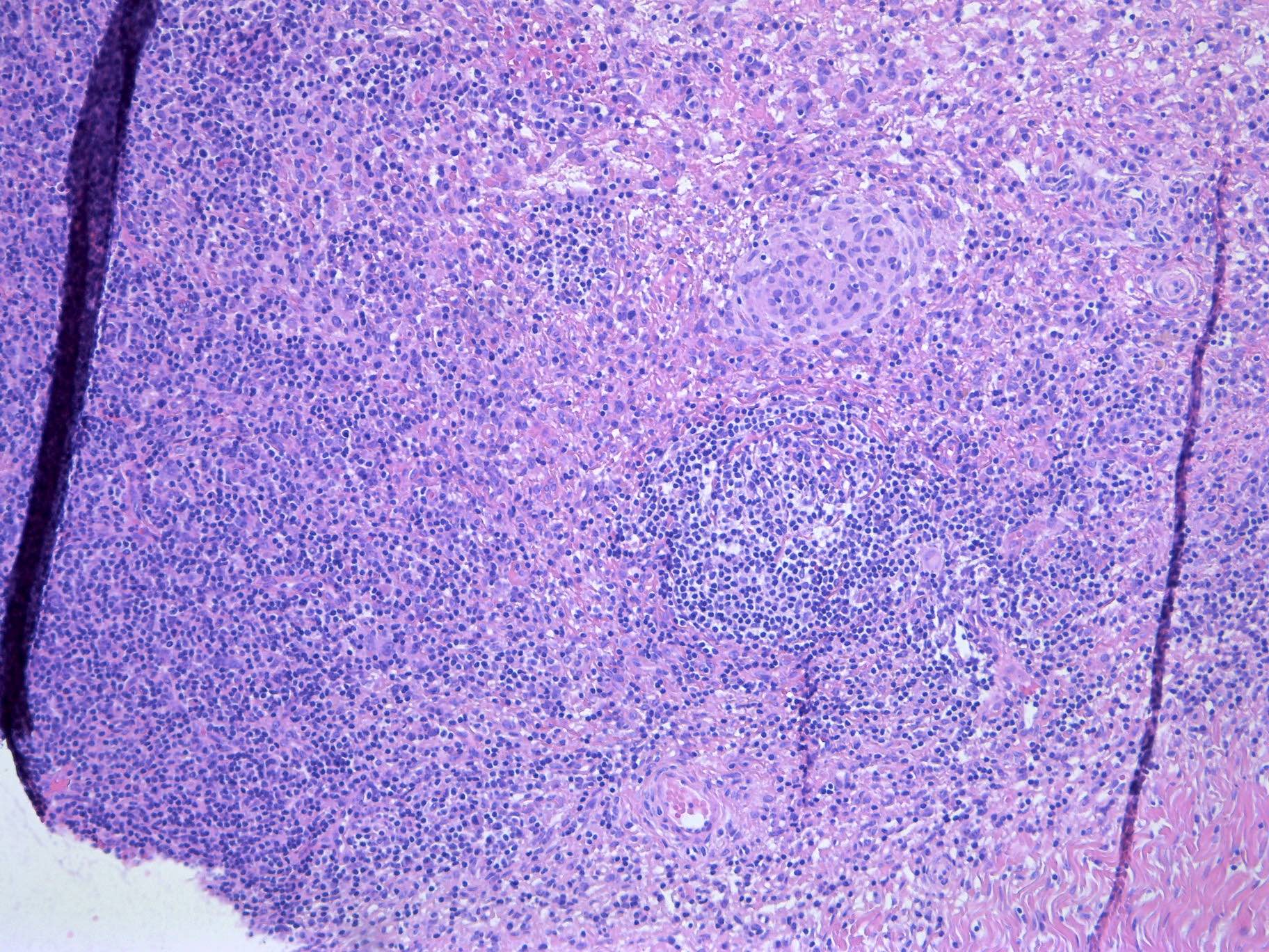
硬脑膜病变的组织学检查显示小而成熟的淋巴细胞伴浆细胞分化,以及被边缘区细胞浸润的残存反应性生发中心。上图显示的是哪种组织学表现?
- 弥漫性大 B 细胞淋巴瘤
- 滤泡植入(滤泡殖民化)
- 滤泡性淋巴瘤
- 地图样坏死
- 星空现象
练习题答案 #2
B.滤泡植入(滤泡殖民化)。图像显示残存的反应性生发中心被边缘区 B 细胞浸润并部分取代,这是边缘区淋巴瘤的特征性表现。选项 C 不正确,因为滤泡性淋巴瘤是一种独立的 B 细胞淋巴瘤,以肿瘤性滤泡生长方式为特征,通常表达 CD10 和 BCL6。选项 D 不正确,因为未见地图样坏死,且这种惰性淋巴瘤中也少见此表现。选项 A 不正确,因为弥漫性大 B 细胞淋巴瘤由成片的大异型淋巴样细胞构成,而本图中未见此类细胞。选项 E 不正确,因为星空现象是高度增殖的淋巴瘤,尤其是伯基特淋巴瘤的特征,本病变中未见。
参考文献:硬脑膜 MALT 淋巴瘤
- MALT lymphoma of the dura: extranodal, dural based, low grade B cell lymphoma of mucosa associated lymphoid tissue, usually with monotypic plasmacytic differentiation; adults, median age in the sixth decade, M:F = 1:5–7; MZL is the most common lymphoma of the dura and more common than intraparenchymal MALT lymphoma.
- MRI may show single or multiple plaque-like dural thickening mimicking meningioma; WHO 5th edition essential criteria: dural based, marginal zone-like features and B cell markers with an FDC meshwork (CD21 / CD23 / CD35+). Follicular colonization, entrapped meningothelium and occasional light chain amyloidosis; plasma cells often IgG4+.
- Molecular: TNFAIP3 inactivation is more common with plasmacytic differentiation (including 3 IgG4+ cases); activating NOTCH2 mutations mostly with monocytoid appearance, often with TBL1XR1 inactivation; IGH::MALT1 reported in 1 case.
- The topic includes 12 microscopic / IHC images, a sample pathology report, differential diagnosis and 2 practice questions; the same update also covers DIP, acne vulgaris, chromoblastomycosis and NUTM1 rearranged sarcoma (summaries only).
Scope: full text of the PathologyOutlines blog post “12 October 2026: Textbook Updates” (5 topic summaries and the featured image), plus the full topic page “MALT lymphoma of the dura” (all sections, 12 on-site images with captions, sample pathology report, differential diagnosis, practice questions and answers). The other 4 topics are included as blog summaries only.
Textbook Updates · 12 October 2026
We have posted updates of the following topics:
CNS & pituitary tumors > Lymphoma & histiocytic tumors > MALT lymphoma of the dura
by Giovanni Martino, M.D., Flavia Adotti, M.D., Antonio d’Amati, M.D., Martina Mandarano, M.D., Ph.D., Marco Gessi, M.D.
Topic summary: Extranodal, dural based, low grade B cell lymphoma of mucosa associated lymphoid tissue (MALT); can have plasmacytic differentiation. Mutations in TNFAIP3, NOTCH2 and TBL1XR1 genes and other chromosomal aberrations have been reported. Unlike other extranodal marginal zone lymphomas, no known pathogenetic mechanism has been described to date. Compression symptoms; most commonly headache, seizures and focal neurological defects. Indolent clinical course; most patients benefit from local treatment strategies.
Lung > Interstitial lung diseases associated with smoking > Alveolar macrophage pneumonia (desquamative interstitial pneumonitis)
by Akira Yoshikawa, M.D., Andrey Bychkov, M.D., Ph.D., Sakda Sathirareuangchai, M.D.
Topic summary: Rare form of idiopathic interstitial pneumonia characterized by accumulation of intra-alveolar macrophages with intracytoplasmic, finely granular brown pigment. Most cases are associated with cigarette smoking; may also occur in association with connective tissue disease, medications, inhalational exposures, aspiration or hereditary disorders. Diagnosis is based on clinical features, radiology and histopathology. Most common symptom is dyspnea, followed by nonproductive cough. Generally favorable prognosis with good response to treatment.
Skin nontumor > Other diseases of skin appendages > Acne vulgaris
by Andrew Kulbiski, M.D., Jayden Ray, D.O., Sepideh Nikki Asadbeigi, M.D.
Topic summary: Acne vulgaris is a common inflammatory skin condition that primarily affects the face and trunk. Clinical lesions range from noninflamed comedones to inflamed papules, pustules, nodules and cysts. Multifactorial pathogenesis involving increased sebum production, follicular hyperkeratinization, Cutibacterium acnes overgrowth and a resulting inflammatory immune response. In the Western world, acne vulgaris is highly prevalent among adolescents. Topical benzoyl peroxide, topical retinoids and topical antibiotics are first line treatments.
Skin nontumor > Infectious disorders > Chromoblastomycosis
by Peyker Temiz, M.D.
Topic summary: Chromoblastomycosis is an infrequent fungal infection that occurs sporadically in tropical and subtropical areas, caused by various darkly pigmented (dematiaceous) fungi. Granulomatous tissue reaction with neutrophilic abscess is seen. Primarily affects rural workers since it is caused by a variety of fungi found in soil, wood and decaying plant material. Most common in middle aged men. Infection usually appears on exposed skin, especially the lower legs and arms. Infection develops slowly, lasts for many years and is resistant to treatment.
Soft tissue > Uncertain differentiation > NUTM1 rearranged sarcoma
by Divya Bansal, M.B.B.S., M.D., Sunil Pasricha, M.B.B.S., M.D.
Topic summary: NUTM1 rearranged sarcoma is an emerging group of aggressive soft tissue sarcomas defined by chromosomal rearrangements involving the NUTM1 (NUT midline carcinoma family member 1) gene on chromosome 15q14, distinct from the more well known NUT carcinoma. Extremely rare entity. Rapidly growing, usually deep seated, often painful or palpable mass. Most common in the somatic soft tissues, thoracic region, gastrointestinal tract and body cavity. Metastasis is seen in 50% of adult cases at presentation; overall survival: 13.4 months.
Featured Image
Topic full text: MALT lymphoma of the dura
Authors: Giovanni Martino, M.D., Flavia Adotti, M.D., Antonio d'Amati, M.D., Martina Mandarano, M.D., Ph.D., Marco Gessi, M.D. · Editorial Board Member: Jared T. Ahrendsen, M.D., Ph.D. · Last staff update: 6 October 2026 (update in progress)
Definition / general
- Extranodal, dural based, low grade B cell lymphoma of mucosa associated lymphoid tissue (MALT)
- Can have plasmacytic differentiation
Essential features
- Small to medium sized marginal zone B cell lymphoma arising in the dura
- B cell phenotype (CD20, CD79a) nonspecific for other lymphoma subtypes
- Usually, plasmacytic differentiation with monotypic plasma cell component
- Indolent clinical course; most patients benefit from local treatment strategies
Terminology
- Extranodal marginal zone lymphoma of mucosa associated lymphoid tissue
- MLD (MALT lymphoma of the dura)
ICD coding
- ICD-O: 9699/3 - MALT lymphoma of the dura
- ICD-10: C88.4 - extranodal marginal zone B cell lymphoma of mucosa associated lymphoid tissue (MALT lymphoma)
- ICD-11: 2A85.3 - extranodal marginal zone B cell lymphoma, primary site excluding stomach or skin
Epidemiology
- Rare disease; incidence unknown (Acta Neurol Belg 2023;123:1189)
- Adult, median age = sixth decade
- M:F = 1:5 - 7 (WHO)
- Marginal zone lymphoma (MZL) is the most common lymphoma of the dura
- More common than intraparenchymal MALT lymphoma (Clin Lymphoma Myeloma Leuk 2018;18:34, Cancer Med 2020;9:663)
Sites
- Dural based
- Intracranial dura most commonly
- Extracranial dura (spinal cord) less frequently (Mod Pathol 2011;24:355, Am J Clin Pathol 2020;154:124)
- Multifocal disease reported in ~23% of patients; systemic recurrence is uncommon (Leuk Lymphoma 2017;58:882)
Pathophysiology
- Unlike other extranodal marginal zone lymphomas, no known pathogenetic mechanism has been described in this setting to date
- Mutations in TNFAIP3, NOTCH2 and TBL1XR1 genes, along with other chromosomal aberrations, have been reported (see Molecular / cytogenetics description) (Oncotarget 2016;7:43052)
Etiology
- Still not completely known
- Correlation with chronic inflammation has not yet been demonstrated (unlike MALT lymphomas of other sites, e.g., gastric)
- Onset of this disease in the setting of multiple sclerosis, Chlamydia psittaci infection and hepatitis C have been reported in single cases (Leuk Res 2012;36:e155, Hum Pathol 2011;42:738, World J Oncol 2020;11:122)
Clinical features
- Compression symptoms; most commonly
- Headache
- Seizures
- Focal neurological defects
- References: Hum Pathol 2025;156:105683, Cancer 2020;126:2811
Diagnosis
- Diagnosis requires histological examination (J Clin Oncol 2005;23:5718)
- WHO 5th edition diagnostic criteria
- Essential: dural based, marginal zone-like features and B cell markers with follicular dendritic cells (FDC) meshwork (CD21 / CD23 / CD35+)
- Desirable: FISH analysis and gene rearrangement clonality assay, if not already demonstrated with immunophenotype
Laboratory
- Cerebrospinal fluid may show neoplastic cells in case of leptomeningeal infiltration (Neurology 2006;66:1763)
Radiology description
- MRI findings (Neurology 2006;66:1763)
- Single or multiple plaque-like thickening of the dura
- Can mimic meningioma
- Extra-axial lesions show gadolinium enhancement
- Underlying parenchymal vasogenic edema, hyperostosis or bone erosion have been reported
Prognostic factors
- Good prognosis with complete remission (better than parenchymal based MZL) (Am J Clin Pathol 2020;154:124)
- 3 year progression free survival: 89% (Leuk Lymphoma 2017;58:882)
Case reports
- 42 year old woman with positive history of MZL of the parotid gland, developed intracranial relapse 4 years later (Acta Neurol Belg 2023;123:1189)
- 49 year old woman with primary MZL of the dura mimicking meningioma (Surg Neurol Int 2024;15:113)
- 52 year old woman with primary MZL of the dura, initially misdiagnosed as meningioma, confirmed after progression and craniotomy (Acta Neurol Belg 2023;123:1189)
- 63 year old woman with massive kappa light chain amyloidosis associated with MZL of the dura (J Neurosurg 2002;96:368)
- 66 year old woman presented with 2 simultaneous intracranial MZL, located in 2 different regions (Front Oncol 2023;12:927086)
- 83 year old woman with MZL of the dura and lymphomeningothelial lesions (Int J Surg Pathol 2023;31:879)
Treatment
- Local treatment strategies usually preferred over systemic treatment (J Clin Oncol 2005;23:5718)
- Surgical resection
- Adjuvant radiation or chemotherapy in variable combination (Clin Lymphoma Myeloma Leuk 2018;18:34, Cancer 2020;126:2811)
Gross description
Frozen section description
- Intraoperative frozen section may be helpful in exclusion of more aggressive neoplasms or meningioma
- Small mature lymphocytes are typically observed with or without plasmacytic differentiation
- Few or no mitoses are reported
Microscopic (histologic) description
- Mass forming proliferation of small lymphocytes (Am J Clin Pathol 2020;154:124)
- Morphology (J Clin Oncol 2005;23:5718, Leuk Lymphoma 2017;58:882)
- Prominent plasmacytic differentiation
- Monocytoid appearance (clear cytoplasm)
- Growth patterns
- Vaguely nodular
- Diffuse
- Reactive germinal centers variably present
- Follicular colonization may be observed
- Small amount of large cell (< 5%)
- Plasma cell component (usually in limited quantity in comparison to parenchymal based MZL) (Am J Clin Pathol 2020;154:124)
- Entrapped meningothelium
- Rarely, Virchow-Robin spaces and brain parenchyma infiltration
- Occasional light chain amyloidosis (J Clin Oncol 2005;23:5718)
Microscopic (histologic) images
Contributed by Giovanni Martino, M.D. and Martina Mandarano, M.D., Ph.D.
Cytology description
- Varying numbers of mature lymphocytes, lymphoplasmacytes and plasma cells are observed in tissue smears; in some cases, nuclear inclusions such as Dutcher bodies are reported and may be a clue for diagnosis
- Most reported cases show no or very limited cerebrospinal fluid involvement; therefore, cerebrospinal fluid cytology examination may be of limited value in this lymphoma (Am J Clin Pathol 2020;154:124)
Positive stains
- B lineage marker: CD20, CD79a (J Clin Oncol 2005;23:5718)
- MUM1 / IRF4 (variable)
- BCL2
- Kappa and lambda chains: to highlight eventual light chain restriction
- Low proliferation index (Ki67) (Clin Lymphoma Myeloma Leuk 2018;18:34)
- As in primary cutaneous MZL (PCMZL), plasma cells are often IgG4+ (without IgG4 related disease) (Mod Pathol 2011;24:355)
- CD21, CD23 and CD35 to point out the possible presence of disrupted follicular dendritic cell meshwork (follicular colonization)
- Congo red can reveal the amyloid deposition (apple green birefringence) (J Neurosurg 2002;96:368)
- References: Cancer 2020;126:2811, Hum Pathol 2025;156:105683
Negative stains
- CD5, CD23, cyclin D1, CD10, BCL6 (J Clin Oncol 2005;23:5718)
- EBER (J Clin Oncol 2005;23:5718)
- Cytokeratins
- PR, SSTR2A
- STAT6, S100, MelanA, PRAME, SOX10
Molecular / cytogenetics description
- IG genes clonality assays to confirm the clonal nature of the proliferation
- Overall, similar genomic landscape of other MZL subtypes, except chromosome 3 and 18 are less frequently involved (Oncotarget 2016;7:43052)
- Characteristically, mutually exclusive genetic alterations correlate with distinct morphophenotypes
- Monoallelic and biallelic inactivation of TNFAIP3 are more common in cases exhibiting plasmacytic differentiation, including 3 IgG4+ cases (Oncotarget 2016;7:43052)
- Activating NOTCH2 mutations are mostly reported in cases with monocytoid appearance
- NOTCH2 mutated cases generally display also inactivating TBL1XR1 mutations
- Other recurrent mutations in KLHL6 and MLL2
- Chromosomal aberrations such as losses at 1p36.32 (encompassing TNFRSF14, PRDM16, RPL22 and CAMTA1 genes) and gains at 6p25.3 (covering the IRF4 gene)
- IGH::MALT1 fusion [t(14;18)(q32;q21)] has been reported in 1 case (J Hematop 2008;1:131)
Sample pathology report
- Dural based lesion, CNS (e.g., frontal lobe), biopsy / excisional surgery:
- Localization of mature B cell neoplasm with plasmacytic differentiation, consistent with extranodal marginal zone lymphoma of mucosa associated lymphoid tissue (see comment)
- Comment: Staging workup (imaging and bone marrow biopsy) is recommended to exclude systemic disease and to confirm primary dural localization.
Differential diagnosis
- With other small to medium B cell lymphomas, such as (J Neurosurg 2002;96:368, J Clin Oncol 2005;23:5718):
- Lymphoplasmacytic lymphoma:
- Morphophenotype overlaps; IgM monoclonal component and MYD88 L265P mutation required
- Chronic lymphocytic leukemia / small lymphocytic lymphoma (CLL / SLL):
- Mantle cell lymphoma:
- Follicular lymphoma:
- Lymphoplasmacytic lymphoma:
- Plasma cell neoplasms in cases of extreme plasmacytic differentiation:
- Differential diagnosis can be very tricky, although isolated extraosseous plasma cell neoplasms are very rare
- FISH analyses may be helpful to distinguish the different diseases (Semin Diagn Pathol 2025;42:150944)
- Meningiomas:
- In selected cases, radiologic features like contrast enhancement, bone erosion and the absence of calcification may be helpful in the diagnosis of lymphoma (Neuroradiology 2021;63:1215)
- Histology is mandatory for a certain diagnosis
- IgG4 related disease in its lesion forming variant, when a prominent plasmacytic component is reported:
- In these cases, the IgG4 / IgG ratio should be carefully evaluated, as in the IgG4 related disease consensus guidelines (Mod Pathol 2012;25:1181)
- Plasma cells in IgG4 related disease are polyclonal
- Cases with oligoclonal or monoclonal plasma cells should be carefully evaluated with clinical and serological data, as an overlap between MALT lymphoma and IgG4 related disease has been reported in rare cases
- Meningeal melanoma or metastatic dissemination of cutaneous melanoma:
- Some cases may be considered in cases of plasmacytoid-like morphological variants; in these cases, an appropriate immunohistochemical panel is mandatory to render the correct diagnosis
- In selected cases, a differential diagnosis with dural based solid tumors should be considered:
- Hypercellular solitary fibrous tumor (SFT):
- Hypercellular SFT may be diagnosed with STAT6 immunohistochemistry and a careful examination of the stromal component
- Small cell sarcomas, such as Ewing sarcoma:
- May also be considered
- Immunohistochemical (e.g., CD99) and appropriate molecular testing are mandatory to correctly render the diagnosis
- Hypercellular solitary fibrous tumor (SFT):
Additional references
Practice question #1
Which of the following histologic findings is most characteristic of marginal zone lymphoma (MZL) of the dura?
- Diffuse brain parenchymal invasion
- Extensive coagulative necrosis
- Follicular colonization by neoplastic marginal zone B cells
- High grade cytologic atypia with brisk mitotic activity
- Perivascular pseudorosettes
Practice answer #1
C. Follicular colonization by neoplastic marginal zone B cells. Follicular colonization is a well recognized feature of MZL and may be seen in dural cases. Answer D is incorrect because dural MZL is a low grade lymphoma with bland cytology and low mitotic activity. Answer B is incorrect because extensive necrosis is not a typical finding and should prompt consideration of a different diagnosis. Answer A is incorrect because the tumor usually remains confined to the dura and lacks significant brain parenchymal invasion. Answer E is incorrect because perivascular pseudorosettes are characteristic of ependymoma and are not seen in MZL.
Reference: MALT lymphoma of the dura
Practice question #2
Histologic examination of a dural based lesion shows small mature lymphocytes with plasmacytic differentiation and residual reactive germinal centers infiltrated by marginal zone cells. Which histologic finding is demonstrated in the image above?
- Diffuse large B cell lymphoma
- Follicular colonization
- Follicular lymphoma
- Geographic necrosis
- Starry sky pattern
Practice answer #2
B. Follicular colonization. The image shows residual reactive germinal centers infiltrated and partially replaced by marginal zone B cells, a characteristic feature of marginal zone lymphoma. Answer C is incorrect because follicular lymphoma is a distinct B cell lymphoma characterized by a neoplastic follicular growth pattern and typically expresses CD10 and BCL6. Answer D is incorrect because geographic necrosis is not present and is uncommon in this indolent lymphoma. Answer A is incorrect because diffuse large B cell lymphoma is composed of sheets of large atypical lymphoid cells, which are not seen in this image. Answer E is incorrect because a starry sky pattern is characteristic of highly proliferative lymphomas, particularly Burkitt lymphoma, and is not present in this lesion.
Reference: MALT lymphoma of the dura