- NUTM1 重排肉瘤是一组新近认识的侵袭性软组织肉瘤,以 15q14 上 NUTM1 基因重排为定义特征,有别于更为人熟知的 NUT 癌;发病年龄范围广,常累及躯体软组织与胃肠道。
- 组织学多样:纤维肉瘤样、圆形细胞、上皮样 / 横纹肌样及透明变 / 巢状模式,可有石棉样纤维样胶原;NUT 蛋白免疫组化常阳性,确诊依赖分子检测(FISH / NGS / RNA 测序),融合伙伴包括 MGA、MXD4 等。
- 临床侵袭:半数成人就诊时已转移,总生存期约 13.4 个月;专题含数十幅镜下、免疫组化与分子图,以及鉴别诊断与练习题。
收录范围:PathologyOutlines「NUTM1 rearranged sarcoma」专题页全文(全部栏目、站内镜下 / 免疫组化 / 分子图像及图注、鉴别诊断、练习题等)。该专题列入 2026-10-12 教科书更新;硬脑膜 MALT 淋巴瘤专题已于本站 2026-10-13 单独成篇。
定义 / 概述
- NUTM1 重排肉瘤是一组新近认识的侵袭性软组织肉瘤,以涉及染色体 15q14 上的 NUTM1 (NUT 中线癌家族成员 1)基因的染色体重排为定义特征,有别于更为人熟知的 NUT 癌
要点
- 一组新近认识的侵袭性软组织肉瘤
- 发病年龄范围广,无性别倾向,无易感因素
- 常累及躯体软组织和胃肠道
- 组织学形态多样,包括纤维肉瘤样、圆形细胞、上皮样 / 横纹肌样及透明变 / 巢状模式,有时可见石棉样纤维样胶原,NUT 蛋白免疫反应阳性
- 定义特征为 NUTM1 重排,最常与 MAD 家族基因(MXD4, MXD1, MXI1, MGA)融合,而非 BRD3 / 4 (后者是 NUT 癌的特征性融合伴侣)
术语
- NUT 重排肉瘤
ICD 编码
- ICD-O: 8800/3 - 肉瘤,非特指型
- ICD-10: C49.9 - 结缔组织和软组织恶性肿瘤,未特指
- ICD-11: 2B5K - 未特指的恶性软组织肿瘤,或其他或未特指部位的骨或关节软骨肉瘤
流行病学
- 极为罕见的肿瘤类型,文献中仅有少量病例报道(约 70 例)
- 发病年龄范围广:1 - 78 岁(Semin Diagn Pathol 2025;42:150941)
- 通常累及中年成人(中位年龄:48 岁)
- 曾报道 1 例发生于 13 个月大男童的病例(Biomedicines 2024;12:1709)
- 女性略多见(男:女 = 1:1.7)
- 无已知地域倾向;世界各地均有报道
部位
- 最常见于躯体软组织、胸部、胃肠道和体腔(Semin Diagn Pathol 2025;42:150941, Mod Pathol 2021;34:1547)
- 其他部位包括下颌骨、肾、膀胱、阴茎、卵巢、子宫、头皮、眶区、脑及中枢神经系统(顶叶和硬脑膜)(Genes Chromosomes Cancer 2022;61:542, Semin Diagn Pathol 2025;42:150941)
- 儿童病例主要累及软组织,位于胸部;尚无腹部或盆腔病例的记载(Semin Diagn Pathol 2025;42:150941)
病理生理
- NUTM1 基因位于染色体 15q,正常情况下在睾丸中表达,在精子发生过程中促进组蛋白乙酰化
- 最常见的融合伴侣:来自 MYC 相关因子 X(MAX)二聚化(MAD)转录家族的基因(例如 MXD1, MXD4, MXI1 [或 MXD2]和 MGA),而非 BRD3 或 BRD4 (常见于 NUT 癌)
- MAD::NUTM1 融合蛋白被推测可导致基因表达失调,可能通过阻断分化和促进异常增殖发挥作用
- 不同于 BRD4::NUT,其可能不形成超大结构域,也不依赖 BET 蛋白,这可能解释其对 BET 抑制剂的耐药性
- 罕见融合伴侣包括 BCORL1, ZNF532, NSD3, ONECUT, BRD3 和 BRD4 (Am J Surg Pathol 2018;42:636, Am J Surg Pathol 2024;48:930)
- 无已知前驱病变
病因
- 目前尚不明确
示意图 / 表格
供图:Bhagat Singh Lali 医学博士
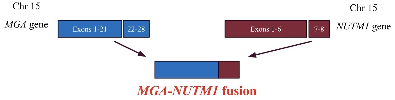
托管于其他服务器的图像:

临床特征
- 肿块生长迅速,通常位于深部,常伴疼痛或可触及
- 症状取决于解剖部位(例如,中枢神经系统肿瘤可引起头痛和偏瘫;胃肠道肿瘤可引起梗阻、腹胀、体重减轻、腹痛;胸部肿瘤可引起气促;眶部原发肿瘤可引起上睑下垂),也可在影像学检查中发现肿块
诊断
- 必要诊断标准
- 部分透明变的背景中可见单一形态的梭形、上皮样及横纹肌样细胞
- 存在以下证据: NUTM1 重排
实验室检查
- 目前尚不明确
影像学描述
- 无特异性表现
- 影像学通常显示具有侵袭性的软组织肿块,呈不均匀强化,伴坏死及浸润性边缘(Mod Pathol 2021;34:1547)
- 18F-FDG PET / CT:显示显著的 FDG 摄取,可识别转移性病变
影像学图像
供图:Divya Bansal 医学与外科学学士、医学博士,Himanshi Diwan 医学博士,Sunil Pasricha 医学与外科学学士、医学博士及 Rakesh Oberoi 医学博士
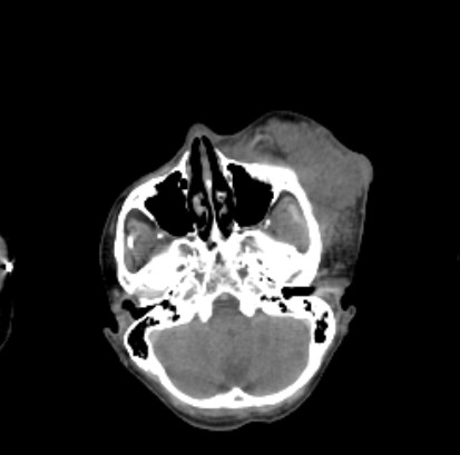
托管于其他服务器的图像:


预后因素
- 总生存期:13.4 个月
- 成人病例表现出侵袭性行为(Semin Diagn Pathol 2025;42:150941)
- 50% 的病例就诊时可见转移:淋巴结转移、远处转移(肺、骨、肝)或腹膜播散
- 就诊时无转移者:最终 66% 的患者发生局部复发或远处转移
- 排除随访时间 < 1 年的病例后,60% 的死亡发生于 2 年内
- 肿瘤伴有 MGA 重排:75% 死于本病,80% 出现局部复发或转移
- 肿瘤伴有 MXD1 / MXD4 / MXI1 重排:56% 死于本病,75% 出现局部复发或转移
- 儿童病例的临床病程侵袭性较低
- 就诊时 78% 无转移,其中 42% 最终发生局部复发或转移
- 已报道的 6 例随访至少 1 年的病例中,仅报道 1 例死亡(17%)(Biomedicines 2024;12:1709)
- 事实上,该患者生存了 25 年后才死于本病(Biomedicines 2024;12:1709)
- 儿童 NUT 重排肉瘤可能代表一个独特的亚群
病例报告
- 13 个月大男童,患有 NSD3::NUTM1 融合肉瘤,生存期较长(Biomedicines 2024;12:1709)
- 10 岁男童及 10 岁女童,患有 MGA::NUTM1 融合的高级别梭形细胞肉瘤(Cold Spring Harb Mol Case Stud 2018;4:a003194)
- 29 - 73 岁女性,患有 NUTM1 / NUTM2B 重排的腹内肉瘤,伴 ESR1 过表达,可作为治疗靶点(Mod Pathol 2025;38:100816)
- 34 岁女性,盆腔巨大肿块(Genes Chromosomes Cancer 2018;57:557)
- 55 岁女性,右肾肿块(J Immunother Precis Oncol 2025;8:113)
治疗
- 对于局限性病变,行广泛局部切除并确保切缘阴性
- 全身治疗的临床经验有限
- 以曲贝替定或异环磷酰胺为基础的方案,尤其是联合多柔比星(软组织肉瘤治疗的基础药物),可能是合理的一线治疗方法(Semin Diagn Pathol 2025;42:150941)
- BET 抑制剂可能对 NUT 重排肉瘤无效
- 正在研究的其他策略包括 EP300 组蛋白乙酰转移酶抑制剂和组蛋白去乙酰化酶抑制剂(伏立诺他)
临床图像
托管于其他服务器的图像:

大体描述
- 边界清楚,切面实性、鱼肉状,呈白褐色至灰色(Semin Diagn Pathol 2025;42:150941)
- 可见出血、旋涡状、囊性及质脆区域
- 肿瘤大小为 1 至 22.5 cm
- 深部肿瘤通常较大
大体图像
托管于其他服务器的图像:

冰冻切片描述
- 目前尚不明确
镜下(组织学)描述
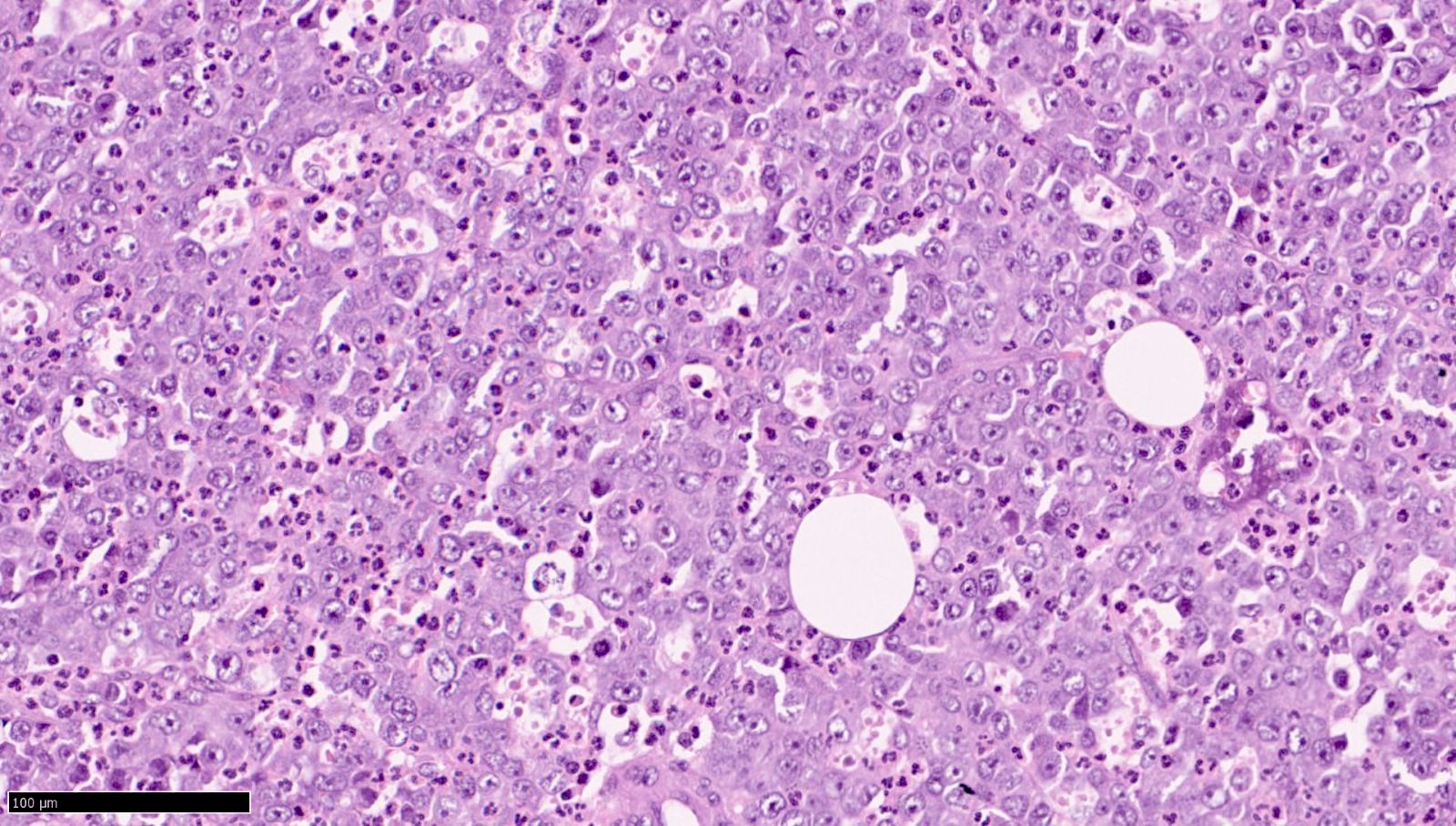
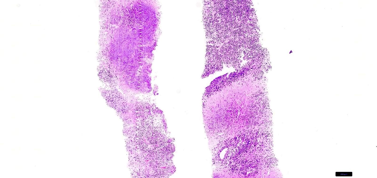
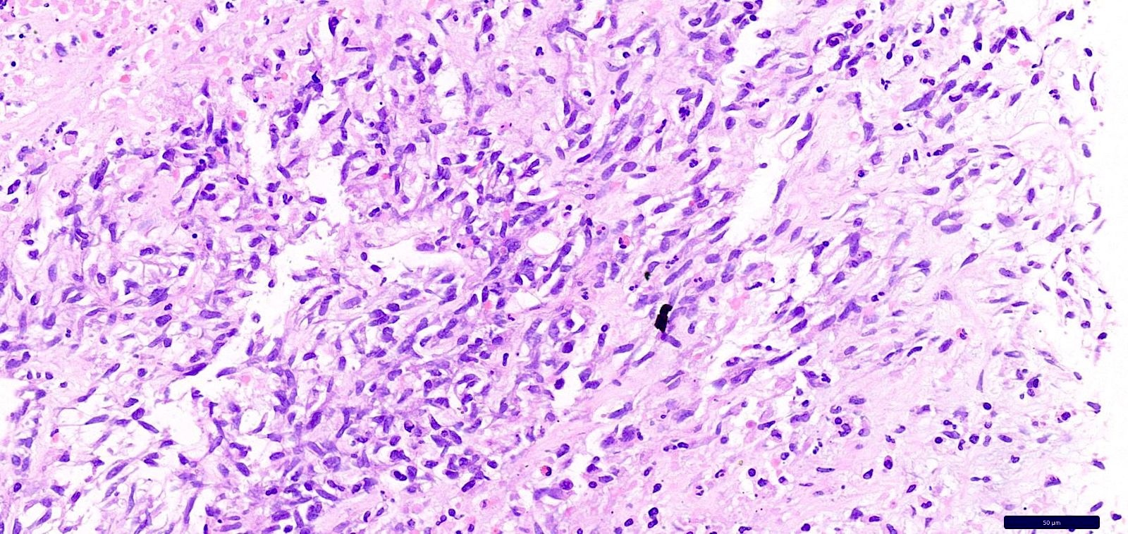
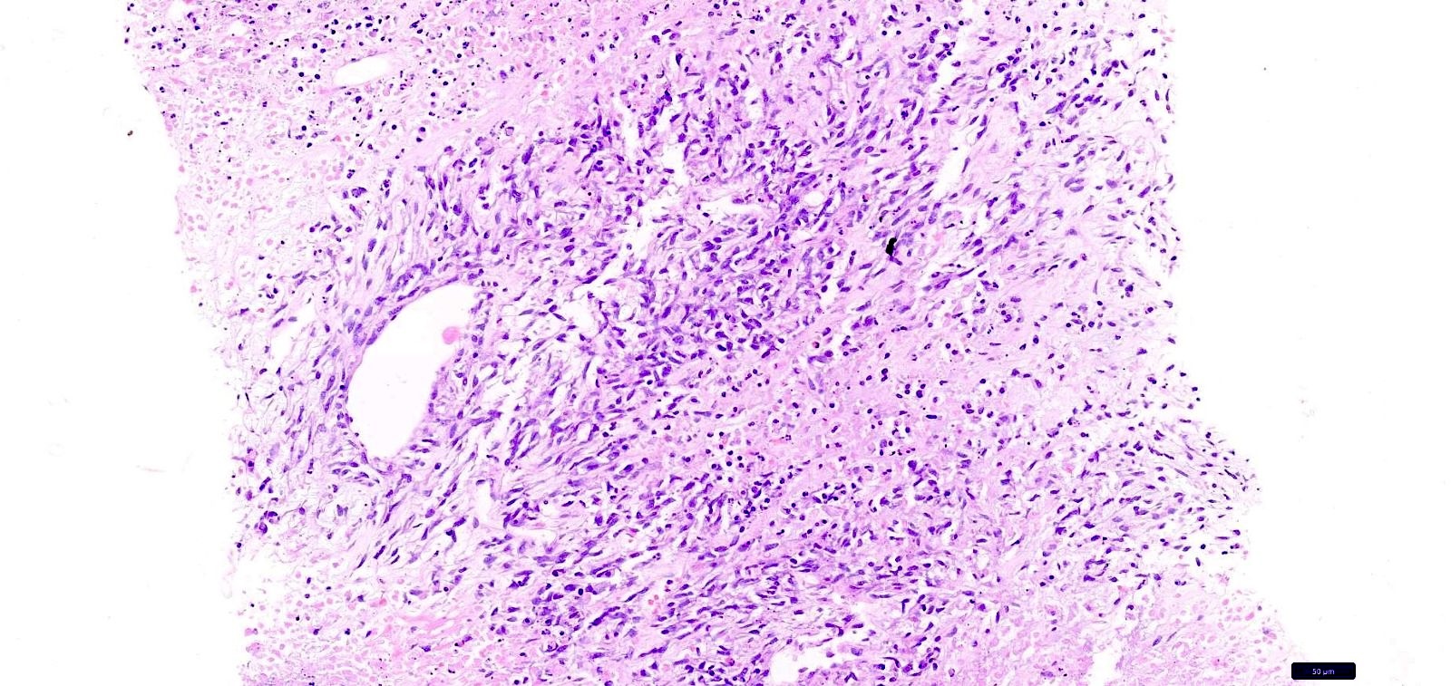
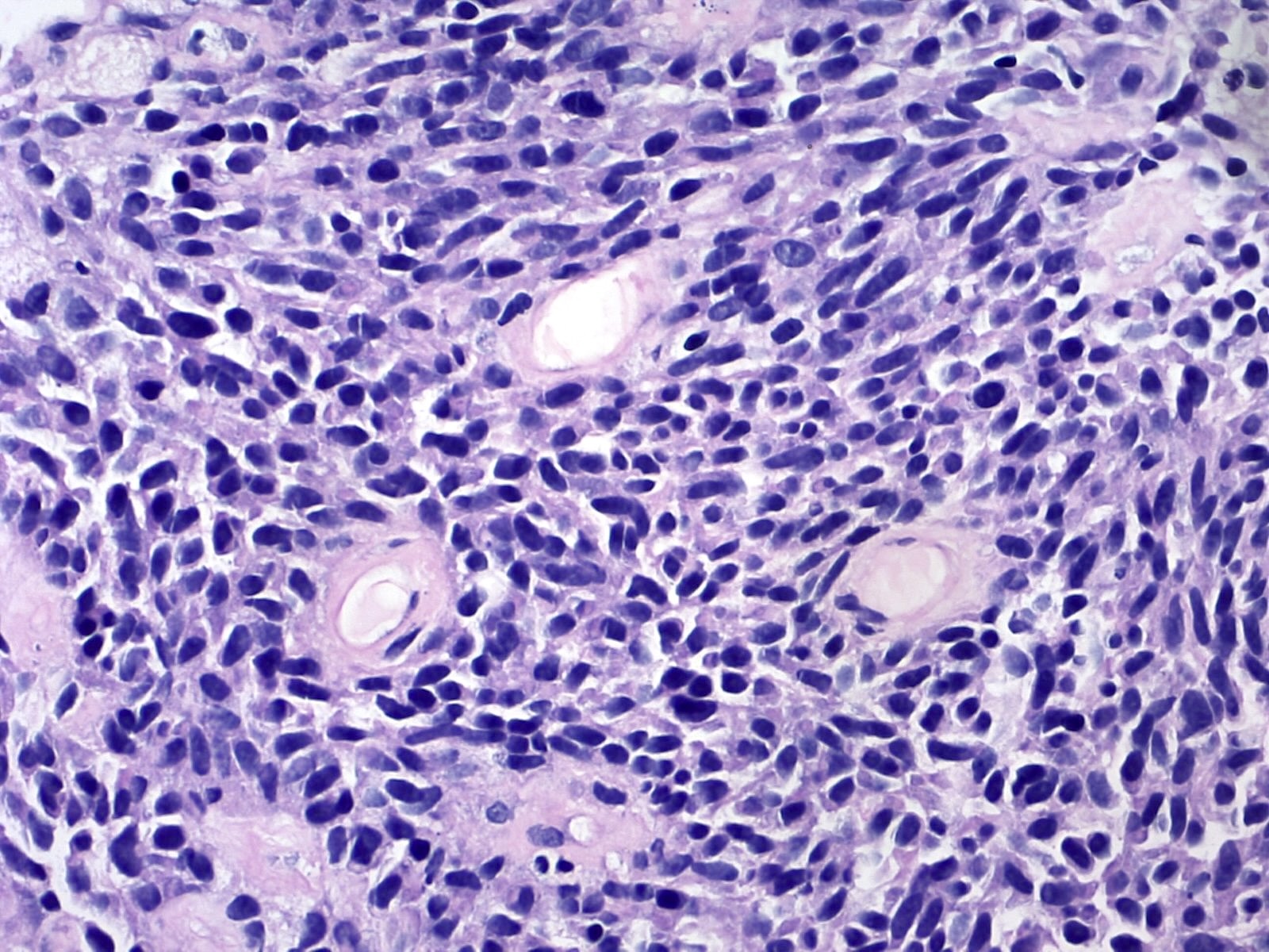
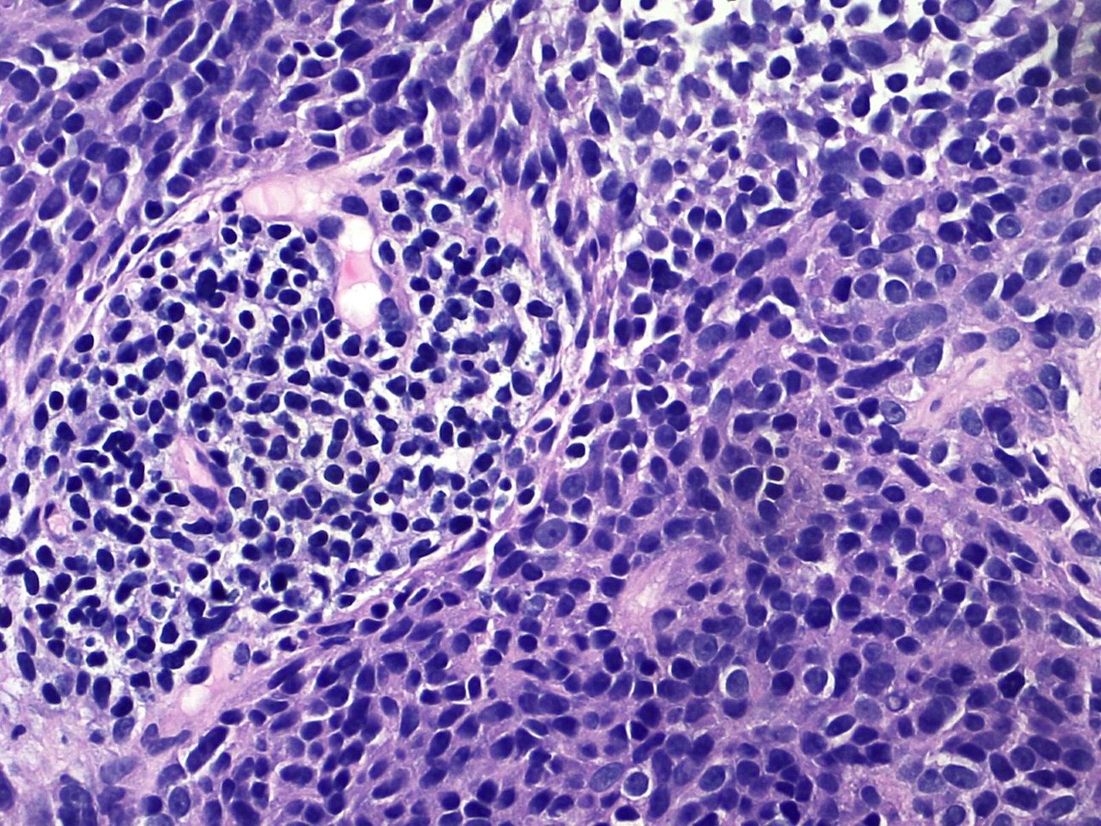
- 肿瘤细胞形态一致,核多形性极轻,符合融合驱动性病变的特征(Semin Diagn Pathol 2025;42:150941)
- 单个肿瘤可表现出 1 种或多种生长模式,包括
- 纤维肉瘤样模式:形态一致的梭形细胞呈交错束状或鱼骨状排列
- 圆形细胞模式:原始圆形细胞呈片状增生
- 上皮样 / 横纹肌样模式:上皮样至横纹肌样细胞呈片状增生
- 透明变 / 巢状模式:条索状和巢状细胞嵌于丰富的透明变间质中,或呈紧密簇状 / 腺泡状排列,间质稀少
- 肿瘤细胞可呈片状、大小不等的巢状、条索状、小梁状或单列排列
- 横纹肌样细胞常呈松散生长
- 可见微囊性间隙(Mod Pathol 2025;38:100729)
- 间质:透明变至黏液样,或极少
- 亦有骨样组织样基质的描述(Mod Pathol 2021;34:1547)
- 石棉样纤维 / 胶原菊形团(24% 的病例)
- 罕见特征:破骨细胞样巨细胞及菊形团形成
- 肿瘤坏死:程度不一
- 核分裂活性:低至活跃
- 胃肠道肿瘤:弥漫浸润黏膜下层、固有肌层及浆膜下层;亦可见黏膜受累(Mod Pathol 2021;34:1547)
- 肿瘤血管可呈血管外皮瘤样,或为厚壁透明变血管,或为肿瘤细胞环绕的拱廊状小血管
- 部分肿瘤区域可见怪异细胞,胞质呈微空泡状,异型性显著,具有类似霍奇金细胞的特征(Mod Pathol 2025;38:100816)
- MGA 重排肿瘤:常有胶原斑块 / 石棉样胶原、微囊性间隙及梭形细胞形态
- MXD4 重排肿瘤:常有上皮样或横纹肌样特征
镜下(组织学)图像
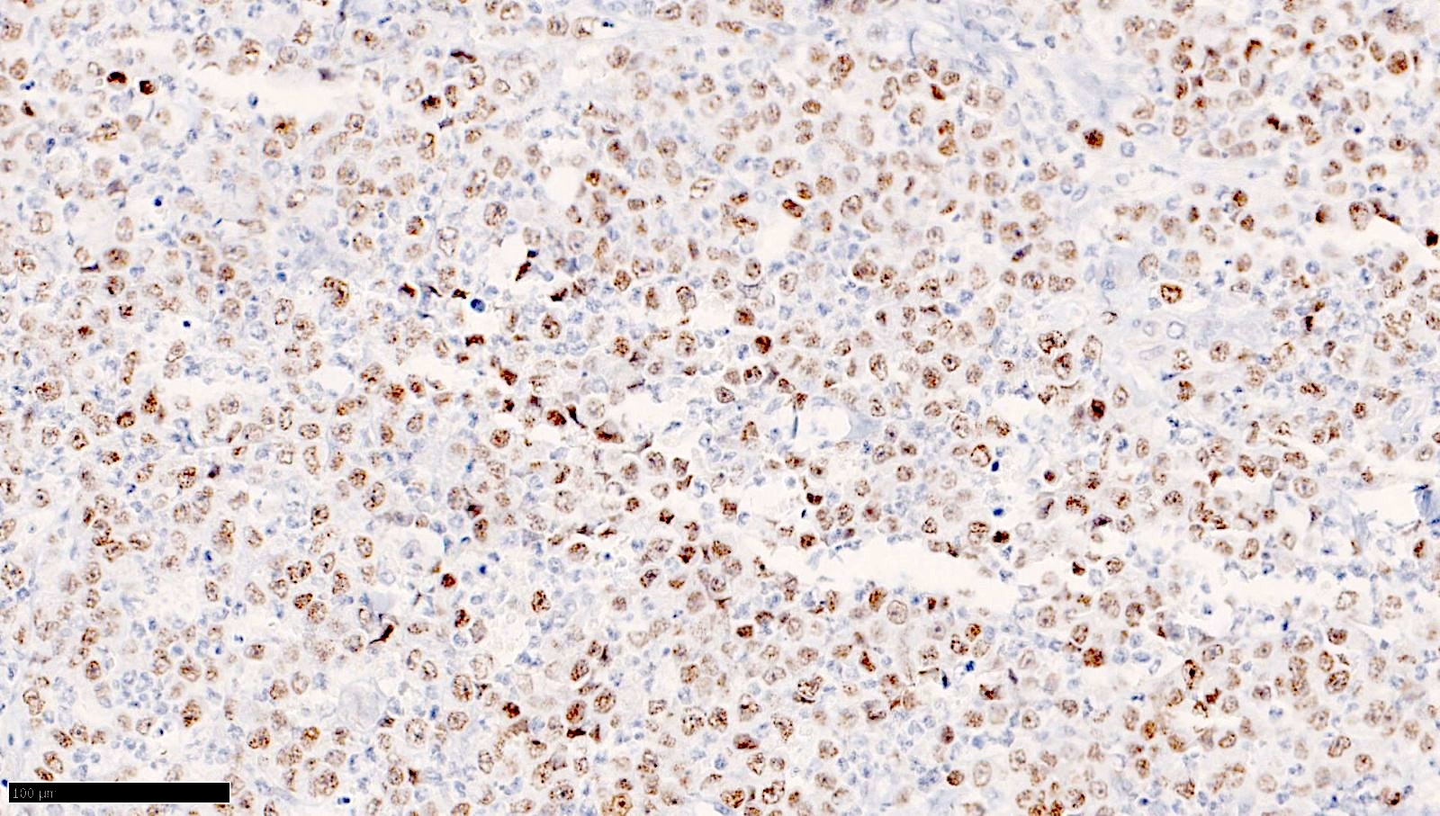
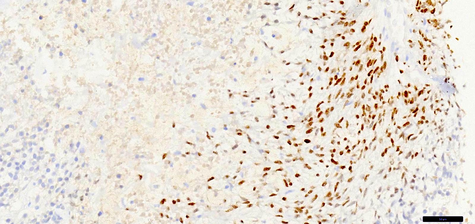
供图:Divya Bansal 医学博士、Sunil Pasricha 医学博士、Bhagat Singh Lali 医学博士及 Shobhna Sharma 医学博士
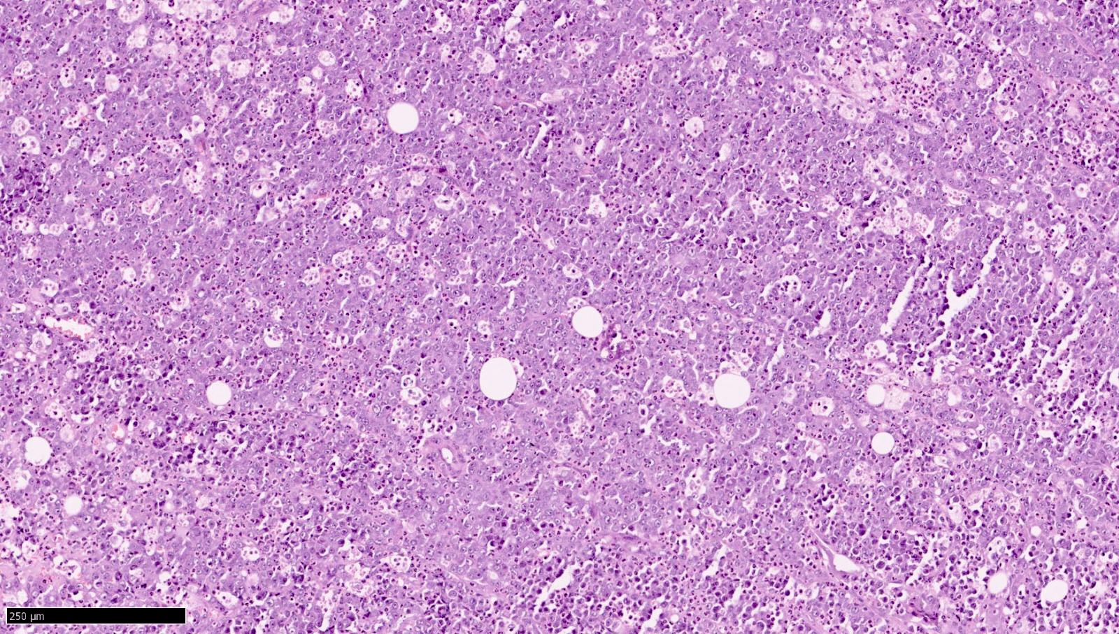
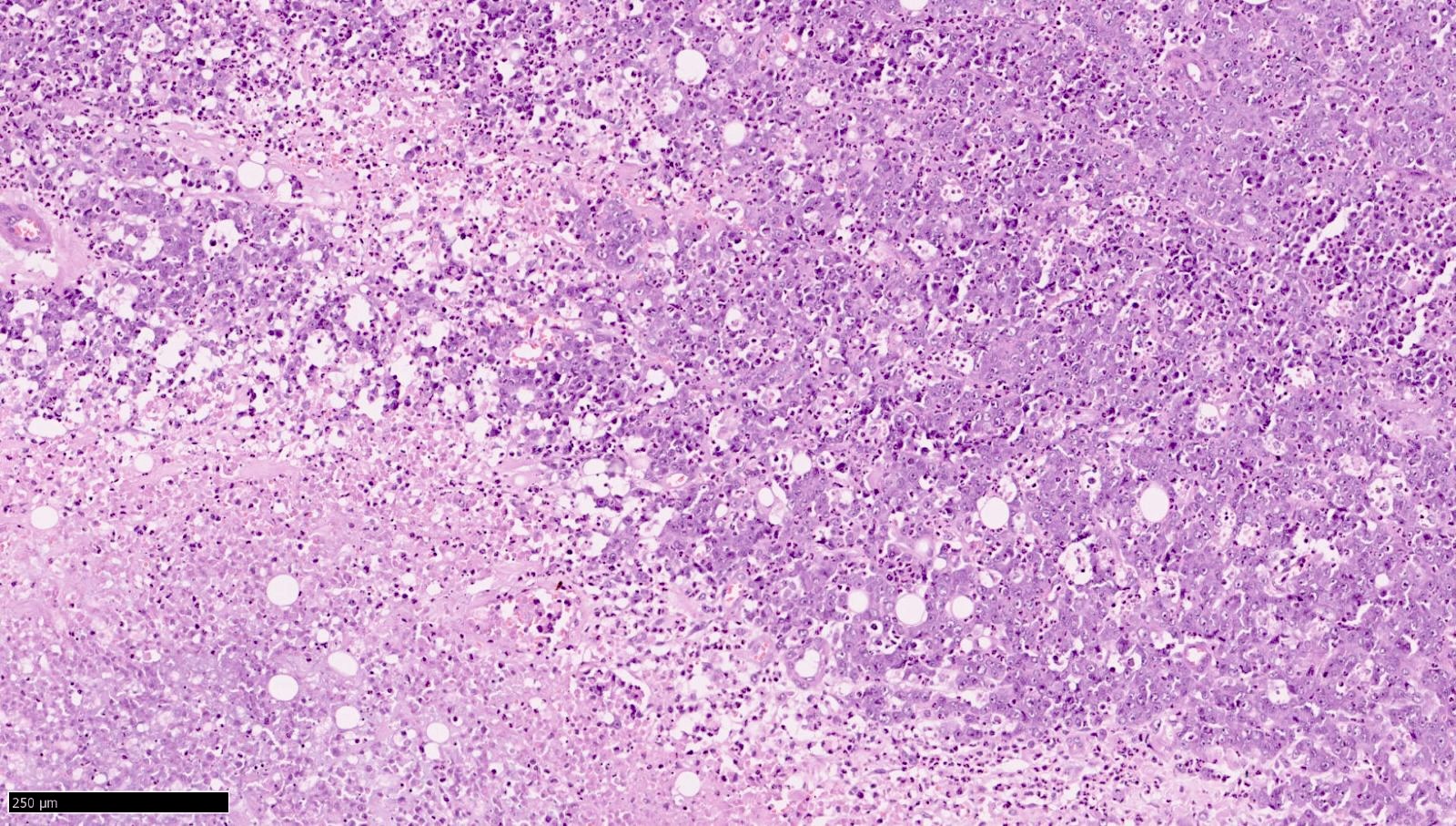
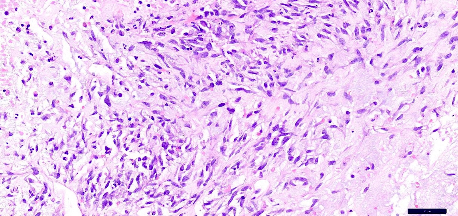
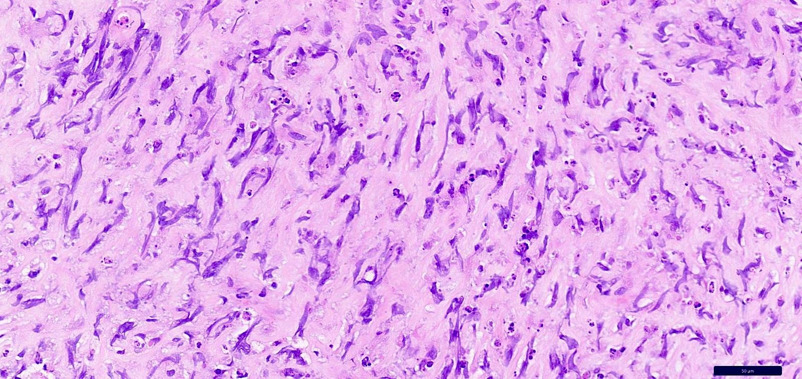
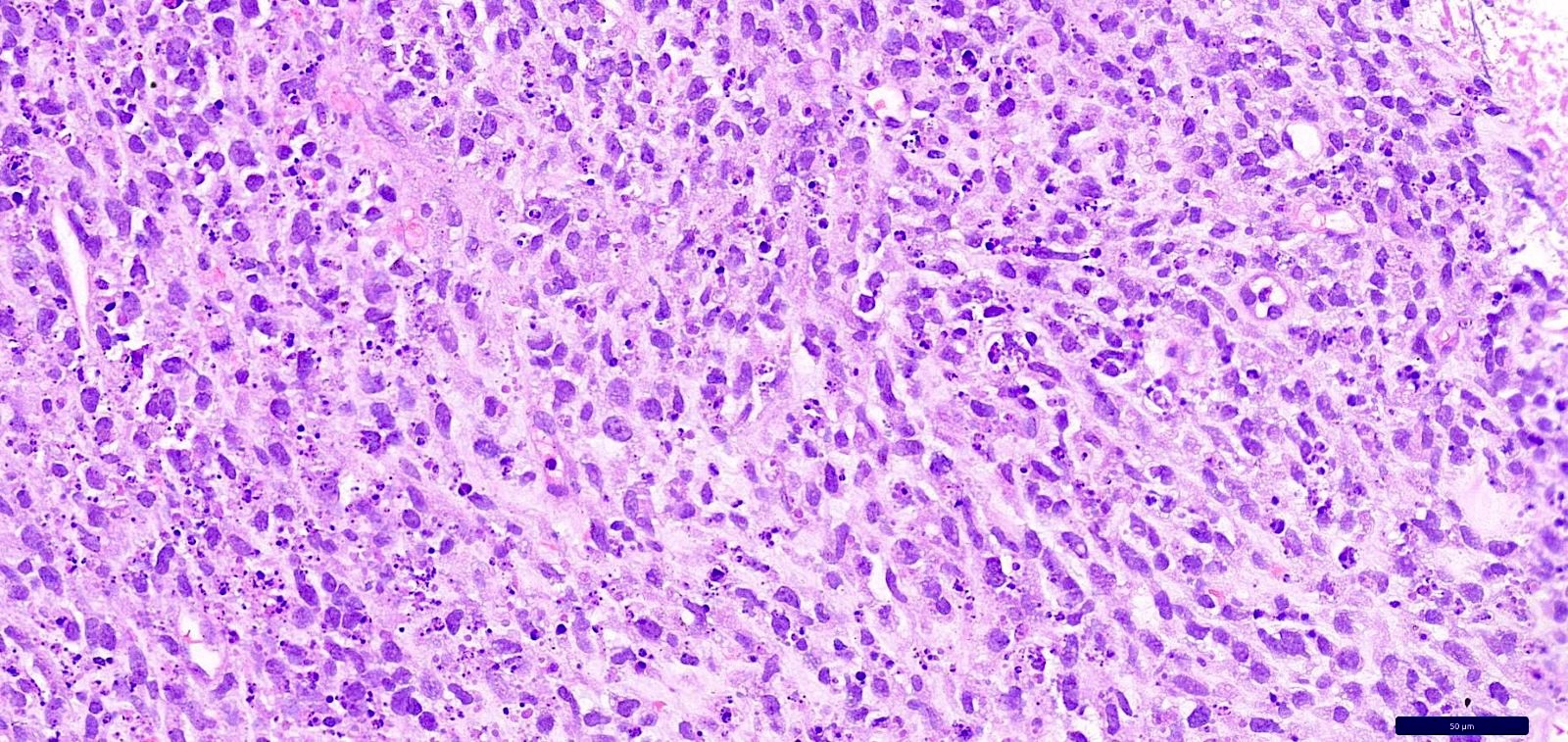
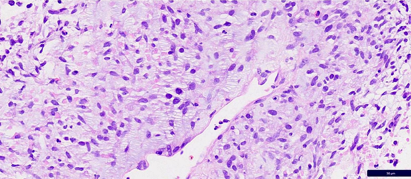
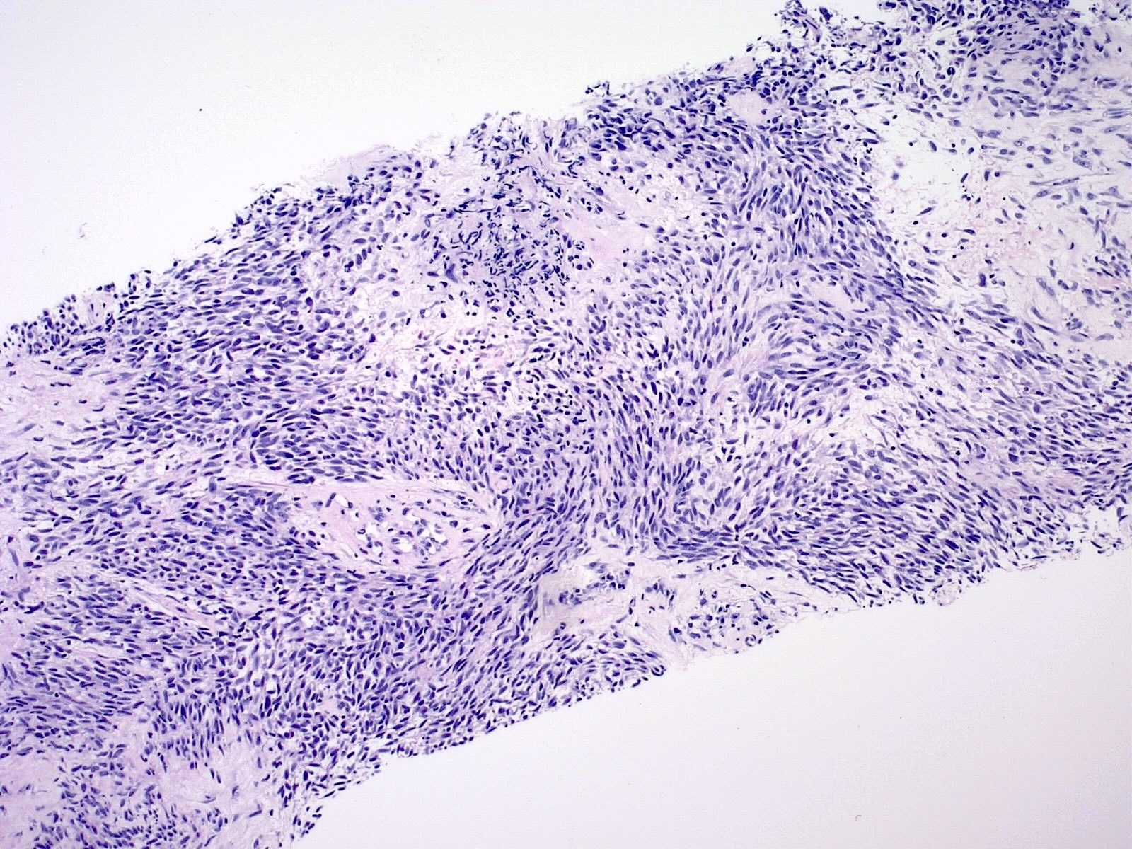
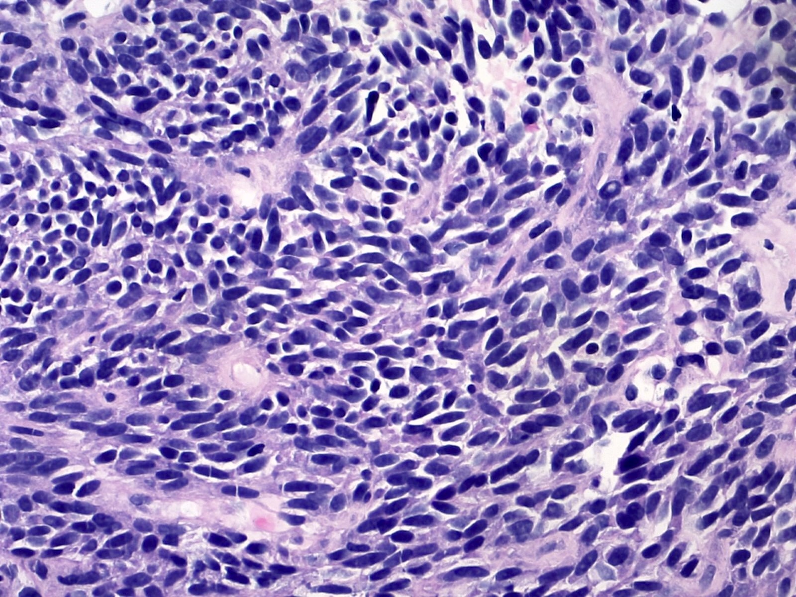
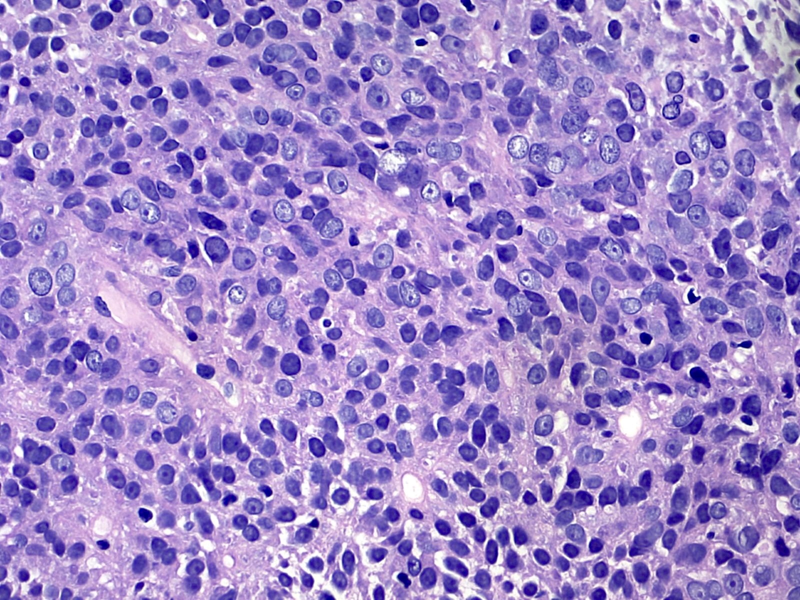
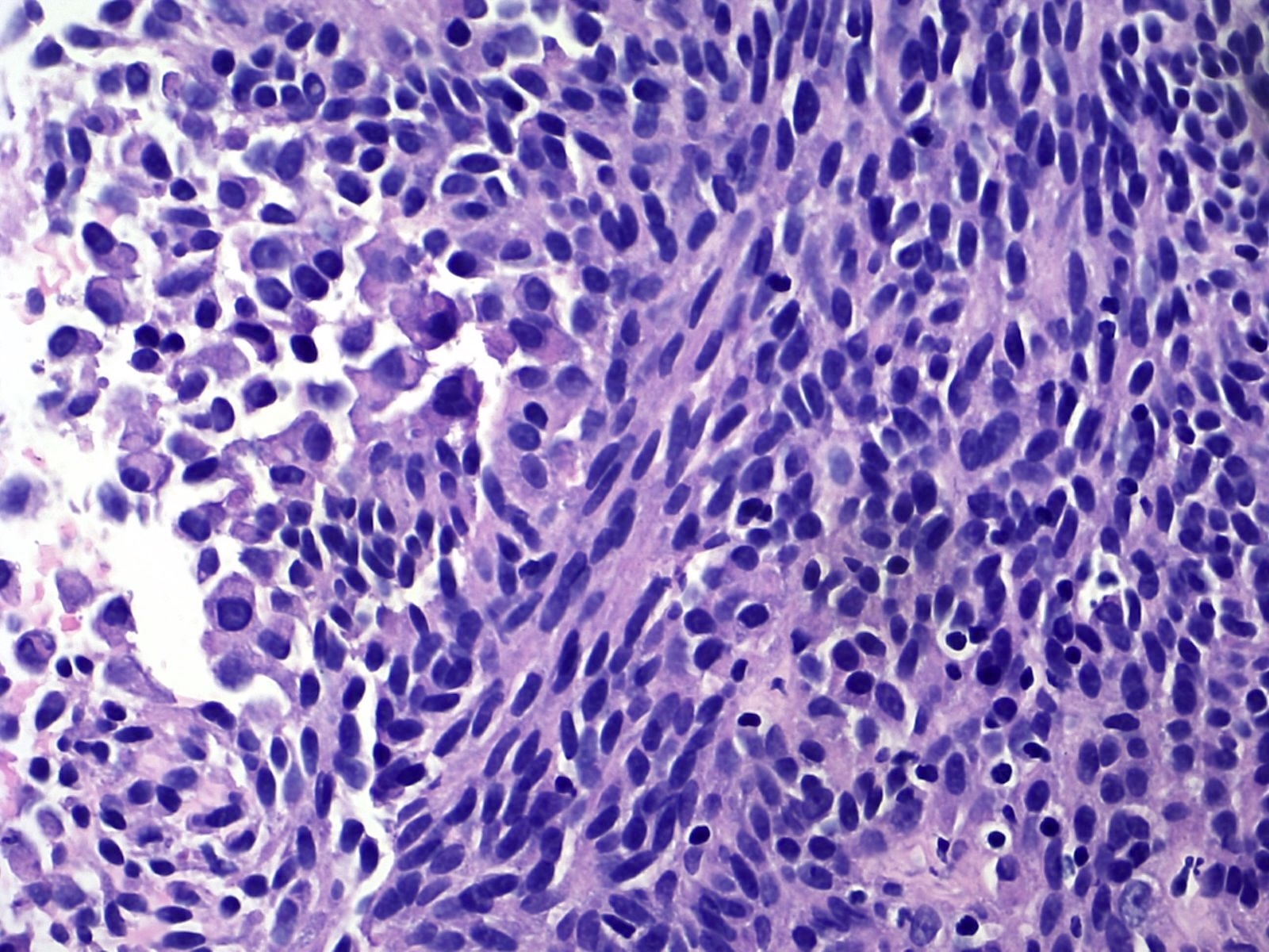
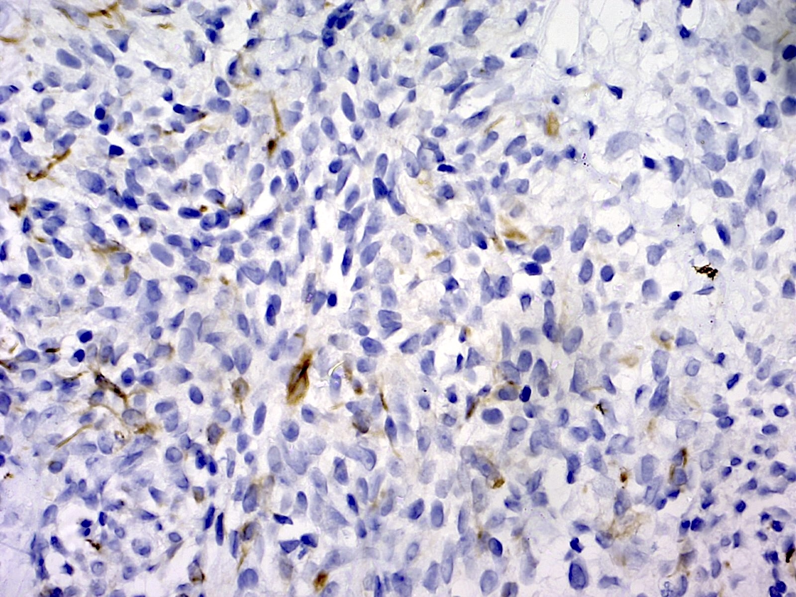
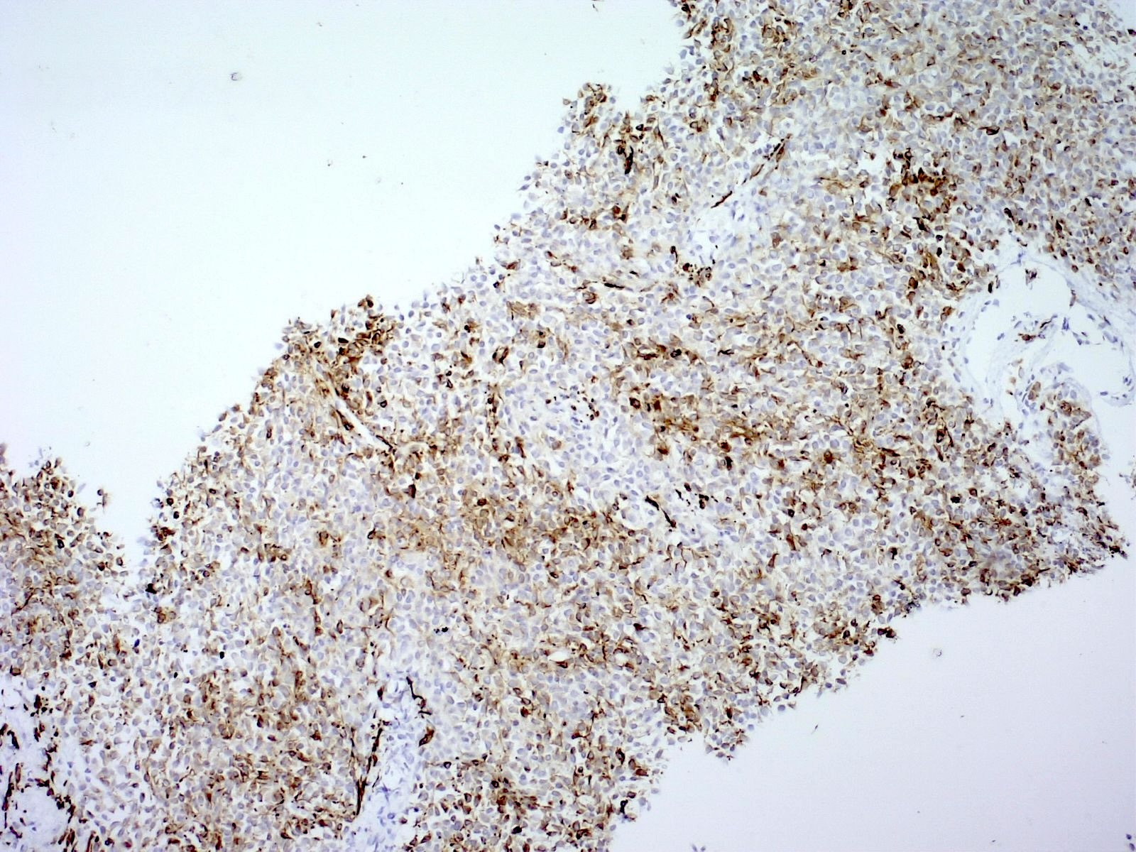
细胞学描述
- 肿瘤细胞单个散在或成簇分布(Diagn Cytopathol 2022;50:E244)
- 小圆形肿瘤细胞
- 横纹肌样细胞
- 核质比高,核仁小而不明显,染色质粗糙
- 偶可见核塑形
- 可见少量体积较大的双核肿瘤细胞
- 局限性
- 多种肿瘤均可呈小圆形细胞形态,伴或不伴横纹肌样细胞;通常需要一组免疫组织化学标记,至少用于缩小鉴别诊断范围
- 细胞蜡块不可或缺,不仅用于免疫组织化学检查,也用于分子诊断
阳性染色
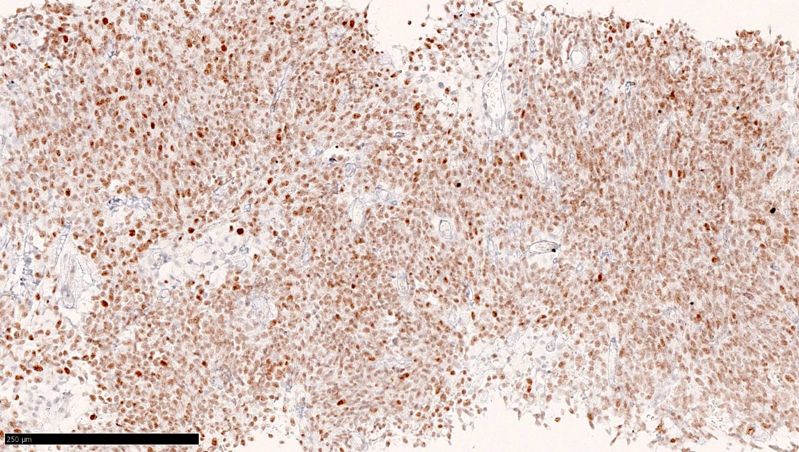
- NUT
- 单克隆 NUT C52B1 抗体的敏感性接近 100%
- 免疫表达呈斑片状至弥漫性(不同肿瘤之间及同一肿瘤内部均有差异)、弱至强的斑点状核染色模式
- 近期文献报道, NUT 免疫组织化学 阴性见于 1 例伴有 NSD3::NUTM1 融合的肿瘤及另外 2 例分别伴有 MXD1::NUTM1 和 BCORL1::NUTM1 融合的肿瘤(Mod Pathol 2019;32:764, Am J Surg Pathol 2018;42:636)
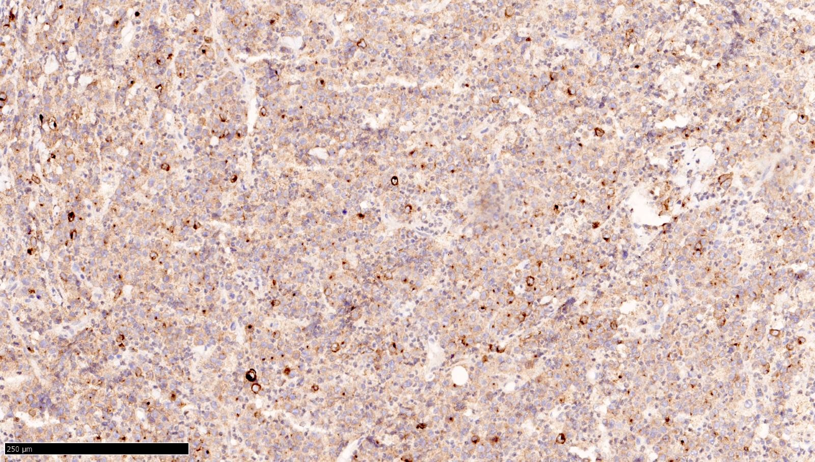
- CD99:可为局灶至弥漫性,通常较弱
- 表达不一,通常仅见于少量至局灶细胞(Semin Diagn Pathol 2025;42:150941)
- 少数病例呈强阳性或弥漫阳性
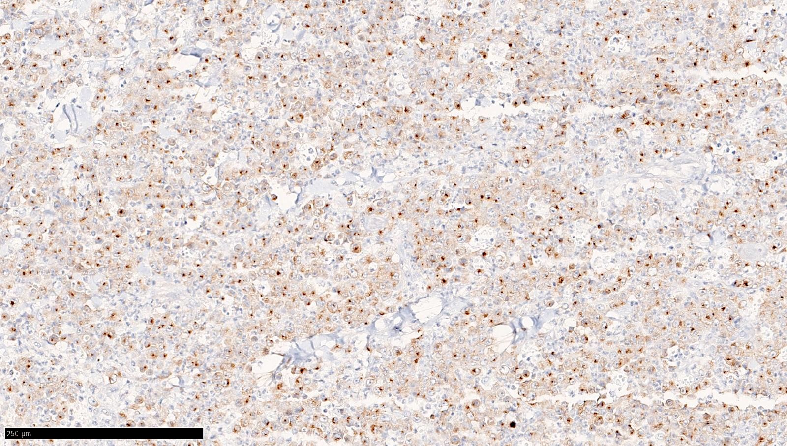
- 若有表达, 角蛋白(keratins) 可在部分 MXD1 / MXD4 / MXI1 重排肿瘤中呈阳性
- 通常不表达于伴有 MGA::NUTM1 的肿瘤
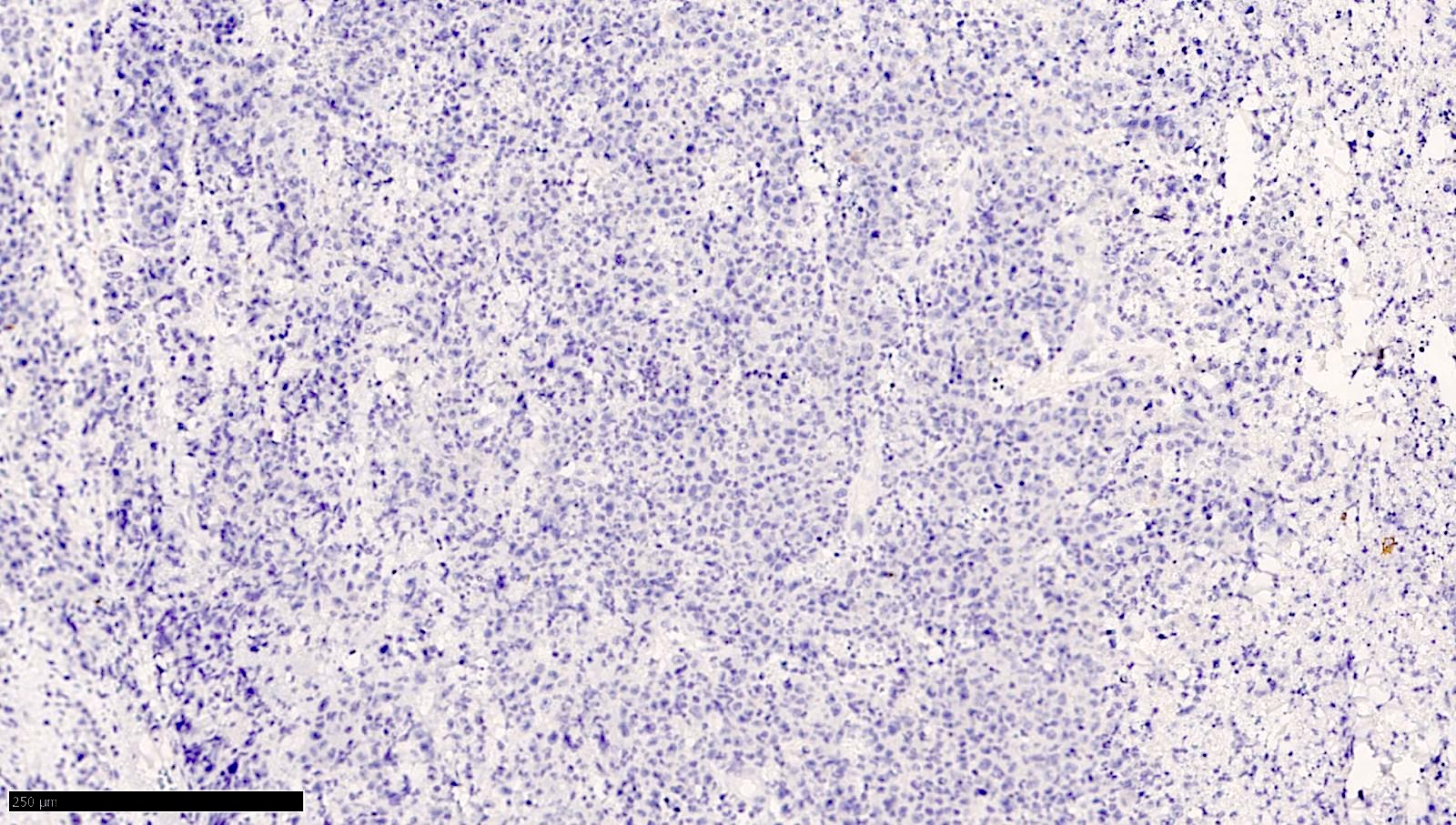
阴性染色
- 肌生成素(Myogenin), MyoD1
- 钙视网膜蛋白(Calretinin)
- HMB45, MelanA
- ALK, pan-TRK, BRAF V600E
- MDM2
- STAT6
- CD117, DOG1
- PAX8
- SATB2
- SS18::SSX
- SMARCB1 (INI1) 和 SMARCA4 (BRG1) 表达:保留
- H3K27me3 表达保留,或呈马赛克模式 / 斑片状缺失(Biomedicines 2024;12:1709, Histopathology 2023;83:712)
- 参考文献: Semin Diagn Pathol 2025;42:150941
电镜描述
- 目前尚不明确
分子 / 细胞遗传学描述
- FISH 检测 NUTM1 基因重排:使用针对 NUTM1 基因在 15q14 位点断裂点的探针
- 局限性:FISH 阴性而 NUT 免疫组织化学阳性时,必须进一步使用覆盖全长 NUTM1 基因的探针进行 FISH 检测,或采用测序方法,优先选择 RNA 测序
- RNA 测序:识别具体的融合伴侣,建议用于明确亚分类
- 局限性:部分 NGS 平台提供的融合检测面板覆盖范围有限
- 基于 DNA 的甲基化谱分析:形成独特的甲基化簇,不同于 NUT 癌及未分化小圆形细胞和梭形细胞肉瘤的甲基化簇
- 融合涉及 MYC / MAX / MXD 转录家族的基因(例如 MXD1, MXD4, MXI1 [或 MXD2]和 MGA)
- NUTM1 断裂点:外显子 2、3 和 5
- 最常见的融合伴侣: MXD4 和 MGA,其次为 MXI1 和 MXD1
- 胃肠道: MXD4 融合更常见
- 软组织: MGA 融合更常见
- MGA::NUTM1 见于除 2 例肿瘤之外的所有儿童病例(NSD3::NUTM1 和 ONECUT::NUTM1)
- 值得关注的是, BRD3这一见于 NUT 癌的伴侣基因,亦曾在 1 例大腿肿瘤中报道(Am J Surg Pathol 2018;42:636)
分子 / 细胞遗传学图像
托管于其他服务器的图像:


病理报告示例
- 盲肠,切除标本:
- 高级别肉瘤,伴 MXD4::NUTM1 融合(见备注)
- 备注:肿瘤由原始小圆形至上皮样细胞组成,呈实性片状排列,其间散布横纹肌样细胞。核分裂活性高(达 15/10 个高倍视野[HPF]),伴局灶坏死。免疫组织化学显示肿瘤细胞 NUT 呈弥漫强核表达。广谱细胞角蛋白(AE1 / AE3)局灶阳性。SMARCA4 和 SMARCA2 表达保留。FISH 检测 NUTM1 重排阳性(分离探针)。RNA 测序证实存在 MXD4::NUTM1 基因融合。上述所见可诊断为 NUTM1 重排肉瘤,这是一种有别于 NUT 癌的侵袭性间叶性肿瘤。伴有 NUT 重排的胃肠道肉瘤患者预后不良。
鉴别诊断
- 梭形细胞形态
- 单一形态的滑膜肉瘤:
- 新型 SS18::SSX 和 SSX C 端 免疫染色:对滑膜肉瘤具有很高的敏感性和特异性
- SS18::SSX1, SSX2 或 SSX4 可用于确诊滑膜肉瘤
- 孤立性纤维性肿瘤(SFT):
- 弥漫性 STAT6 表达见于 SFT(始终阴性者为 NUT 重排肉瘤)
- 定义性分子特征为 NAB2::STAT6 融合
- 恶性外周神经鞘瘤(MPNST):
- 鉴别可能存在困难
- 有放射治疗史、1 型神经纤维瘤病,或肿瘤发生于神经纤维瘤或外周神经
- 完全缺失的 H3K27me3 表达强烈支持恶性外周神经鞘瘤
- 保留或呈异质性的 H3K27me3 表达可能需要进一步分子检测
- NUT 免疫组织化学 阴性
- 激酶重排梭形细胞肿瘤:
- 胃肠道间质瘤(GIST):
- 肉瘤样癌:
- 角蛋白(Keratin) 表达不能解决鉴别诊断问题
- 前驱上皮病变、低级别癌或常规型成分支持肉瘤样癌
- 肉瘤样间皮瘤:
- WT1 (偶可见于 NUT 重排肉瘤)
- 肉瘤样间皮瘤可丢失 角蛋白(keratin) 及间皮标记的表达
- 多发浆膜结节及胸膜或腹膜增厚提示间皮瘤
- 替代标记 BAP1 或 MTAP 免疫染色缺失(分别提示 BAP1 和 CDKN2A 基因缺失)支持间皮瘤
- 单一形态的滑膜肉瘤:
- 圆形细胞形态
- 尤文肉瘤:
- 更常见
- NKX2.2 与强弥漫膜性 CD99 表达的组合(NKX2.2 可呈局灶阳性,弱而斑片状的 CD99 表达可见于 NUT 重排肉瘤)
- EWSR1::ETS 融合
- NUT 免疫组织化学 阴性
- 圆形细胞肉瘤,伴有 EWSR1::非ETS 融合:
- 可有不同程度的透明变、黏液透明样或黏液样间质;可局灶表达 角蛋白(keratin)
- EWSR1 / FUS::NFATC2 - EWSR1 重排及扩增可通过 FISH 检出
- EWSR1::PATZ1 共表达肌源性和神经标记
- NUT 免疫组织化学 阴性
- CIC 重排肉瘤:
- BCOR 改变的肉瘤:
- 表达 BCOR, 细胞周期蛋白 D1(cyclin D1) 和 SATB2 (NUT 重排肉瘤的 SATB2 为阴性,尽管数据有限)
- 存在 BCOR 重排或内部串联重复
- 促结缔组织增生性小圆形细胞肿瘤(DSRCT):
- 恒定表达 角蛋白(keratin) 和 结蛋白(desmin) (点状模式)(NUT 重排肉瘤可呈 结蛋白(desmin) 阳性,但通常为局灶性)
- 低分化滑膜肉瘤:
- SS18::SSX 免疫组织化学
- 表达下调的标记为 INI1
- 淋巴瘤:
- 小细胞癌:
- 鉴别可能存在困难; NUT 免疫组织化学 为阴性
- 尤文肉瘤:
- 上皮样 / 横纹肌样及巢状 / 透明变形态
- 硬化性上皮样纤维肉瘤(SEF):
- 具有重叠特征,包括上皮样细胞及透明变 / 硬化性间质(NUT 重排肉瘤可阳性表达 MUC4,甚至可弥漫表达,从而干扰诊断)
- EWSR1::FUS 重排
- NUT 免疫组织化学:阴性
- SWI / SNF 缺陷肿瘤 (包括 上皮样肉瘤 和 恶性横纹肌样瘤):
- 缺失 INI1 或 BRG1 表达
- 角蛋白(Keratin) 染色无助于区分这些肿瘤类型
- 恶性胃肠道神经外胚层肿瘤(GNET) 和 透明细胞肉瘤:
- 骨外骨肉瘤:
- 至少局灶可见单个细胞周围形成明确的花边状骨样组织
- 肌上皮肿瘤:
- 常表达 角蛋白(keratin), S100 蛋白, SOX10, SMA, p63, 肌钙调蛋白(calponin) 和 GFAP
- 部分病例还存在 EWSR1 重排
- 低分化癌:
- NUT 免疫组织化学 阴性
- 黑色素瘤:
- 上皮样间皮瘤:
- WT1 弥漫强阳性
- NUT 免疫组织化学 阴性
- 硬化性上皮样纤维肉瘤(SEF):
- NUT 癌:
- 特征为原始上皮样细胞伴鳞状分化,通常表现为突然角化
- 约 66% 的 NUT 癌中, NUTM1 的融合伴侣为 BRD4,其余肿瘤则涉及 BRD3, NSD3, ZNF532, ZNF592 或 ZNF618
- 结合临床和影像学仍是最有用的手段,但部分肿瘤可能难以分类
- NUTM2 重排肉瘤:
- 与 NUTM1 重排肉瘤具有共同的形态学特征
- NUTM2 融合的肿瘤可呈弱染色或不染色,所用标记为 NUT 免疫染色
- CIC::NUTM1 肉瘤:
练习题 #1
一名 56 岁女性因 5.5 cm 的结肠肿块就诊。活检显示高级别肿瘤,由梭形、原始小圆形至少量横纹肌样细胞组成。免疫组织化学显示肿瘤细胞 NUT 呈弥漫强核染色,广谱细胞角蛋白阴性,SMARCA4(BRG1)和 SMARCB1(INI1)表达保留。FISH 检测 NUTM1 重排阳性。以下哪项诊断最恰当?
- 尤文肉瘤
- 恶性横纹肌样瘤
- NUT 癌
- NUTM1 重排肉瘤
- SMARCA4 缺陷性未分化肿瘤
练习题答案 #1
D.NUTM1 重排肉瘤。临床表现(结肠肿块、中年成人)、组织学形态(梭形、原始小圆形至横纹肌样细胞)、弥漫强 NUT 核染色,以及存在 NUTM1 重排但无鳞状分化,可诊断为 NUTM1 重排肉瘤。选项 A 不正确,因为尤文肉瘤通常表现为膜性 CD99、核性 NKX2.2 阳性,存在 EWSR1::ETS 融合,且 NUT 阴性。选项 B 不正确,因为恶性横纹肌样瘤通常发生于幼儿,表现为 SMARCB1(INI1)表达缺失,且并非以 NUTM1 重排为定义特征。选项 C 不正确,因为 NUT 癌通常呈鳞状分化,常发生于中线结构(纵隔、头 / 颈部),并常涉及 BRD3 / 4 融合,尽管其也可表达 NUT。选项 E 不正确,因为 SMARCA4 缺陷性未分化肿瘤表现为 SMARCA4(BRG1)核表达缺失,而本例肿瘤的表达保留。
参考文献: NUTM1 重排肉瘤
- NUTM1 rearranged sarcoma is an emerging group of aggressive soft tissue sarcomas defined by NUTM1 rearrangements at 15q14, distinct from NUT carcinoma; wide age range; commonly involves somatic soft tissues and GI tract.
- Variable histology (fibrosarcomatous, round cell, epithelioid/rhabdoid, hyalinized/nested; amianthoid fiber-like collagen); NUT IHC often positive; diagnosis requires molecular testing (FISH/NGS/RNA-seq); partners include MGA, MXD4, etc.
- Aggressive course: ~50% of adult cases metastatic at presentation; overall survival ~13.4 months; topic includes numerous microscopic, IHC and molecular images plus differential diagnosis and practice questions.
Scope: full PathologyOutlines topic page “NUTM1 rearranged sarcoma” (all sections, on-site microscopic/IHC/molecular images with captions, differential diagnosis, practice questions). Listed in the 12 October 2026 Textbook Updates; the dura MALT lymphoma topic was published separately on this site on 2026-10-13.
Definition / general
- NUTM1 rearranged sarcoma is an emerging group of aggressive soft tissue sarcomas defined by chromosomal rearrangements involving the NUTM1 (NUT midline carcinoma family member 1) gene on chromosome 15q14, distinct from the more well known NUT carcinoma
Essential features
- Emerging group of aggressive soft tissue sarcomas
- Wide age range, no sex predilection, no predisposing factors
- Commonly involves somatic soft tissues and gastrointestinal tract
- Histology is variable, including fibrosarcomatous, round cell, epithelioid / rhabdoid and hyalinized / nested patterns, sometimes with amianthoid fiber-like collagen, immunoreactivity for NUT protein
- Defined by NUTM1 rearrangement, most commonly fused with MAD family genes (MXD4, MXD1, MXI1, MGA) rather than BRD3 / 4 (which are characteristic fusion partners in NUT carcinoma)
Terminology
- NUT rearranged sarcoma
ICD coding
- ICD-O: 8800/3 - sarcoma, NOS
- ICD-10: C49.9 - malignant neoplasm of connective and soft tissue, unspecified
- ICD-11: 2B5K - unspecified malignant soft tissue tumors or sarcomas of bone or articular cartilage of other or unspecified site
Epidemiology
- Extremely rare entity with few cases reported in literature (~70 cases)
- Wide age range: 1 - 78 years (Semin Diagn Pathol 2025;42:150941)
- Typically affects middle aged adults (median age: 48 years)
- 1 reported case in a 13 month old boy (Biomedicines 2024;12:1709)
- Slight female predilection (M:F = 1:1.7)
- No known geographic predilection; reported worldwide
Sites
- Most common in the somatic soft tissues, thoracic region, gastrointestinal tract and body cavity (Semin Diagn Pathol 2025;42:150941, Mod Pathol 2021;34:1547)
- Other locations include the mandible, kidney, bladder, penis, ovary, uterus, scalp, orbital area, brain and central nervous system (parietal lobe and dura) (Genes Chromosomes Cancer 2022;61:542, Semin Diagn Pathol 2025;42:150941)
- Pediatric cases primarily involve soft tissue and are located in the thoracic region; cases in the abdomen or pelvis have not been documented (Semin Diagn Pathol 2025;42:150941)
Pathophysiology
- NUTM1 gene, located on chromosome 15q, is normally expressed in the testis and promotes histone acetylation during spermatogenesis
- Most common fusion partners: genes from the MYC associated factor X (MAX) dimerization (MAD) transcription family (e.g., MXD1, MXD4, MXI1 [or MXD2] and MGA) rather than BRD3 or BRD4 (commonly seen in NUT carcinoma)
- MAD::NUTM1 fusion protein is hypothesized to dysregulate gene expression, potentially through blocking differentiation and promoting abnormal proliferation
- Unlike BRD4::NUT, it may not form megadomains or require BET proteins, potentially explaining resistance to BET inhibitors
- Rare partners include BCORL1, ZNF532, NSD3, ONECUT, BRD3 and BRD4 (Am J Surg Pathol 2018;42:636, Am J Surg Pathol 2024;48:930)
- No known precursor lesion
Etiology
- Unknown at this time
Diagrams / tables
Contributed by Bhagat Singh Lali, M.D.
Images hosted on other servers:
Clinical features
- Rapidly growing, usually deep seated, often painful or palpable mass
- Symptoms depend on the anatomic site (e.g., headache and hemiparesis for CNS tumors; obstruction, abdominal distension, weight loss, abdominal pain for GI tumors; shortness of breath for thorax tumors; ptosis in orbital primary) or with a mass discovered on imaging studies
Diagnosis
- Essential diagnostic criteria
- Monomorphic spindled, epithelioid and rhabdoid cells in a partially hyalinized background
- Evidence of NUTM1 rearrangement
Laboratory
- Unknown at this time
Radiology description
- No specific findings
- Imaging typically reveals an aggressive, heterogeneously enhancing soft tissue mass with necrosis and infiltrative margins (Mod Pathol 2021;34:1547)
- 18F-FDG PET / CT: demonstrates intense FDG avidity, identifies metastatic disease
Radiology images
Contributed by Divya Bansal, M.B.B.S., M.D., Himanshi Diwan, M.D., Sunil Pasricha, M.B.B.S., M.D. and Rakesh Oberoi, M.D.
Images hosted on other servers:
Prognostic factors
- Overall survival: 13.4 months
- Adult cases demonstrate aggressive behavior (Semin Diagn Pathol 2025;42:150941)
- Metastasis is seen in 50% of cases at presentation: lymph node, distant metastasis (lung, bones, liver) or peritoneal spread
- Without metastasis at presentation: eventually 66% of patients developed local recurrence or distant metastasis
- Excluding cases with a follow up period of < 1 year, 60% of deaths occurred within 2 years
- Tumors with MGA rearrangements: 75% died of disease, 80% with local recurrence or metastasis
- Tumors with MXD1 / MXD4 / MXI1 rearrangements: 56% died of disease, 75% with local recurrence or metastasis
- Pediatric cases follow a less aggressive clinical course
- At presentation, 78% had no metastasis, out of which 42% eventually developed local recurrence or metastasis
- Of 6 reported cases with at least 1 year of follow up, only 1 death (17%) was reported (Biomedicines 2024;12:1709)
- In fact, this patient survived 25 years before succumbing to the disease (Biomedicines 2024;12:1709)
- Pediatric NUT rearranged sarcomas may represent a distinct subset
Case reports
- 13 month old boy with NSD3::NUTM1 fusion sarcoma and prolonged survival (Biomedicines 2024;12:1709)
- 10 year old boy and 10 year old girl with MGA::NUTM1 fusion high grade spindle cell sarcoma (Cold Spring Harb Mol Case Stud 2018;4:a003194)
- 29 - 73 year old women with NUTM1 / NUTM2B rearranged intraabdominal sarcomas with ESR1 overexpression as a therapeutic target (Mod Pathol 2025;38:100816)
- 34 year old woman with a large pelvic mass (Genes Chromosomes Cancer 2018;57:557)
- 55 year old woman with a right kidney mass (J Immunother Precis Oncol 2025;8:113)
Treatment
- Wide local excision with negative margins for localized disease
- Clinical experience with systemic therapies is limited
- Trabectedin based or ifosfamide based regimens, especially in combination with doxorubicin (the backbone of soft tissue sarcoma), may offer a reasonable approach as frontline therapy (Semin Diagn Pathol 2025;42:150941)
- BET inhibitors may not be effective in NUT rearranged sarcomas
- Other strategies under investigation include EP300 histone acetyltransferase inhibitors and histone deacetylase inhibitors (vorinostat)
Clinical images
Images hosted on other servers:
Gross description
- Circumscribed with a solid, fleshy, white-tan to gray cut surface (Semin Diagn Pathol 2025;42:150941)
- May show hemorrhagic, whorled, cystic and friable areas
- Tumor sizes range from 1 to 22.5 cm
- Deeply located tumors are generally larger
Gross images
Images hosted on other servers:
Frozen section description
- Unknown at this time
Microscopic (histologic) description
- Tumor cells display uniform cytomorphology with minimal nuclear pleomorphism, in keeping with a fusion driven process (Semin Diagn Pathol 2025;42:150941)
- Single tumor may display 1 or multiple growth patterns, including
- Fibrosarcomatous pattern: uniform spindled cells arranged in intersecting fascicles or a herringbone pattern
- Round cell pattern: sheet-like proliferation of primitive round cells
- Epithelioid / rhabdoid pattern: sheet-like proliferation of epithelioid to rhabdoid cells
- Hyalinized / nested pattern: cords and nests of cells embedded in an abundant hyalinized stroma or tight clusters / alveolar pattern with scant stroma
- Tumor cells can be arranged in sheets, variably sized nests, cords, trabeculae or single filing pattern
- Rhabdoid cells often display a discohesive growth
- Microcystic spaces can be seen (Mod Pathol 2025;38:100729)
- Stroma: hyalinized to myxoid or minimal
- Osteoid-like matrix has also been described (Mod Pathol 2021;34:1547)
- Amianthoid fibers / collagen rosettes (24% of cases)
- Rare features: osteoclast-like giant cells and rosette formation
- Tumor necrosis: variable
- Mitotic activity: low to brisk
- Gastrointestinal tumors: diffuse infiltration of submucosa, muscularis propria and subserosa; mucosal involvement can also be seen (Mod Pathol 2021;34:1547)
- Tumor vasculature, hemangiopericytomatous or thick walled hyalinized vessels or arcades of smaller vessels cuffed by tumor cells
- Monstrous cells with microvacuolar cytoplasm, severe atypia and features reminiscent of Hodgkin cells are present in some tumor areas (Mod Pathol 2025;38:100816)
- MGA rearranged tumors: often have collagen plaques / amianthoid collagen, microcystic spaces and spindle morphology
- MXD4 rearranged tumors: often have epithelioid or rhabdoid features
Microscopic (histologic) images
Contributed by Divya Bansal, M.D., Sunil Pasricha, M.D., Bhagat Singh Lali, M.D. and Shobhna Sharma, M.D.
Cytology description
- Singly scattered or clusters of tumor cells (Diagn Cytopathol 2022;50:E244)
- Small round tumor cells
- Rhabdoid cells
- High N:C ratio, small inconspicuous nucleoli and coarse chromatin
- Occasional nuclear molding can be seen
- Rare larger binucleated tumor cells can be seen
- Limitations
- Small round cell morphology with or without rhabdoid cells can be seen in many tumors; a panel of immunohistochemical markers is usually required to at least narrow the differential diagnosis
- Cell blocks are essential, not only for immunohistochemistry studies but also for the purpose of molecular diagnostics
Positive stains
- NUT
- Near 100% sensitivity for the monoclonal NUT C52B1 antibody
- Patchy to diffuse (varies between and within tumors), weak to strong, speckled nuclear pattern of immunoexpression
- Recent papers reported negative NUT IHC in a tumor with NSD3::NUTM1 fusion and 2 other tumors harboring MXD1::NUTM1 and BCORL1::NUTM1 fusions (Mod Pathol 2019;32:764, Am J Surg Pathol 2018;42:636)
- CD99: may be focal to diffuse, usually weak
- Variably expressed, typically rare to focal cells (Semin Diagn Pathol 2025;42:150941)
- Rare cases, strong or diffuse
- If expressed, keratins may be positive in a subset of MXD1 / MXD4 / MXI1 rearranged tumors
- Not typically expressed in tumors with MGA::NUTM1
Negative stains
- Myogenin, MyoD1
- Calretinin
- HMB45, MelanA
- ALK, pan-TRK, BRAF V600E
- MDM2
- STAT6
- CD117, DOG1
- PAX8
- SATB2
- SS18::SSX
- SMARCB1 (INI1) and SMARCA4 (BRG1) expression: intact
- H3K27me3 expression is retained or displays a mosaic pattern / patchy loss (Biomedicines 2024;12:1709, Histopathology 2023;83:712)
- Reference: Semin Diagn Pathol 2025;42:150941
Electron microscopy description
- Unknown at this time
Molecular / cytogenetics description
- FISH for NUTM1 gene rearrangement: probe for NUTM1 gene breakpoint at the 15q14 locus
- Limitation: negative FISH, positive NUT IHC must be further investigated by FISH using a probe spanning the full length of the NUTM1 gene or by a sequencing method, preferably RNA sequencing
- RNA sequencing: identifies specific fusion partners and is recommended for definitive subclassification
- Limitation: fusion panel coverage offered by some of the NGS platforms
- DNA based methylation profiling: distinct methylation cluster differing from those of NUT carcinoma and undifferentiated small round cell and spindle cell sarcomas
- Fusions involving genes from the MYC / MAX / MXD transcription family (e.g., MXD1, MXD4, MXI1 [or MXD2] and MGA)
- NUTM1 breakpoints: exon 2, 3 and 5
- Most common fusion partners: MXD4 and MGA, followed by MXI1 and MXD1
- Gastrointestinal tract: MXD4 fusions are more common
- Soft tissue: MGA fusions are more common
- MGA::NUTM1 has been found in all pediatric cases except 2 tumors (NSD3::NUTM1 and ONECUT::NUTM1)
- Interestingly, BRD3, a partner gene found in NUT carcinoma, has been reported in a tumor in the thigh (Am J Surg Pathol 2018;42:636)
Molecular / cytogenetics images
Images hosted on other servers:
Sample pathology report
- Cecum, resection:
- High grade sarcoma with MXD4::NUTM1 fusion (see comment)
- Comment: The tumor is composed of primitive small round to epithelioid cells arranged in solid sheets with interspersed rhabdoid cells. Mitotic activity is high (up to 15/10 high power fields [HPF]) with focal necrosis. By immunohistochemistry, the tumor cells show diffuse and strong nuclear expression of NUT. Pancytokeratin (AE1 / AE3) is focally positive. SMARCA4 and SMARCA2 expression are retained. FISH for NUTM1 rearrangement is positive (breakapart probe). RNA sequencing confirms an MXD4::NUTM1 gene fusion. These findings are diagnostic of NUTM1 rearranged sarcoma, an aggressive mesenchymal neoplasm distinct from NUT carcinoma. The prognosis for patients with NUT rearranged gastrointestinal sarcomas is poor.
Differential diagnosis
- Spindle cell morphology
- Monomorphic synovial sarcoma:
- Novel SS18::SSX and SSX C terminus immunostains: highly sensitive and specific for synovial sarcoma
- SS18::SSX1, SSX2 or SSX4 are diagnostic of synovial sarcoma
- Solitary fibrous tumor (SFT):
- Diffuse STAT6 expression in SFT (consistently negative in NUT rearranged sarcoma)
- Defining molecular feature is NAB2::STAT6 fusion
- Malignant peripheral nerve sheath tumor (MPNST):
- Distinguishing can be challenging
- History of radiation therapy, neurofibromatosis type 1 or a tumor arising in a neurofibroma or peripheral nerve
- Complete loss of H3K27me3 strongly supports malignant peripheral nerve sheath tumor
- Intact or heterogeneous H3K27me3 expression may necessitate molecular testing
- NUT IHC negative
- Kinase rearranged spindle cell neoplasm:
- Gastrointestinal stromal tumor (GIST):
- Sarcomatoid carcinoma:
- Keratin expression cannot resolve the differentials
- Precursor epithelial lesion, low grade carcinoma or a conventional component supports sarcomatoid carcinoma
- Sarcomatoid mesothelioma:
- WT1 (can be rarely seen in NUT rearranged sarcoma)
- Sarcomatoid mesothelioma may lose expression of keratin and mesothelial markers
- Multiple serosal nodules and thickened pleura or peritoneum suggest mesothelioma
- Loss of surrogate markers BAP1 or MTAP immunostains (indicating loss of BAP1 and CDKN2A genes, respectively) supports mesothelioma
- Monomorphic synovial sarcoma:
- Round cell morphology
- Ewing sarcoma:
- Round cell sarcoma with EWSR1::non-ETS fusions:
- CIC rearranged sarcoma:
- BCOR altered sarcoma:
- Desmoplastic small round cell tumor (DSRCT):
- Poorly differentiated synovial sarcoma:
- SS18::SSX IHC
- Downregulation of INI1
- Lymphoma:
- Small cell carcinoma:
- Distinguishing can be challenging; NUT IHC is negative
- Epithelioid / rhabdoid and nested / hyalinized morphology
- Sclerosing epithelioid fibrosarcoma (SEF):
- SWI / SNF deficient neoplasms (including epithelioid sarcoma and malignant rhabdoid tumor):
- Malignant gastrointestinal neuroectodermal tumor (GNET) and clear cell sarcoma:
- Extraskeletal osteosarcoma:
- Unequivocal lace-like osteoid formation around individual cells, at least focally
- Myoepithelial tumors:
- Poorly differentiated carcinoma:
- NUT IHC negative
- Melanoma:
- Epithelioid mesothelioma:
- NUT carcinoma:
- Characterized by primitive epithelioid cells with squamous differentiation, typically as abrupt keratinization
- In ~66% of NUT carcinomas, the NUTM1 fusion partner is BRD4, with the remaining tumors harboring BRD3, NSD3, ZNF532, ZNF592 or ZNF618
- Clinical and radiologic correlation remains the most useful tool, though some tumors may be difficult to classify
- NUTM2 rearranged sarcomas:
- Shared morphologic features with NUTM1 rearranged sarcomas
- NUTM2 fusions may show weak / absent staining for the NUT immunostain
- CIC::NUTM1 sarcomas:
Practice question #1
A 56 year old woman presented with a 5.5 cm colonic mass. Biopsy shows a high grade neoplasm composed of spindled, primitive small round to few rhabdoid cells. The tumor cells show diffuse, strong nuclear staining for NUT by immunohistochemistry, are pancytokeratin negative and retain SMARCA4 (BRG1) and SMARCB1 (INI1) expression. FISH for NUTM1 rearrangement is positive. Which of the following is the most appropriate diagnosis?
- Ewing sarcoma
- Malignant rhabdoid tumor
- NUT carcinoma
- NUTM1 rearranged sarcoma
- SMARCA4 deficient undifferentiated tumor
Practice answer #1
D.NUTM1 rearranged sarcoma. The clinical presentation (colonic mass, middle aged adult), histology (spindled, primitive small round to rhabdoid), diffuse strong NUT nuclear staining and presence of NUTM1 rearrangement without squamous differentiation are diagnostic of NUTM1 rearranged sarcoma. Answer A is incorrect because Ewing sarcoma typically shows membranous CD99, nuclear NKX2.2 positivity, EWSR1::ETS fusion and NUT negativity. Answer B is incorrect because malignant rhabdoid tumors typically occur in young children, show loss of SMARCB1 (INI1) expression and are not defined by NUTM1 rearrangement. Answer C is incorrect because NUT carcinoma typically shows squamous differentiation, commonly occurs in midline structures (mediastinum, head / neck) and often involves BRD3 / 4 fusions, though it can also express NUT. Answer E is incorrect because SMARCA4 deficient undifferentiated tumors show loss of SMARCA4 (BRG1) nuclear expression, whereas this tumor showed retained expression.
Reference: NUTM1 rearranged sarcoma