- PFA 是最常见、临床侵袭性最强的室管膜瘤亚组;6q 缺失(6q-,可伴或不伴 1q+)界定超高危 PFA,复发常为终末事件。
- 累积性 6q 缺失见于近半数 PFA 相关死亡,且历次复发中 6q- 比例递增;拷贝数谱显示所有 6q- 病例均丢失 LATS1 位点(含 10/60 例部分 6q 缺失),无论全臂还是部分缺失结局均差。
- 6q- 肿瘤 Ki-67 更高、YAP 表达升高、TEAD2 相关转录活性增强;TEAD2 高表达预示无进展生存差,并富集于未分化祖细胞样亚群(UEC-A)。
- 在 6q- 细胞系中敲入 LATS1 可抑制增殖;TEAD 棕榈酰化抑制剂 VT107 与 PROTAC 降解剂 H122 在体外对 6q- 有选择性活性。附 6 幅图。
收录范围:Acta Neuropathologica 开放获取全文(CC BY 4.0)摘要、引言、方法、统计学、结果、讨论与要点,6 幅图及图注;数据可用性、参考文献、致谢、基金、作者信息、声明与补充材料未收录,方括号数字为原文参考文献序号。
摘要
后颅窝 A 组(PFA)是最常见且临床侵袭性最强的室管膜瘤(EPN)亚组,尽管采用强化多模式治疗,复发通常仍意味着终末事件。超高危 PFA 室管膜瘤的特征为 6 号染色体长臂缺失(6q-),可伴或不伴 1q 获得(1q +)。尽管驱动 1q+ 的分子机制已得到阐明,6q- PFA 的病理生物学仍知之甚少。在此,我们表明,PFA 室管膜瘤中的 6q 缺失导致 LATS1(大肿瘤抑制激酶 1)缺失,从而使 Hippo 信号通路失调并增强 YAP/TEAD 活性。我们对原发和复发 PFA 室管膜瘤开展整合分析,结合临床、多组学及功能检测,以明确 6q- PFA 室管膜瘤的生物学及治疗敏感性。累积性 6q 缺失与近半数 PFA 室管膜瘤相关死亡有关。拷贝数谱分析发现,所有 6q- 病例均反复出现 LATS1 位点缺失,其中包括具有部分 6q 缺失的 10/60 例。在 6q- PFA 细胞系中恢复 LATS1 表达可重建其蛋白水平并抑制增殖。肿瘤分析显示 Hippo 通路明显紊乱,伴 YAP 表达升高及 TEAD2-相关转录活性增强。TEAD2 棕榈酰化抑制剂 VT107 和蛋白水解靶向嵌合体(PROTAC)降解剂 H122 在体外显示出对 PFA 6q- 的选择性活性。在此前描述的未分化、祖细胞样 PFA 室管膜瘤细胞群中,也进一步观察到 TEAD2 上调。总之,6q 缺失界定了一个与极差结局相关的侵袭性 PFA 生物学亚群。鉴定出 LATS1 是反复丢失的抑癌基因,提示 Hippo 通路失调是恶性行为的核心驱动因素,我们还将 YAP/TEAD 信号确定为潜在治疗靶点。
引言
室管膜瘤(EPN)是儿童第二常见的恶性脑肿瘤,包含多种分子实体 [32, 33]。后颅窝 A 组(PFA)室管膜瘤是最常见的亚组,累及年龄最小的儿童,且结局最差 [32, 40]。尽管现代治疗力求最大程度切除肿瘤并随后进行局部放疗 [42],约 50% 的病例仍会复发,而这些儿童中仅有 25% 的生存期超过 5 年 [40, 56]。表现出染色体不稳定性的 PFA 室管膜瘤——通常为 1q 获得(1q +)和/或 6 号染色体长臂缺失(6q-)——预后尤其不良,构成一个超高危群体,现行标准治疗干预极少能够使其治愈 [5]。
我们此前假设,某些经分子定义的室管膜瘤亚组注定具有不良结局 [39]。此后,我们提供了来自大型国际队列的证据,显示在 PFA 室管膜瘤复发和进展过程中,1q+ 和 6q- 的频率增加。首次复发时,多达三分之二的患者出现高危拷贝数改变,而初发时这一比例为五分之一 [10]。这种肿瘤生物学的演变或可解释为何复发与如此不良的结局相关。
近期关于 1q+ 在 PFA 进展中生物学作用的研究强调,位于 1q 上的 MDM4, 作为 P53 的下游负调控因子,可模拟 P53 突变的作用 [15]。此外,同样位于 1q 上的 UCK2, 是一个潜在的治疗薄弱点,可能使 1q+ PFA 室管膜瘤对化疗药物 5-氟尿嘧啶(5-FU)敏感。这一假设的治疗干预目前正成为北美(ACNS2422)和欧洲开展生物学驱动的 5-FU 联合放疗临床试验的重点。
相比之下,6q- PFA 室管膜瘤预后不良的生物学基础仍未明确。在此,我们使用一个既有的国际数据集,研究 6q- PFA 室管膜瘤的肿瘤生物学。我们假设,与 6q- 相关的不良结局源于抑癌基因 LATS1 的丢失,进而导致 Hippo 通路失调,并可通过抑制 YAP/TEAD 信号提供可药物干预的靶点。
方法
队列详情
配对原发和复发肿瘤样本的拷贝数变异(CNV)数据,已在我们此前的多机构研究中通过 DNA 甲基化谱分析获得 [10]。该数据集包括 101 例经 DNA 甲基化分型的复发性 PFA 室管膜瘤患儿。
对于原始多机构队列之外的补充实验,我们使用来自科罗拉多儿童医院的 75 例 PFA 室管膜瘤(25 例 6q- 和 50 例野生型)的存档福尔马林固定石蜡包埋(FFPE)材料进行免疫组织化学检测。对来自科罗拉多儿童神经肿瘤脑肿瘤样本库的 14 份速冻肿瘤样本进行了 Xenium 原位空间转录组分析。整体 RNAseq、免疫组织化学及 Xenium 样本分别在常规手术或尸检期间获取,并获得机构批准及患者同意(COMIRB 95–500)。
整体 RNA 测序分析
使用原发及复发 PFA 数据集(GSE226961、GSE243682、GSE327676)进行整体 RNA-seq 分析 [10, 13]。从速冻材料中分离 RNA,并在 Illumina NovaSeq 6000 平台上测序。使用 STAR [8] 对测序数据进行比对,使用 Cufflinks [50] 生成转录本定量结果(以每百万读段中每千碱基的片段数,FPKM 表示)。通过 GenePattern 门户,使用各样本的 RNA-seq FPKM 数据进行单样本基因集富集分析(ssGSEA)[4, 45]。
生存分析
采用 Kaplan–Meier 法进行单因素分析,评估 6 号染色体长臂全部或部分缺失对从原发疾病及复发开始计算的生存的影响。我们还采用相同方法评估基因表达和转录因子靶基因富集评分(ssGSEA)对生存的影响。将无进展生存作为时间依赖变量建模,以捕捉复发时 CNV 变化及相关生物学效应的影响。这一方法处理了 PFA 复发时 1q 获得和/或 6q 缺失对结局的重大影响,这些改变会显著混杂总生存分析。
LATS1 敲入实验
LATS1 开放阅读框(ORF)敲入表达载体通过 GeneCopia 设计并购得。使用 pLKO 系统生成慢病毒 LATS1 载体(hLATS1)及相应对照载体(Ctl),其中将 LATS1 和对照载体与 psPAX、pMD2.G 病毒包装质粒及 TransIT-LT1 试剂(Mirus Bio)一同转染 HEK293FT 细胞。对于体外实验,在含 15% FBS 和 1% 青霉素/链霉素的 Opti-MEM 中收集病毒上清液。在我们实验室此前建立并表征的两株 PFA 6q- 细胞系(MAF-811;MAF-928)中进行 LATS1 ORF 敲入 [1]。我们使用了三株具有 PFA 6q WT 的比较细胞系:两株幕上 ZFTA 融合型室管膜瘤细胞系(MAF-1329 [19] 和 DFKZ-EP1NS [30])以及一株儿童胶质母细胞瘤(GBM)细胞系 SF188 [9]。所有细胞系均常规进行 DNA 指纹鉴定及支原体检测。本研究中,细胞系在标准组织培养条件下扩增,并在补充胎牛血清(FBS)的培养基中以单层生长,体外实验所用细胞采用含 8 μg/mL 聚凝胺的病毒上清液进行转导。通过蛋白质印迹分析确认 LATS1 敲入。
TEAD 棕榈酰化抑制剂 VT107 与 TEAD PROTAC 降解剂 H122 的体外比较药物筛选
我们按照此前用于确定室管膜瘤选择性药物敏感性的方法 [9],在 PFA 6q- 细胞系 MAF-811 和 MAF-928 以及一组用于比较的儿童脑肿瘤细胞系中,筛选 TEAD 棕榈酰化抑制剂 VT107(MedChemExpress,货号 HY-134957)[48] 和 TEAD PROTAC(蛋白水解靶向嵌合体)降解剂 H122(MedChemExpress,货号 HY-172113)[27] 的作用。这些细胞系包括室管膜瘤 PFA 6q WT 细胞系 DKFZ-BT232 [47]、ZFTA 融合型室管膜瘤细胞系 MAF-1329 和 DFKZ-EP1NS,以及非室管膜瘤的儿童高级别胶质瘤(HGG)细胞系(弥漫性内生型脑桥胶质瘤 DIPG-IV 和胶质母细胞瘤 SF188)和髓母细胞瘤(MED)细胞系(DAOY、ONS76、FH-411和 D458),均按此前所述方法维持培养 [9]。除半贴壁悬浮细胞系 FH-411 和 D458 外,其余细胞系均以单层方式扩增。
将细胞系以约 50% 融合度接种于 96 孔板,并以一系列剂量的 VT107(范围 = 10μM 至 0.6 nM)和 H122(范围 = 1000 nM 至 0.06 nM)处理 72 h。按此前所述方法,采用氚标记胸腺嘧啶核苷掺入测定细胞增殖 [9]。使用 Prism 统计软件(GraphPad Software, Inc.,加利福尼亚州拉霍亚)对治疗剂量曲线及相应的 IC-50 值建模。
对一系列 H122 剂量(1000 nM 至 8 nM)处理 4 h 后的 PFA 6q- 细胞系提取蛋白,通过蛋白质印迹分析确定 H122 PROTAC 对 TEAD 的特异性降解。为确定 PFA 6q- 细胞系的基线内源性 TEAD 降解速率,在使用蛋白质翻译抑制剂环己酰亚胺(100μγ/ml;Sigma 货号 C-6679)处理后 4 h 和 24 h 提取蛋白。
蛋白质印迹
按此前所述方法进行蛋白质印迹 [52]。所用一抗为抗 LATS1(1:1000,Proteintech #17,049–1-AP)、抗磷酸化 YAP(S127)(Cell Signaling,#4911)、泛 TEAD(Cell-signaling #13,295)、DNMT1(Cell Signaling #5032)及抗 β-actin。使用 ImageJ 软件进行灰度定量分析。
YAP 免疫组织化学
使用此前描述的技术 [13],对 YAP(1:200,抗原,Santa Cruz sc-101199)进行标准免疫组织化学检测,进行抗原修复(10 mM 柠檬酸钠,pH 6.0,110 °C,10 min),并以苏木精复染。使用主观评分量表(范围 0–4)进行盲法评分(AMD)。
Xenium 原位空间转录组分析
使用 Xenium 原位空间转录组技术(10X Genomics)分析 PFA 6q-(n=5)和 PFA WT(n=9)肿瘤细胞亚群中 TEAD2 的空间表达。将速冻样本包埋于最佳切割温度(OCT)包埋剂中,使用冷冻切片机(Leica CM1520)切成 10μM 厚的切片,并贴附于 Xenium 载玻片上。按照制造商说明,在 Xenium Analyzer(仪器软件 v3.3.0.0.;分析软件 v 3.3.0.1)上处理载玻片。Xenium Analyzer 在 15 轮连续解码和成像循环中检测并扩增 RNA、DNA 和蛋白质探针,同时保持组织完整性,并生成具有单细胞和单分子分辨率的完整组织切片图像。合成了包含 480 个基因的定制基因探针面板,其中包括此前在室管膜瘤 [13,14,15] 及其他儿童脑肿瘤诊断类别 [7, 37, 57] 中确定的细胞亚群标志基因(补充表 1)。RNA 探针杂交过夜,经连接后,按照制造商说明(10× Genomics CG000578、CG000580、CG000613)使用滚环扩增进行扩增。所有孵育步骤均使用 10× Genomics 提供的试剂,在 XT96 PCR 仪(VWR)上进行。随后淬灭自发荧光,并加入用于细胞分割的形态学染色,采用细胞核染色(DAPI)(样本 812–1、821–1、928–1、723–1 和 723–2),或采用细胞质(18S RNA、alphaSMA 和波形蛋白)及细胞膜(ATP1A1、CD45、E-钙黏蛋白)染色细胞分割混合试剂,以改善细胞分割(样本 928–2、897–1、897-X、987–1、911–1、1536–1、1536–2、459–1 和 459–2)。随后将载玻片装入 Xenium Analyser,进行全切片组织概览扫描及区域选择。标记组织区域后,仪器上的循环解码和成像持续 20 – 48 h。图像采集后,在仪器上分析数据,进行探针解卷积、细胞分割及基于图的探针聚类。在 Xenium Explorer(4.1.0)软件中审核质量控制、图像热图和运行数据。Xenium 分析后,对 Xenium 载玻片进行 H&E 染色,并进行全切片扫描(PhenoImager HT:20× 物镜,0.5 微米分辨率)。使用 QuPath v0.5.1 将图像转换为 OME. TIFFs 文件,导入 Xenium Explorer 并与组织图像对齐。
通过超几何分析,根据细胞簇与单细胞及单细胞核 RNAseq [10, 14] 和 Visium 空间转录组 [13] 所鉴定的 PFA 肿瘤性及非肿瘤性亚群的基因表达特征的重叠情况,确定细胞簇身份。随后,对每个样本,将肿瘤细胞簇归并为五类(纤毛型(CEC)、转运型(TEC)、间充质型(MEC)、未分化 A 型(UEC)及增殖性未分化 A 型(UECprolif.)室管膜瘤细胞。随后提取各细胞簇中每个细胞的 TEAD2 和 LATS1 转录本计数,用于样本间及亚群间的比较分析。Xenium 空间转录组数据可从 GEO(GSE327676)获取。
统计学
使用 R、GraphPad Prism 和 Excel(Microsoft)软件进行统计分析。采用 log-rank 检验比较 Kaplan–Meier 生存曲线,除非另有说明,p<0.05 视为具有统计学意义。
结果
6 号染色体长臂的累积性缺失是近 50% PFA 死亡的基础
我们重新分析了此前发表的原发及复发室管膜瘤国际队列数据 [10],以更详细地了解高危拷贝数变化,重点关注 6 号染色体长臂缺失(图 1)。与 1q+ 相比,6q- 在首次诊断时相对少见,单独出现于 4%(3/75)的肿瘤,与 1q+ 共同出现时占 8%(6/75)。其分布与一个大型独立队列的数据一致 [33](图 1a)。具有 6q- 的肿瘤比例随历次复发逐渐增加,首次复发时占 34%(25/73),第二次复发时占队列的 47%(8/17),第三次及以后复发时占 69%(9/13)。单独 6q- 和 1q+/6q- 共现的比例均随时间上升(图 1a)。相比之下,1q+ 较早出现——在首次复发时累及 48%(36/75)的病例,此后保持相对稳定;到第三次及以后复发时,100%(5/5)的 1q+ 均与 6q- 共同出现(图 1b)。
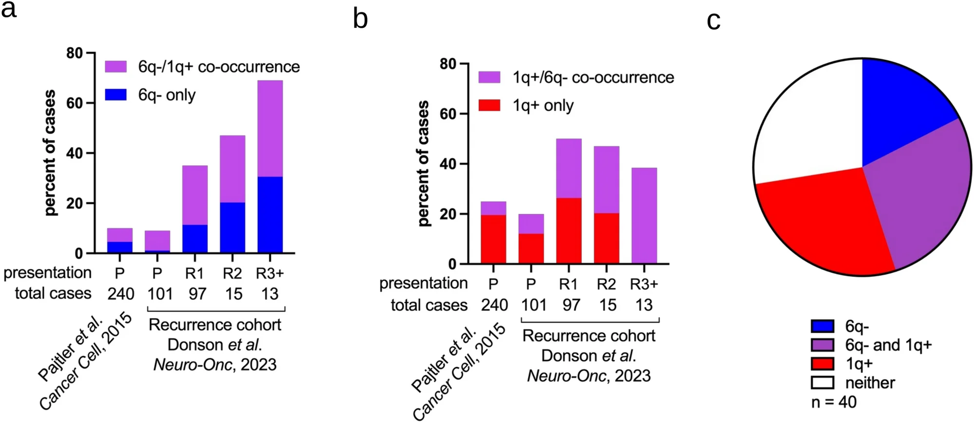
与此前阐述 1q+ 在室管膜瘤中高危行为的研究一致 [2, 5, 25, 29, 38],具有 1q+(单独存在或合并 6q-)的患者占全部死亡的 55%(22/40)。然而,6q- 也与致死结局密切相关;具有 6q-(单独存在或与 1q + 共现)的肿瘤也占全部死亡的 45%(18/40),凸显了其对该队列死亡的重要贡献(图 1c)。
全臂及部分 6q- 室管膜瘤均丢失 LATS1 位点,且无论缺失范围如何,结局均不良
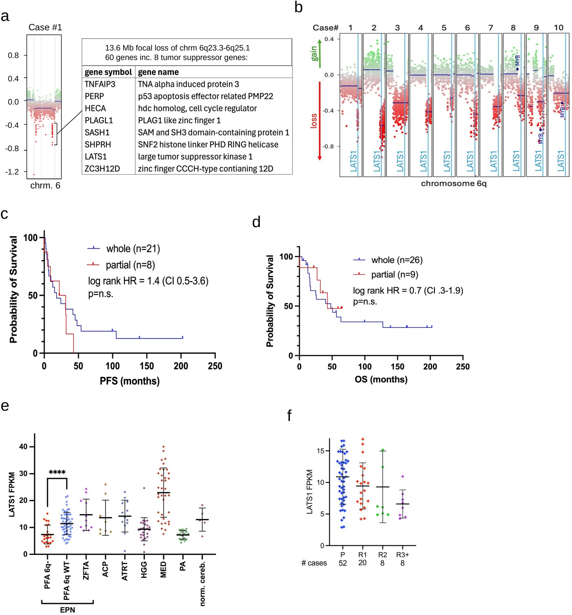
为探索 6q- PFA 室管膜瘤的侵袭性表型,我们尝试确定驱动 6q- 表型的一个或多个基因。首先,我们检查了一份尸检样本(病例−1),发现其 6q 存在局灶性缺失区域,跨越 q23.3-q25.1,覆盖 13.6 兆碱基(图 2a)。该区域的 60 个基因中,有八个是已记载的抑癌基因 [55]:TNFAIP3、PERP, HECA、PLAGL1、SASH1、SHPRH、LATS1 和 ZC3H12D。其中,LATS1 和 PLAGL1 此前已被发现与室管膜瘤相关。大肿瘤抑制激酶 1(LATS1)是多种人类恶性肿瘤中已确立的抑癌基因 [55],在一项关于肿瘤驱动因素的泛癌研究中,在 PFA 6q- 最小缺失区的八个抑癌基因中,其缺失发生率最高(1.5%)[3]
这一 PFA 6q- 局灶性缺失区域也包含抑癌基因 PLAGL1,但在室管膜瘤背景下,PLAGL1 被报道为癌基因,因此不符合所提出的通过 6q- 丧失抑癌基因功能的机制。具体而言,在一类新的儿童幕上神经上皮肿瘤亚群中发现了导致 PLAGL1 转录本高表达的 PLAGL1 融合,其中很大比例的病例在组织病理学上被诊断为室管膜瘤 [44]。此外,一项较新的研究表明,在一部分幕上室管膜瘤中,PLAGL1 转录活性升高是 ZFTA 融合的结果 [24]。
为了解这一区域的重要性,我们复查了队列中所有 6q- 病例由 DNA 甲基化数据推导的拷贝数图谱。60 例 6q- 病例中,10 例(16.7%)仅显示部分缺失。值得关注的是,所有这些部分缺失区域均涵盖病例−1 所见的 6q 局灶性缺失区域,该区域包括 LATS1 位点(图 2b),提示在这些 6q 部分缺失病例中,它是与 PFA 6q- 表型相关的主要基因。
随后,我们比较了 6q 部分缺失与全臂缺失肿瘤的生存情况。从获得 6q- 时起计算的无进展生存在两组之间无显著差异(log-rank HR = 1.6,95% CI 0.4–4.5;部分缺失为 26.65 个月,全臂缺失为 18 个月;图 2c)。两组总生存均较差,中位总生存期均约为 50 个月(图 2d)。
整体 RNA-seq 数据分析证实,与 6q 野生型(n=58)PFA 室管膜瘤相比,6q-(n=18)中的 LATS1 转录本表达显著降低(倍数变化 = 1.5;双侧 p=2×10⁻⁵),6q- PFA 室管膜瘤中 LATS1 的表达也低于所检查的任何其他脑肿瘤类型(图 2e)。与 PFA 历次复发中 6q 缺失增加相呼应(图 1a),我们观察到 LATS1 转录本表达在历次复发中下降(图 2f)。然而,与 6q 缺失不同,在我们的整体 RNAseq 队列中,按高于或低于 LATS1 表达中位数将样本二分后进行无进展生存(PFS)分析,无论在全部病例(n=88)中,还是排除具有混杂性高危临床变量(1q 获得、6q 缺失、1 岁以下婴儿)的病例后(n=40),LATS1 转录本表达均与无进展生存无关。
LATS1 敲入可恢复 6q- PFA 细胞系的蛋白表达并抑制增殖
为确定 6q- 与 6q 野生型 PFA 室管膜瘤之间细胞增殖的差异,我们重新分析了此前研究中以最高高倍视野计数定量的 Ki-67 增殖指数 [10]。6q- 肿瘤显示 Ki-67 水平显著升高(6q- n=17,6q WT n=36,双侧 p = 0.0005),支持 6q- PFA 室管膜瘤细胞本身具有更强增殖表型的假设(图 3a)。
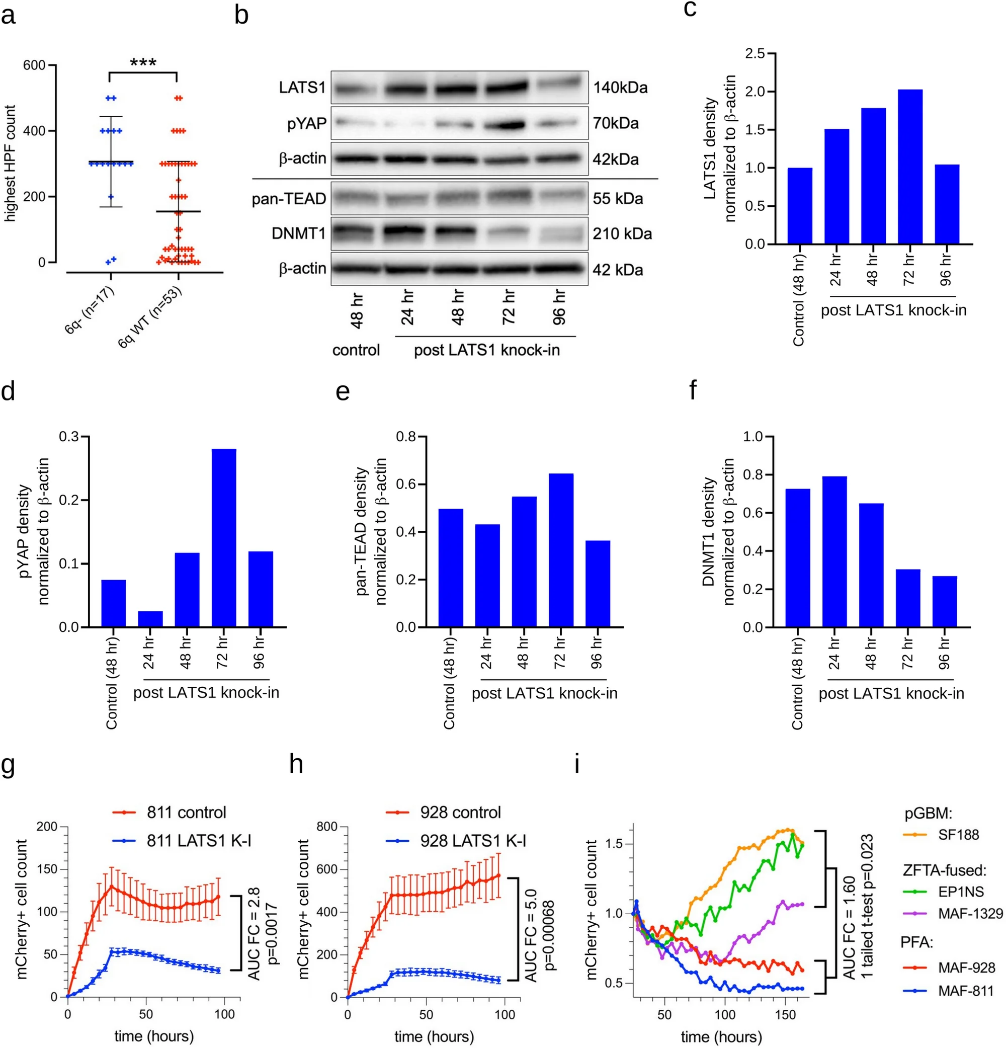
鉴于 6q-、Ki-67 指数升高与不良预后之间的关联 [5],我们研究了 LATS1 丢失是否在功能上促进肿瘤生长。我们进行了 LATS1 慢病毒敲入实验,并通过蛋白质印迹定量 LATS1 蛋白表达,以 β-肌动蛋白进行归一化。在 6q- PFA 室管膜瘤细胞系 MAF-811 中,LATS1 蛋白在敲入后 24 h 显著增加,并持续升高至 72 h(图 3b 和 c),始终高于对照水平。
此外,与对照相比,恢复 LATS1 使两株独立的 6q- PFA 室管膜瘤细胞系(MAF-811 和 MAF-928)的增殖显著减少(图 3g,h)。而且,与其他已建立的儿童脑肿瘤细胞系(ZFTA 融合型室管膜瘤 EP1NS 和 MAF-1329,以及儿童 GBM 细胞系 SF188)相比,LATS1-重建的 6q- 室管膜瘤细胞显示出最受限制的生长模式,细胞增殖迅速下降,而 6q 野生型细胞系则维持稳定增殖(图 3i)。
6 号染色体长臂缺失抑制 LATS1 并激活 Hippo 通路
LATS1 是 Hippo 信号通路的关键负调控因子,该通路最初被发现参与控制器官大小、细胞命运及增殖;LATS1 通过 S127 磷酸化及随后在细胞质中降解 YAP,阻止其进入细胞核并在核内结合、激活 TEAD 转录因子 [18, 28]。它在多种恶性肿瘤中发挥抑癌基因作用,包括肺癌、乳腺癌、胃癌、肝癌、肾癌、结直肠癌及血液系统恶性肿瘤 [17, 34]。
其他几种室管膜瘤变异型也存在 Hippo 失调。一种罕见的儿童幕上室管膜瘤由导致 Hippo 通路失调的 YAP1 融合驱动 [33]。NF2 突变型脊髓室管膜瘤因 NF2 与 LATS1 相互作用受损而表现出 Hippo 失调 [36];同样,包含 NF2 位点的 22 号染色体缺失也可能破坏 LATS1 活性 [23, 31, 36]。除体内神经祖细胞中敲低 LATS1 后形成室管膜瘤样肿瘤外,Eder 等还表明,在该模型中激活 YAP1 可产生室管膜瘤样肿瘤 [11]。因此,我们假设 PFA 6q- 中 LATS1 丢失通过解除对 YAP/TEAD 活性的抑制,使 Hippo 信号失调,并促进其核转位。
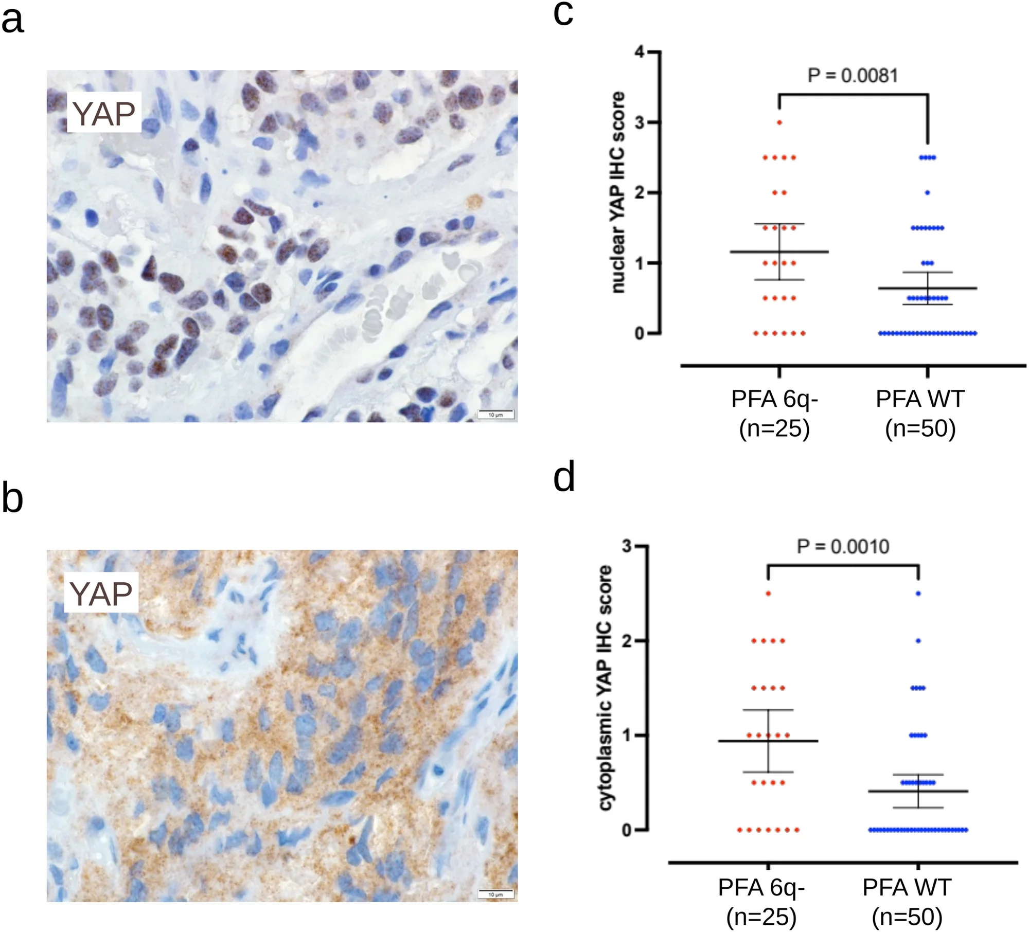
为检验这一假设,我们在一个包含 75 例 PFA 室管膜瘤(25 例 6q- 和 50 例野生型)的队列中,通过免疫组织化学检测 YAP 表达。6q- 肿瘤的细胞核和细胞质 YAP 表达均显著更高(分别为 p = 0.0081 和 p = 0.0010;图 4),与 LATS1 减少病例中的 Hippo 通路激活一致。我们进一步假设,核内 YAP 增加驱动 TEAD 表达,而细胞质 YAP 升高可能反映 LATS1 介导的降解减少。在 PFA 6q- 细胞系 MAF-811 中敲入 LATS1 后,磷酸化 YAP(S127)增加,表明 LATS1 对 PFA 中 Hippo 通路的功能性影响(图 3b,d)。
6q- PFA 室管膜瘤中 TEAD2 表达上调并预测不良生存
随后,我们检查了 Hippo 信号级联的主要效应因子 TEAD 转录因子在 PFA 6q- 中的参与情况。对整体 RNA-seq 数据的重新分析显示,与 6q 野生型(n=60)PFA 室管膜瘤相比,6q-(n=23)中 TEAD2 RNA 表达显著更高(FC=1.63;p=2.08×10⁻⁶)(图 5a)。与 6q- 和 TEAD2 RNA 增加之间的这一负相关一致,我们观察到 PFA 历次复发中 TEAD2 表达升高(图 5b),而 6q 缺失也日益增加(图 1a)。PFA 6q- 中 TEAD4 也显著更高(FC=1.47;p=0.035),而 TEAD1 和 TEAD3 则无差异表达。在功能上,LATS1 敲入未显著改变泛 TEAD 蛋白表达(图 3b,e),这与 TEAD 作为 Hippo 通路转录因子、在与核内 YAP 结合后被激活的作用一致。
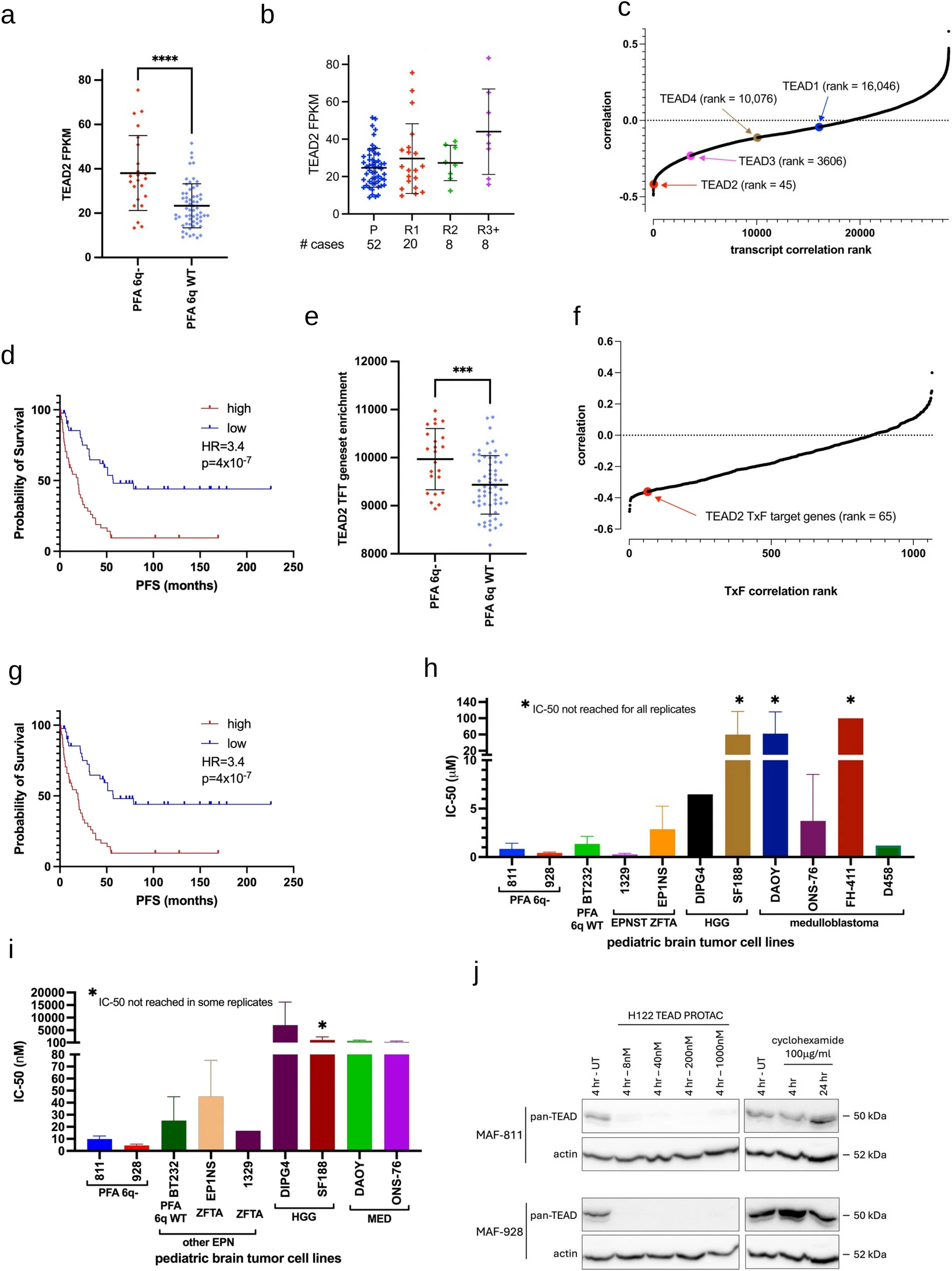
为确定 TEAD1-4 基因表达的相对预后关联,我们在来自任一次发病的 79 例复发性 PFA 中,将所有转录本的基因表达值与进展时间(TTP)进行相关性分析。TEAD2 在与进展时间负相关的总计 28,595 个转录本中排名第 45(图 5c)。高于中位数的 TEAD2 靶基因表达与不良生存相关(HR = 3.2;p = 4×10⁻⁷)(图 5d)。TEAD3(HR = 1.8;p = 0.017)和 TEAD4(HR = 1.9;p = 0.013)也呈类似趋势,即高表达与更差结局相关。
分子特征数据库(MSigDB;加州大学圣迭戈分校和 Broad 研究所)包含转录因子靶标(TFT)基因集(n=1066),其中包括 TEAD2,但不包括 TEAD1、3 或 4。与 6q 野生型(n=60)PFA 室管膜瘤相比,6q- 病例(n=23)的 TEAD2 TFT 单样本基因集富集(ssGSE)评分更高(p = 3.44×10⁻⁴)(图 5e)。PFA 6q- 病例中过表达最显著的 TEAD2 TFT 包括 DNMT1(DNA 甲基转移酶 1)(p=0.0013)。DNMT1 在癌症中常过表达,通过启动子高甲基化驱动抑癌基因沉默 [41],并使分化基因沉默及维持干细胞样状态 [35]。我们检查了 LATS1 敲入对 TEAD2 转录靶标 DNMT1(DNA 甲基转移酶 1)[22] 的影响,结果显示 LATS1 敲入后第 3 和第 4 天,其蛋白表达下降(图 3b,f)。
与上述单个基因表达的分析类似,我们在复发性 PFA(n=79)中对所有 TFT 基因集富集与 TTP 进行相关性分析,显示 TEAD2 在全部 1066 个 TFT 中,按基因集富集负相关程度排名第 65(图 5f)。Kaplan–Meier 分析证实,较高的 TEAD2 靶基因表达富集(ssGSEA)与不良生存相关(n=88)(HR = 2.80;p = 3.6×10⁻⁶)(图 5g)。
PFA 6q- 细胞对 TEAD 棕榈酰化抑制及 PROTAC 介导的 TEAD 降解敏感
我们在 PFA 6q- 细胞系 MAF-811 和 MAF-928 中,以及 PFA 6q WT 细胞系 DKFZ-BT232、幕上 ZFTA::RELA 融合型室管膜瘤 EP1NS 和 1329 及一组此前使用过的用于比较的儿童脑肿瘤细胞系中,开展了泛 TEAD 棕榈酰化抑制剂 VT017 [48] 的体外剂量曲线分析,以进一步确定其对 PFA 6q- 的选择性作用 [9]。通过 3H-胸腺嘧啶核苷掺入测定的 VT107 IC-50 值显示,两株 PFA 6q- 细胞系均敏感,呈现高纳摩尔浓度范围的敏感性(MAF-811 = 845 nM;MAF-928 = 420 nM)(图 5h)。用于比较的儿童脑肿瘤细胞系对 VT107 的敏感性各异,在所测试的广泛剂量范围内(0.61 nM 至 10μM),多株细胞系未达到 IC-50 剂量(图 5h)。PFA 6q WT 细胞系 DKFZ-BT232 的 IC-50 值(1.36 μM)高于 PFA 6q- 细胞系,但与非室管膜瘤的儿童高级别胶质瘤(HGG)和髓母细胞瘤(MED)比较细胞系相比,该 IC-50 仍显示其对药物具有相对较高的敏感性。总体而言,除 PFA 6q- 外,BT232 和 ZFTA::RELA 融合型幕上室管膜瘤细胞系也表现敏感,提示室管膜瘤各亚型普遍依赖 TEAD 活性,并提示 Hippo 信号失调的参与,而已知该信号失调可驱动 YAP 融合型幕上室管膜瘤 [33]。
蛋白水解靶向嵌合体(PROTAC)正在肿瘤领域开展临床试验,代表了一种改变治疗范式的转录因子靶向治疗方法,与小分子抑制剂相比具有多项优势,包括在更低剂量下提高效率以及克服耐药性的能力 [46]。这些化合物也为选择性、快速敲低靶蛋白提供了强大的实验室工具。我们在上述 PFA 6q- 及比较细胞系组中测试了近期开发的泛 TEAD 靶向 PROTAC 降解剂 H122 [27]。PFA 6q- 显示出最低的 H122 IC-50 值(MAF-811 = 9.8 nM;MAF-928 = 4.5 nM)(图 5i),约比使用 VT107 获得的值低 100 倍。与 VT107 类似,非室管膜瘤的 HGG 和 MED 细胞系的 H122 IC-50 显著更高。PFA 6q WT DKFZ-BT232 的 IC-50(25.1 nM)高于 PFA 6q- 细胞系,ZFTA::RELA 融合型幕上室管膜瘤细胞系亦如此,与使用 VT107 抑制 TEAD 时所见一致,再次提示室管膜瘤整体上存在潜在的 Hippo 通路失调。从机制上看,H122 处理的 PFA 6q- 细胞系在 4 h 后显示 TEAD 蛋白完全降解,而抑制蛋白质翻译(100μγ/ml 环己酰亚胺)后直至 24 h 均未见内源性 TEAD 降解(图 5j),这证实了 H122 PROTAC 的靶向作用,同时也表明 PFA 6q- 细胞系具有完整的泛素化降解通路,而该通路对 PROTAC 的疗效至关重要。
TEAD2 在未分化室管膜瘤细胞亚群(UEC-A)中富集
基于我们此前关于 PFA 室管膜瘤肿瘤内异质性的研究 [13, 14, 16],我们检查了不同肿瘤细胞亚群中的 TEAD2 表达。在早期单细胞分析中,我们界定了一类具有神经干细胞样特征和祖细胞潜能的未分化室管膜瘤细胞类型(UEC)群体 [13]。
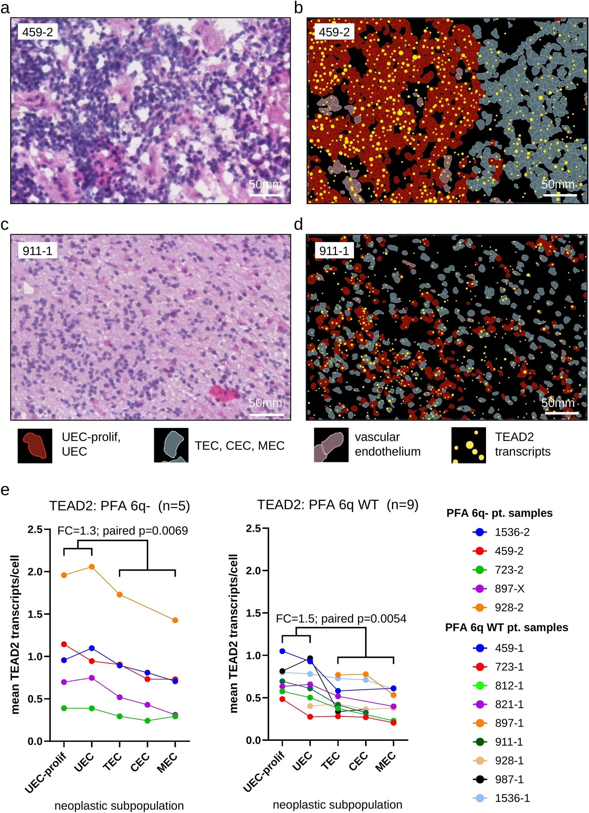
使用 Xenium 原位空间转录组技术,我们测定了 14 张 PFA 组织切片(5 份 PFA 6q- 和 9 份 PFA 6q WT 患者样本)中单个细胞的 TEAD2 和 LATS1 表达(细胞总数 1,096,706)。根据此前在室管膜瘤 [13,14,15] 及其他儿童脑肿瘤诊断类别 [7, 37, 57] 中鉴定的细胞亚群,使用包含 480 个定制探针的面板进行亚细胞分辨率的原位空间转录组分析(补充表 1)。将组织切片的细胞分割与相应的定制探针表达相结合,通过基于图的聚类识别细胞类型。将标志基因与室管膜瘤单细胞 RNAseq(scRNAseq)及 Visium 空间转录组的肿瘤性和非肿瘤性细胞亚群标志物进行比较,为 Xenium 细胞簇作注释 [13, 14],并将肿瘤细胞归入相应的主要 PFA 肿瘤细胞亚群:未分化室管膜瘤细胞 UEC-A(包括增殖性 UEC-A 亚群(UEC-A prolif.))、具有室管膜分化的转运型 TEC 和纤毛型 CEC 室管膜瘤细胞,或及具有间充质分化的 MEC 室管膜瘤细胞。无论在 PFA 6q- 还是 PFA 6q WT 中,UEC 和 UEC-A 增殖性亚群的 TEAD2 表达均显著高于其他肿瘤细胞亚群(TEC、CEC 和 MEC)(图 6e)。PFA 6q- 中 LATS1 总体低于 PFA 6q WT,但在 UEC 与其他群体之间未显示差异表达(补充图 1)。此前已证明,TEAD 的功能获得性活性可驱动神经祖细胞库扩增 [6]。与此一致,我们的数据表明,TEAD——尤其是 TEAD2——是 UEC-A 增殖的基础,支持了我们的假设,即 UEC-A 细胞代表一个促进 PFA 室管膜瘤侵袭性行为的增殖性祖细胞群体。
讨论
在本研究中,我们表明,6q- 在历次复发中逐渐累积,是 PFA 室管膜瘤中的不良预后事件。在任何一次复发时出现 6q- 均与生存缩短相关,并占近半数死亡。通过分析 6 号染色体长臂部分缺失与全臂缺失,我们证明 LATS1 缺失是其基础,导致 Hippo 通路失调及下游 TEAD 家族转录因子——尤其是 TEAD2——激活。临床前测试显示,PFA 6q- 细胞系对 TEAD 抑制敏感。空间转录组进一步将 TEAD2 过表达定位于未分化、干细胞样 UEC-A 祖细胞。我们的发现支持这样一个模型:染色体 6q- 导致 LATS1 失活,从而释放 YAP,增强 TEAD2 转录活性,以及这一干细胞样肿瘤细胞群体。因此,Hippo 通路失调成为侵袭性、复发性 PFA 室管膜瘤的一个核心特征。
与在初发及较早复发时出现的 1q 获得不同,6q 缺失出现较晚,并随历次复发稳步增加,提示存在不同的进化压力及互补的侵袭性机制。这或可解释为何同时具有 1q+ 和 6q- 的肿瘤结局最差 [5, 15]。既往研究主要聚焦于 1q 获得,其通过 MDM4 过表达模拟 p53 抑制,并可能被 5-FU 靶向 [15]。我们的数据通过将 6q 缺失确定为同样重要的、较晚出现的快速进展驱动因素,扩展了这一范式。
Hippo 通路已被发现参与多种恶性肿瘤,并调控癌症的标志性特征,其中许多已被证实参与 PFA 室管膜瘤,包括血管生成、增殖、上皮-间充质转化、炎症和免疫逃逸 [13, 16, 28, 51, 53]。本研究与结直肠癌的相关研究一致,后者发现罕见的“复苏干细胞”在癌症治疗应答中出现,并受 Hippo 通路组分调控 [12, 49]。同样,我们的数据为将 TEAD2 高表达的 UEC-A 细胞确定为潜在的复苏样祖细胞提供了初步研究基础,与此前关于肿瘤胎儿样重编程的观察一致 [10, 13, 16, 43]。未来研究应进一步表征 UEC-A 群体,并加深对其作为潜在治疗靶点在高危 PFA 室管膜瘤中作用的理解。
此前已有研究提示,LATS1 丢失可能是幕上室管膜瘤的一种致瘤机制。具体而言,是在探索 YAP1 融合型幕上疾病的背景下。Eder 及其同事描述了一种小鼠模型,在双条件敲除小鼠的 NEX-Cre 谱系中,删除 YAP1 的负调控因子 LATS1 和 LATS2 激酶会导致室管膜瘤样肿瘤 [11]。这一实验证据有力支持了我们的结论,即 Hippo 通路失调是室管膜瘤共有的致癌机制,进一步强化了开发 Hippo 靶向治疗的依据。
从临床角度看,我们的发现具有重要意义。PFA 初发时 6q- 罕见,意味着应谨慎解读其在该时间点的意义,例如回顾性 E-HIT2000 队列中的情况 [21]。然而,所有关于复发性 PFA 的研究均应按 6q- 状态分层。看来,接受手术 ± 放疗的 PFA 室管膜瘤有时可能治愈,或常常经过多年才再次复发,而 6q- 室管膜瘤并非如此。在体外恢复 LATS1 信号的能力提示,在 6q- PFA 室管膜瘤中靶向 YAP/TEAD 相互作用或更广泛的 Hippo 通路可能可行。此外,TEAD2 在未分化 UEC-A 细胞中的富集,也提示了基于分化的治疗策略的可能性。
目前 Hippo 通路调节的治疗前景令人鼓舞。相关方法包括破坏 YAP-TEAD 结合、抑制 TEAD 调控的转录、靶向转录机器以及阻断 YAP-TEAD 靶基因 [20]。Yap 等报道了一项 Hippo 药物 VT3989 治疗间皮瘤的 1/2 期试验,具有较高的部分缓解率和疾病稳定率 [54]。目前计划开展 3 期试验。VT3989 是 VT107 的类似物,后者在我们的研究中表现出 PFA 6q- 特异性的抗增殖活性。因此,这一方法对 PFA 6q- 以及可能的 YAP1 融合型幕上疾病具有近期转化潜力。目前,超过 40 种 PROTAC 正在肿瘤学临床试验中进行评估,其标志是 FDA 近期批准 vepdegestrant 用于晚期或转移性乳腺癌 [26]。PFA 6q- 细胞系对 TEAD PROTAC H122 的高度敏感性,为进一步开展这一突破性治疗策略的临床前测试提供了依据。鉴于伴 6q- 的复发性 PFA 室管膜瘤预后极差,优先推动这些发现的临床转化至关重要。
未来研究应描绘所有室管膜瘤亚型中 Hippo 通路失调的完整谱系,包括上游调控因子及其他 YAP/TEAD 靶标。临床前 6q-/LATS1 模型对于评估候选抑制剂至关重要,但与此同时,我们必须迫切考虑如何将这一新的生物学知识转化应用。不过,我们相信,将 6q 状态纳入前瞻性临床试验,将完善风险分层,并指导复发性疾病的治疗。
总之,6 号染色体长臂缺失及其导致的 LATS1 缺失,成为 PFA 室管膜瘤复发和不良结局的关键分子驱动因素,通过 Hippo 通路失调及 TEAD2 介导的增殖程序发挥作用。这些认识揭示了超高危室管膜瘤的一条新生物学轴,并为针对患儿开发 Hippo 靶向治疗方法提供了依据。
要点
(1)抑癌基因 LATS1 位于 PFA 6q- 的染色体最小缺失区内
(2)LATS1 缺失驱动 Hippo 通路失调和 YAP/TEAD 激活。
(3)恢复 LATS1 表达或抑制 TEAD 可在体外抑制生长,揭示了一条可靶向的通路。
- PFA is the most frequent and clinically aggressive ependymoma subgroup; chromosome 6q loss (6q-, with or without 1q+) defines ultra-high-risk PFA, where relapse is typically terminal.
- Cumulative 6q loss accounted for nearly half of PFA deaths and rose in successive recurrences; copy number profiling showed loss of the LATS1 locus in all 6q- cases (including 10/60 with partial 6q deletion), with poor outcome irrespective of extent of loss.
- 6q- tumours had higher Ki-67, elevated YAP and increased TEAD2-related transcriptional activity; high TEAD2 predicted poor PFS and was enriched in an undifferentiated progenitor-like population (UEC-A).
- LATS1 knock-in suppressed proliferation of 6q- cell lines; the TEAD palmitoylation inhibitor VT107 and PROTAC degrader H122 showed 6q- selective activity in vitro. Six figures.
Scope: open-access full text (CC BY 4.0) from Acta Neuropathologica — abstract, introduction, methods, statistics, results, discussion and key points, with 6 figures and legends; data availability, references, acknowledgements, funding, author information, declarations and supplementary material are not included. Bracketed numbers are the original reference numbers.
Abstract
Posterior Fossa A (PFA) is the most frequent and clinically aggressive ependymoma (EPN) subgroup, with relapse typically representing a terminal event despite intensive multimodal therapy. Ultra-high-risk PFA EPNs are characterised by chromosome 6q loss (6q-) with or without chromosome 1q gain (1q +). While the molecular mechanisms driving 1q+ have been elucidated, 6q- PFA pathobiology remains poorly understood. Here, we show that loss of 6q in PFA EPN results in deletion of LATS1 (Large Tumour Suppressor Kinase 1), thereby dysregulating the Hippo signalling pathway and enhancing YAP/TEAD activity. Integrated analyses of primary and recurrent PFA EPNs, incorporating clinical, multiomic, and functional assays, were performed to define the biology and therapeutic susceptibility of 6q- PFA EPNs. Cumulative 6q loss was associated with nearly half of PFA EPN-related deaths. Copy number profiling identified the LATS1 locus as recurrently deleted across all 6q- cases, including 10/60 with partial 6q deletions. Restoration of LATS1 expression in 6q- PFA cell lines re-established protein levels and suppressed proliferation. Tumour analyses showed marked Hippo pathway disruption with elevated YAP expression and TEAD2-related transcriptional activity. TEAD2 palmitoylation inhibitor VT107, and Proteolysis Targeted Chimera (PROTAC) degrader H122, exhibited PFA 6q- selective activity in vitro. TEAD2 upregulation was further observed in a previously described undifferentiated, progenitor-like population of PFA EPN cells. In conclusion, 6q loss defines an aggressive biological subset of PFA associated with dismal outcomes. Identification of LATS1 as a recurrently lost tumour suppressor implicates Hippo pathway dysregulation as a central driver of malignancy, and we identify YAP/TEAD signalling as a potential therapeutic target.
Introduction
Ependymoma (EPN) is the second most common malignant brain tumour in childhood and comprises multiple molecular entities [32, 33]. Posterior fossa A (PFA) EPN is the most frequent subgroup, affects the youngest children, and carries the poorest outcomes [32, 40]. Despite modern therapy targeting maximal tumour resection followed by focal radiotherapy [42], relapse occurs in around 50% of cases, and only 25% of these children survive beyond 5 years [40, 56]. PFA EPNs showing chromosomal instability—typically gain of chromosome 1q (1q +) and/or loss of chromosome 6q (6q-) exhibit an especially dismal prognosis, forming an ultra-high-risk group for whom current standard-of-care interventions are rarely curative [5].
We previously hypothesised that certain molecularly defined EPN subgroups are destined to have poor outcomes [39]. Since then, we have presented evidence from a large, international cohort showing that 1q+ and 6q- increase in frequency during PFA EPN recurrence and progression. Up to two-thirds of patients acquire a high-risk copy number alteration by first relapse, compared to one-fifth at initial presentation [10]. This evolution of tumour biology may explain why recurrence is associated with such dismal outcomes.
Recent work into the biology of 1q+ in PFA progression has highlighted the role of MDM4, located on chromosome 1q, as a negative downstream regulator of P53, mimicking a P53 mutation [15]. In addition, UCK2, also on 1q, represents a potential therapeutic weakness that may render 1q+ PFA EPN sensitive to the chemotherapy agent 5-Fluorouracil (5-FU). This hypothesised therapeutic intervention is now a focus for the development of biology-driven clinical trials of 5-FU and radiotherapy in both North America (ACNS2422) and Europe.
In contrast, the biological basis for the poor prognosis associated with 6q- PFA EPN remains undefined. Here, we investigate the tumour biology of 6q- PFA EPN using an established international dataset. We hypothesise that the adverse outcome associated with 6q- results from the loss of the tumour-suppressor gene LATS1, leading to dysregulation of the Hippo pathway and offering a druggable target through inhibition of YAP/TEAD signalling.
Methods
Cohort details
Matched primary and recurrent tumour sample copy-number variation (CNV) data was previously obtained by DNA methylation profiling in our prior multi-institutional study [10]. This dataset included 101 children with recurrent, DNA methylation-classified PFA EPNs.
For additional experiments beyond the original multi-institutional cohort, immunohistochemistry was performed on archival FFPE material from 75 PFA EPNs (25 6q- and 50 wild type), which were obtained from Children’s Colorado Hospital. Xenium in situ spatial transcriptomics was performed on 14 snap-frozen tumour samples from the Colorado paediatric neuro-oncology brain tumour bank. Bulk RNAseq, Immunohistochemistry and Xenium samples were obtained during routine surgery or autopsy respectively, with institutional approval and patient consent (COMIRB 95–500).
Bulk RNA sequencing analysis
Bulk RNA-seq analysis was performed on a dataset of primary and recurrent PFA (GSE226961, GSE243682, GSE327676)[10, 13]. RNA was isolated from snap-frozen material and sequenced on the Illumina NovaSeq 6000 platform. Sequencing data were aligned using STAR [8], and transcript quantification (in fragments per kilobase per million reads, FPKM) was generated with Cufflinks [50]. Single-sample gene-set enrichment analysis (ssGSEA) was performed on individual RNA-seq FPKM data using the GenePattern portal [4, 45].
Survival analysis
Univariate analyses using the Kaplan–Meier method were employed to assess the effect of whole or partial loss of chromosome 6q on survival from both primary disease and recurrence. We also evaluated the impact of gene expression and transcription-factor target gene enrichment score (ssGSEA) on survival using the same method. Progression free survival was modelled as a time-dependent variable to capture the effect of CNV changes and associated biological effects at recurrence. This approach addresses the significant outcome impact of 1q gains and/or 6q losses at recurrence in PFA, which significantly confound overall survival analyses.
LATS1 knock-in experiments
LATS1 open reading frame (ORF) knock-in expression vector was designed and purchased through GeneCopia. Lentiviral LATS1 vector (hLATS1) and corresponding control vector (Ctl) were generated using the pLKO system, where LATS1 and control vectors are transfected into HEK293FT cells with psPAX and pMD2.G viral packaging plasmids and TransIT-LT1 Reagent (Mirus Bio). For in vitro assays, viral supernatant was collected in Opti-MEM with 15% FBS and 1% pen/strep. LATS1 ORF knock-in was performed in two PFA 6q- cell lines previously established and characterized in our laboratory (MAF-811; MAF-928)[1]. We used three comparator cell lines with PFA 6q WT: two supratentorial EPN ZFTA-fused cell lines (MAF-1329 [19] and DFKZ-EP1NS [30]) and a paediatric glioblastoma (GBM) cell line SF188 [9]. All cell lines were subjected to routine authentication by DNA fingerprinting and mycoplasma testing. For this study cell lines were propagated under standard tissue culture conditions and grown in fetal bovine serum (FBS)-supplemented media as monolayers, Cells used in in vitro experiments were transduced with viral supernatant using 8 μg/mL polybrene. LATS1 knock-in was confirmed by Western blot analysis.
In vitro comparative TEAD palmitoylation inhibitor VT107 and TEAD PROTAC degrader H122 drug screen
We screened the effect of TEAD palmitoylation inhibitor VT107 (MedChemExpress. Cat. no. HY-134957) [48] and TEAD PROTAC (proteolysis-targeting chimera) degrader H122 (MedChemExpress. Cat. no. HY-172113) [27] in PFA 6q- cell lines MAF-811 and MAF-928 and a panel of comparator paediatric brain tumour cell lines as previously described to identify ependymoma selective drug sensitivity [9]. These included EPN PFA 6q WT cell line DKFZ-BT232 [47], EPN ZFTA-fused cell lines MAF-1329 and DFKZ-EP1NS and non-EPN paediatric high-grade glioma (HGG) cell lines (diffuse intrinsic pontine gliomas DIPG-IV and glioblastoma SF188) and medulloblastoma (MED) cell lines (DAOY, ONS76, FH-411and D458) that were maintained as described previously [9]. Cell lines were propagated as monolayers apart from semi-adherent suspension cell lines FH-411 and D458.
Cell lines were plated at ~50% confluence in 96-well plates and treated with a range of doses of VT107 (range = 10μM to 0.6 nM) and H122 (range = 1000 nM to 0.06 nM) for 72 h. Cell proliferation was measured using tritiated thymidine incorporation as previously described [9]. Treatment dose curves and associated IC-50 values were modelled using Prism statistical software (GraphPad Software, Inc., La Jolla, CA).
H122 PROTAC-specific degradation of TEAD was determined by Western blot analysis of protein extracted from PFA 6q- cell lines treated with a range of H122 doses (1000 nM to 8 nM) for 4 h. Determination of the baseline endogenous TEAD degradation rate in PFA 6q- cell lines was performed by extracting protein at 4 h and 24 h after treatment with protein translation inhibitor cycloheximide (100μγ/ml; Sigma Cat. no. C-6679).
Western blot
Western blots were performed as previously described [52]. Primary antibodies used were anti-LATS1 (1:1000, Proteintech #17,049–1-AP), anti-phospho-YAP (S127) (Cell Signaling, #4911), pan-TEAD (Cell-signaling #13,295), DNMT1 (Cell Signaling #5032) and anti-β-actin. Densitometry was performed using ImageJ software.
YAP immunohistochemistry
Standard immunohistochemistry was performed for YAP (1:200, antigen, Santa Cruz sc-101199) with antigen retrieval (10 mM Sodium Citrate pH 6.0, 110 °C, 10 min) and counterstained with hematoxylin, using techniques described previously [13]. Blinded scoring was performed using a subjective scoring scale (range 0–4) (AMD).
Xenium in-situ spatial transcriptomic analyses
The spatial expression of TEAD2 in PFA 6q- (n=5) and PFA WT (n=9) neoplastic subpopulations was performed using Xenium in situ spatial transcriptomics (10X Genomics). Snap-frozen samples were embedded in Optimal Cutting Temperature (OCT) compound, cryosectioned at 10μM thickness (Leica CM1520 cryostat) and mounted onto Xenium Slides. Slides were processed on a Xenium Analyzer (Instrument software v3.3.0.0.; analysis v 3.3.0.1) following the manufacturer’s instructions. The Xenium Analyzer detected and amplified RNA, DNA, and protein probes in 15 serial decoding and imaging cycles whilst maintaining tissue integrity and producing images of whole tissue sections with single-cell and single-molecule resolution. A custom gene probe panel of 480 genes was synthesized that included marker genes of previously identified cellular subpopulations in ependymoma [13,14,15] and other paediatric brain tumour diagnoses [7, 37, 57] (Supplementary Table 1). RNA probes were hybridized overnight, ligated, and then amplified using rolling circle amplification according to the manufacturer’s instructions (10× Genomics CG000578, CG000580, CG000613). All incubations were performed using reagents provided by 10× Genomics on an XT96 PCR machine (VWR). Autofluorescence was then quenched and morphology stains added for cell segmentation, either nuclear (DAPI) (samples 812–1, 821–1, 928–1, 723–1 and 723–2) or, cytoplasmic (18S RNA, alphaSMA and vimentin protein), and membrane (ATP1A1, CD45, E-cadherin) stain cell segmentation cocktail for improved cell segmentation (samples 928–2, 897–1, 897-X, 987–1, 911–1, 1536–1, 1536–2, 459–1 and 459–2). Slides were then loaded onto the Xenium Analyser for whole-slide tissue overview scanning and region selection. Tissue regions were denoted, and the onboard cyclic decoding and imaging occurred over a 20 – 48 h period. Data were analysed on the instrument after image acquisition, performing probe deconvolution, cell segmentation, and probe graph-based clustering. Quality control, image heatmaps and run data were reviewed in the Xenium Explorer (4.1.0) software. After Xenium analysis, H&E staining was performed on Xenium slides and whole-slide scans were performed (PhenoImager HT: 20× objective, 0.5 micron resolution). Images were converted to OME. TIFFs file using QuPath v0.5.1 and imported to Xenium Explorer and aligned with tissue images.
Cell cluster identities were assigned based on overlap with gene expression signatures from PFA neoplastic and non-neoplastic subpopulations identified by single-cell and single-nuclei RNAseq [10, 14] and Visium spatial transcriptomics [13] using hypergeometric analysis. For each sample, neoplastic clusters were then consolidated into five categories (ciliated (CEC), transportive (TEC), mesenchymal (MEC), undifferentiated-A (UEC) and proliferative undifferentiated-A (UECprolif.) EPN cells. Per cell TEAD2 and LATS1 transcript counts for each cluster were then extracted for comparative inter-sample and inter-subpopulation analyses. Xenium spatial transcriptomic data is available at GEO (GSE327676).
Statistics
Statistical analyses were performed using R, GraphPad Prism, and Excel (Microsoft) software. Kaplan–Meier survival curves were compared with log-rank tests, and p<0.05 was considered statistically significant unless otherwise stated.
Results
Cumulative loss of chromosome 6q underlies almost 50% of PFA deaths
We re-analysed data from our previously published international cohort of primary and recurrent ependymomas [10] to provide a more detailed view of the high-risk copy-number changes, focusing on chromosome 6q- (Fig. 1). Compared with 1q+, 6q- was relatively uncommon at first diagnosis, occurring in 4% (3/75) of tumours in isolation, and 8% (6/75) when co-occurring with 1q+. The distribution was consistent with data from a large independent cohort [33] (Fig. 1a). The proportion of tumours with 6q- increased progressively across recurrences, affecting 34% (25/73) at the first recurrence, 47% (8/17) of the cohort by the second recurrence, and 69% (9/13) of the third and later recurrences. Both isolated 6q- and 1q+/6q- co-occurrence rose over time (Fig. 1a). In contrast, 1q+ appeared early – affecting 48% (36/75) of cases at first recurrence and then remained relatively stable, with 100% (5/5) of 1q+ co-occurring with 6q- by third and later recurrences (Fig. 1b).
Consistent with prior studies delineating the high-risk behaviour of 1q+ in EPN [2, 5, 25, 29, 38], patients with 1q+, either alone or combined with 6q-, accounted for 55% (22/40) of all deaths. However, 6q- was also strongly associated with fatal outcomes; tumours harbouring 6q- (either alone or co-occurring with 1q +) also represented 45% (18/40) of all deaths, underscoring its major contribution to mortality in this cohort (Fig. 1c).
The LATS1 locus is lost in both whole and partial 6q- ependymoma, and outcome is poor irrespective of extent of loss
To explore the aggressive phenotype of 6q- PFA EPN, we sought to identify the gene or genes driving the 6q- phenotype. We first examined an autopsy sample (case−1), which showed a focal region of 6q loss spanning q23.3-q25.1, covering 13.6 megabases (Fig. 2a). Among 60 genes in this region, eight were documented tumour suppressors [55]: TNFAIP3, PERP, HECA, PLAGL1, SASH1, SHPRH, LATS1, and ZC3H12D. Of these, LATS1 and PLAGL1 have been previously linked to EPN. Large Tumour Suppressor Kinase 1 (LATS1) is an established tumour suppressor in a broad range of human malignancies [55] and, of the eight PFA 6q- minimal region of loss tumour suppressors, has the highest incidence of loss (1.5%) in a pan cancer study of tumour drivers [3]
This PFA 6q- focal region of loss also contained tumour suppressor PLAGL1, however PLAGL1 has been reported as an oncogene in the context of EPN, therefore not consistent with the proposed loss of tumour suppressor function via 6q-. Specifically, PLAGL1 fusions, resulting in high expression of PLAGL1 transcripts, have been identified in a novel subset of paediatric supratentorial neuro-epithelial tumours, of which a high proportion were histopathologically diagnosed as EPN [44]. Additionally, a more recent study has shown that increased PLAGL1 transcriptional activity is a result of ZFTA fusion in a subset of supratentorial EPN [24].
To understand the importance of this region, we reviewed the DNA-methylation-derived copy-number plots for all cases of 6q- in our cohort. Of 60 cases with 6q-, 10 (16.7%) demonstrated only partial deletions. Notably, all these areas of partial deletion encompassed the focal region of 6q loss as seen in case−1 that includes that LATS1 locus (Fig. 2b), implicating its role as the principal gene associated with the PFA 6q- phenotype in those cases with partial 6q loss.
We next compared survival between tumours with partial versus whole 6q loss. Progression-free survival from the time of 6q- acquisition showed no significant difference between these two groups (log-rank HR = 1.6, 95% CI 0.4–4.5; 26.65 months for partial loss and 18 months for whole loss; Fig. 2c). Both exhibited poor overall survival, with median overall survival of around 50 months in either group (Fig. 2d).
Analysis of bulk RNA-seq data confirmed markedly reduced LATS1 transcript expression in 6q- (n=18) compared with 6q wild-type (n=58) PFA EPN (Fold change = 1.5; 2-tailed p=2×10⁻⁵), LATS1 in 6q- PFA EPN also had the lowest expression of any brain tumour type examined (Fig. 2e). Mirroring the increased 6q loss seen in successive recurrences of PFA (Fig. 1a) we observed a decrease in the expression of LATS1 transcript in successive recurrences (Fig. 2f). Unlike 6q loss however, LATS1 transcript expression was not associated with progression free survival in our bulk RNAseq cohort where samples were dichotomized to higher or lower than median LATS1 expression for progression free survival (PFS), either in all cases (n=88), or after exclusion of cases with confounding high-risk clinical variables (1q gain, 6q loss, infants under 1 year old)(n=40).
LATS1 knock-in restores protein expression and suppresses proliferation in 6q- PFA cell lines
To identify differences in cellular proliferation between 6q- and 6q wild-type PFA EPN, we reanalysed quantified Ki-67 proliferation indices by highest high-power field count from our prior study [10]. The 6q- tumours exhibited significantly increased Ki-67 levels (6q- n=17, 6q WT n=36, 2-tailed p = 0.0005), supporting the hypothesis that the 6q- PFA EPN cells have an intrinsically more proliferative phenotype (Fig. 3a).
Given the association between 6q-, increased Ki-67 indices, and poor prognosis [5], we investigated whether loss of LATS1 contributes functionally to tumour growth. We performed LATS1 lentiviral knock-in experiments and quantified LATS1 protein expression by western blot, normalised to beta-actin. In the 6q- PFA EPN cell line, MAF-811, LATS1 protein increased significantly at 24 h post-knock-in and continued rising through 72 h (Figs. 3b and c), remaining persistently above control levels.
Furthermore, LATS1 restoration resulted in markedly reduced proliferation in two independent 6q- PFA EPN lines (MAF-811 and MAF-928), compared to controls (Figs. 3g,h). Moreover, compared with other established paediatric brain tumour lines (ZFTA-fused EPNs EP1NS and MAF-1329, and paediatric GBM line SF188), LATS1-reconstituted 6q- EPN cells displayed the most restricted growth patterns with a rapid fall in cellular proliferation, whilst the 6q wildtype lines maintained steady proliferation (Fig. 3i).
Loss of chromosome 6q suppresses LATS1 and activates the Hippo pathway
LATS1 is a key negative regulator of the Hippo signalling pathway originally implicated in controlling organ size, cell fate, and proliferation, through S127 phosphorylation and subsequent degradation of YAP in the cytoplasm, preventing its transit to the nucleus where it binds and activates TEAD transcription factors [18, 28]. It acts as a tumour suppressor in multiple malignancies, including lung, breast, gastric, hepatic, renal, colorectal, and haematologic cancers [17, 34].
Hippo dysregulation exists in several other EPN variants. A rare supratentorial EPN of childhood is driven by a Hippo pathway dysregulating YAP1-fusion [33]. NF2 mutant spinal ependymomas show Hippo dysregulation through the disrupted interaction of NF2 and LATS1 [36], and similarly, loss of chromosome 22 that includes the NF2 locus potentially disrupts LATS1 activity [23, 31, 36]. In addition to the formation of EPN-like tumours following LATS1 knockdown in neural progenitors in vivo, Eder et al. also showed YAP1 activation in this model produced EPN-like tumours [11]. We therefore hypothesised that LATS1 loss in PFA 6q- deregulates Hippo signalling via release of YAP/TEAD activity and promotes their nuclear translocation.
To test this hypothesis, we performed immunohistochemistry for YAP expression on a cohort of 75 PFA EPNs (25 6q- and 50 wild type). Both nuclear and cytoplasmic YAP expression were significantly higher in 6q- tumours (p = 0.0081 and p = 0.0010, respectively; Fig. 4), consistent with Hippo pathway activation in those cases with reduced LATS1. We further hypothesise that increased nuclear YAP drives TEAD expression, whereas elevated cytoplasmic YAP likely reflects reduced LATS1-mediated degradation. The functional impact of LATS1 on the Hippo pathway in PFA is indicated by increased phospho-YAP (S127) upon LATS1 knock-in in PFA 6q- cell line MAF-811 (Fig. 3b,d).
TEAD2 expression is upregulated in 6q- PFA EPN and predicts poor survival
We next examined the involvement of TEAD transcription factors, the primary effectors of the Hippo signalling cascade, in PFA 6q-. Reanalysis of bulk RNA-seq data revealed significantly higher TEAD2 RNA expression in 6q- (n=23) versus 6q wild-type (n=60) PFA EPNs (FC=1.63; p=2.08×10⁻⁶)(Fig. 5a). Consistent with this negative correlation between 6q- and increased TEAD2 RNA, we observed increased TEAD2 expression in successive PFA recurrence (Fig. 5b) whereas 6q is increasingly lost (Fig. 1a). TEAD4 was also significantly higher in PFA 6q- (FC=1.47; p=0.035), whereas TEAD1 and TEAD3 were not differentially expressed. Functionally, LATS1 knock-in did not significantly change pan-TEAD protein expression (Fig. 3b,e), consistent with TEADs role as a transcription factor in the Hippo pathway that is activated once bound to nuclear YAP.
To determine the relative prognostic association of TEAD1-4 gene expression, we correlated all transcripts’ gene expression values with time to progression (TTP) in 79 recurrent PFA from any presentation. TEAD2 ranked 45th out of a total of 28,595 transcripts negatively correlated with time to progression (Fig. 5c). Higher than median TEAD2 target-gene expression was associated with poor survival (HR = 3.2; p = 4×10⁻⁷)(Fig. 5d). Similar trends were observed for TEAD3 (HR = 1.8; p = 0.017) and TEAD4 (HR = 1.9; p = 0.013), where high expression correlated with worse outcomes.
The Molecular Signatures Database (MSigDB; UC San Diego and Broad Institute) includes Transcription Factor Target (TFT) genesets (n=1066) of which TEAD2 is included, but not TEADs 1, 3 or 4. TEAD2 TFT single sample gene set enrichment (ssGSE) scores were higher in 6q- cases (n=23) versus 6q wild-type (n=60) PFA EPNs (p = 3.44×10⁻⁴)(Fig. 5e). The most significantly overexpressed TEAD2 TFTs in PFA 6q- cases included DNMT1 (DNA methyltransferase 1) (p=0.0013). DNMT1 is frequently overexpressed in cancers and drives silencing of tumor suppressor gene via promoter hypermethylation [41] and silencing of differentiation genes and maintenance of a stem-like state [35]. We examined the impact of LATS1 knock-in on TEAD2 transcriptional target DNMT1 (DNA methyltransferase 1)[22], which showed decreased protein expression 3 and 4 days post-LATS1 knock-in (Fig. 3b,f).
As above for individual gene expression, we correlated all TFT geneset enrichments with TTP in recurrent PFA (n=79), which showed TEAD2 was the 65th most negatively correlated TFT geneset enrichment of all 1066 TFTs (Fig. 5f). Kaplan–Meier analysis confirmed that high TEAD2 target-gene expression enrichment (ssGSEA) was associated with poor survival (n=88) (HR = 2.80; p = 3.6×10⁻⁶) (Fig. 5g).
PFA 6q- cells are sensitive to TEAD palmitoylation inhibition and PROTAC-mediated TEAD degradation
We performed dose curve analysis of pan-TEAD palmitoylation inhibitor VT017 [48] in vitro in PFA 6q- cell lines MAF-811 and MAF-928 alongside PFA 6q WT cell line DKFZ-BT232, supratentorial ZFTA::RELA fusion ependymomas EP1NS and 1329 and a panel of comparator paediatric brain tumour cell lines as previously used to further determine PFA 6q- selective effects [9]. IC-50 values of VT107, as measured by 3H-thymidine incorporation, showed sensitivity in both PFA 6q- cell lines, showing high nanomolar sensitivity (MAF-811 = 845 nM; MAF-928 = 420 nM) (Fig. 5h). Comparator paediatric brain tumour cell lines showed variable sensitivity to VT107, with IC-50 doses not achieved in a number of cell lines at the broad dose range tested (0.61 nM to 10μM)(Figs. 5h). PFA 6q WT cell line DKFZ-BT232 had a higher IC-50 value (1.36 μM) than PFA 6q- cell lines, although the IC-50 revealed a relatively high sensitivity to the drug compared to non-ependymoma high-grade glioma (HGG) and medulloblastoma (MED) pediatric comparator lines. Collectively, the sensitivity of BT232 and ZFTA::RELA fusion supratentorial ependymoma cell lines in addition to PFA 6q- suggest that ependymomas subtypes are broadly dependent on TEAD activity and implicate dysregulated Hippo signaling that is already known to drive YAP-fusion supratentorial EPN [33].
Proteolysis targeted chimeras (PROTACs) are in clinical trial in the oncology setting and represent a paradigm-shifting therapeutic approach to target transcription factors with several advantages over small molecule inhibitors, with improved efficiency at lower dosing and their ability to overcome drug resistance [46]. These compounds also provide a powerful laboratory tool for selective and rapid knock-down of target proteins. We tested a recently developed pan-TEAD-targeted PROTAC degrader H122 [27] in the PFA 6q- and comparator cell line panel as described above. PFA 6q- demonstrated the lowest IC-50 values for H122 (MAF-811 = 9.8 nM; MAF-928 = 4.5 nM) (Fig. 5i), approximately 100-fold lower than those obtained using VT107. As for VT107, H122 IC-50 s were significantly higher for non-ependymoma HGG and MED cell lines. PFA 6q WT DKFZ-BT232 had a higher IC-50 (25.1 nM) than PFA 6q- cell lines, as did ZFTA::RELA fused supratentorial ependymoma cell lines, as seen using VT107 TEAD inhibition, again suggesting an underlying Hippo pathway dysregulation in EPN as a whole. Mechanistically, H122-treated PFA 6q- cell lines showed complete degradation of TEAD protein after 4 h and endogenous TEAD degradation was not seen up to 24 h post-protein translation inhibition (100μγ/ml cycloheximide) (Fig. 5j), confirming H122 PROTAC on target effects and also demonstrating that PFA 6q- cell lines have an intact ubiquitination degradation pathway that is critical for PROTAC efficacy.
TEAD2 is enriched in undifferentiated ependymoma cell subpopulations (UEC-A)
Building on our prior work identifying intratumoural heterogeneity in PFA EPN [13, 14, 16], we examined TEAD2 expression within distinct neoplastic subpopulations. In earlier single-cell analyses, we defined an Undifferentiated Ependymoma Cell type (UEC) population with neural stem-like features and progenitor potential [13].
Using Xenium in situ spatial transcriptomics, we measured the expression of TEAD2 and LATS1 in individual cells in 14 PFA tissue sections (5 PFA 6q- and 9 PFA 6q WT patient samples) (1,096,706 total cells). Subcellular resolution in situ spatial transcriptomics was performed using a panel of 480 custom probes based on previously identified cellular subpopulations in EPN [13,14,15] and other paediatric brain tumour diagnoses [7, 37, 57] (Supplementary Table 1). Cell segmentation of tissue sections was combined with corresponding custom probe expression to identify cell types using graph-based clustering. Xenium clusters were annotated by comparison of marker genes with EPN single-cell RNAseq (scRNAseq) and Visium spatial transcriptomic neoplastic and non-neoplastic cell subpopulation markers [13, 14] and neoplastic cells were assigned to correspond with major PFA neoplastic subpopulations: undifferentiated EPN cell UEC-A (including a proliferative UEC-A subpopulation (UEC-A prolif.)), ependymally-differentiated transportive TEC and ciliated CEC EPN cells or and mesenchymally-differentiated MEC EPN cells. Significantly higher TEAD2 expression was seen in UEC and UEC-A proliferative subpopulations than other neoplastic subpopulations (TEC, CEC and MEC) both in PFA 6q- and PFA 6q WT (Fig. 6e). LATS1, was collectively lower in PFA 6q- than PFA 6q WT, but did not show differential expression between UEC and other populations (Supplementary Fig. 1). Gain-of-function activity of TEAD has previously been shown to drive expansion of the neural progenitor pool [6]. Consistent with this, our data indicate that TEADs—particularly TEAD2—underlie UEC-A proliferation, supporting our hypothesis that UEC-A cells represent a proliferative progenitor population contributing to the aggressive behaviour of PFA EPN.
Discussion
In this study, we show that 6q- is progressively accumulated at successive recurrences and is a prognostically adverse event in PFA EPN. 6q- at any recurrence is associated with shortened survival and accounts for nearly half of deaths. Through analysis of partial versus whole chromosome 6q loss, we demonstrate that LATS1 deletion underlies this, leading to Hippo pathway dysregulation and downstream activation of TEAD family transcription factors – particularly TEAD2. Preclinical testing showed PFA 6q- cell lines were sensitive to TEAD inhibition. Spatial transcriptomics further localised TEAD2 over-expression to undifferentiated, stem-like UEC-A progenitor cells. Our findings support a model in which chromosome 6q- leads to LATS1 inactivation, thereby releasing YAP, enhancing TEAD2 transcriptional activity, and this stem-like tumour-cell population. Thus, Hippo pathway dysregulation emerges as a central feature of aggressive, recurrent PFA EPN.
Unlike 1q gain, which appears at presentation and earlier relapses, 6q loss emerges later and increases steadily across recurrences, indicating distinct evolutionary pressures and complementary mechanisms of aggressiveness. This may explain why tumours harbouring both 1q+ and 6q- exhibit the poorest outcomes [5, 15]. Previous research has focused mainly on 1q gain, which mimics p53 suppression through MDM4 overexpression and may be targetable with 5-FU [15]. Our data extend this paradigm by identifying 6q loss as an equally important later driver of rapid progression.
The Hippo pathway has been implicated in many malignancies and regulates cancer hallmarks, many of which have already been implicated in PFA EPN, including angiogenesis, proliferation, epithelial-mesenchymal transition, inflammation and immune evasion [13, 16, 28, 51, 53]. This work aligns with work in colorectal carcinoma, where rare “revival stem cells” emerge in response to cancer therapy and are regulated by Hippo pathway components [12, 49]. Similarly, our data provides initial work to identify TEAD2-high UEC-A cells as potential revival-like progenitors, consistent with prior observations of oncofetal reprogramming [10, 13, 16, 43]. Future studies should aim to further characterise the UEC-A population and improve understanding of its role in high-risk PFA EPN as a potential therapeutic target.
Loss of LATS1 has previously been implicated as a potential tumorigenic mechanism in supratentorial EPN. Specifically, in the context of the exploration of YAP1-fused supratentorial disease. Eder and colleagues described a murine model in which deletion of YAP1's negative regulators LATS1 and LATS2 kinases in NEX-Cre lineage in double conditional knockout mice caused ependymoma-like tumours [11]. This experimental evidence strongly supports our conclusion that Hippo-pathway dysregulation represents a shared oncogenic mechanism in EPN, reinforcing the rationale for developing Hippo-targeted therapies.
Clinically, our findings carry important implications. The rarity of 6q- at PFA presentation means that its significance at that time point, as in the retrospective E-HIT2000 cohort is to be interpreted cautiously [21]. However, all studies on recurrent PFA should be stratified for 6q-. It does appear that PFA ependymoma treated with surgery ± radiation may sometimes be cured or go often years before another relapse while this is not the case in 6q- ependymoma. The ability to restore LATS1 signalling in vitro suggests that targeting YAP/TEAD interactions or the wider Hippo pathway in 6q- PFA EPN may be feasible. Moreover, the enrichment of TEAD2 in undifferentiated UEC-A cells raises the possibility of differentiation-based therapeutic strategies.
The current therapeutic landscape for Hippo pathway modulation is encouraging. Approaches include disrupting YAP-TEAD binding, suppressing TEAD-regulated transcription, targeting transcriptional machinery, and blocking YAP-TEAD target genes [20]. Yap et al. reported a Phase1/2 trial of the Hippo agent VT3989 in mesothelioma, a high rate of partial responses and stable disease [54]. A Phase 3 trial is planned. VT3989 is an analogue of VT107 that demonstrated PFA 6q- specific anti-proliferative activity in our study. This approach, therefore, offers near-term translational potential for PFA 6q- and possibly YAP1-fused supratentorial disease. Over 40 PROTACs are currently in oncology clinical trials, marked by the recent FDA approval of vepdegestrant for advanced or metastatic breast cancer [26]. The high sensitivity of PFA 6q- cell lines to TEAD PROTAC H122 provide rationale for further preclinical testing of this breakthrough therapeutic strategy. Given the dismal prognosis of relapsed PFA EPN with 6q-, prioritising clinical translation of these findings is essential.
Future research should map the full spectrum of Hippo pathway dysregulation in all subtypes of EPN, including upstream regulators and other YAP/TEAD targets. Pre-clinical 6q-/LATS1 models will be crucial for evaluating candidate inhibitors, but we must simultaneously and urgently consider how we translate this new biological knowledge. However, we are confident that integrating 6q status into prospective clinical trials will refine risk stratification and guide therapy in recurrent disease.
In conclusion, loss of chromosome 6q, and consequent LATS1 deletion emerge as a key molecular driver of recurrence and poor outcome in PFA EPN, acting through Hippo pathway dysregulation and TEAD2-mediated proliferative programs. These insights illuminate a new biological axis of ultra-high-risk ependymoma and provide a rationale for developing Hippo-targeted therapeutic approaches for affected children.
Key points
(1) Tumour suppressor LATS1 is in the minimal region of chromosomal loss in PFA 6q-
(2) LATS1 deletion drives Hippo pathway dysregulation and YAP/TEAD activation.
(3) Restoring LATS1 expression or inhibiting TEAD suppresses growth in vitro, revealing a targetable pathway.