- 47 岁女,右叶 0.6 cm 包膜清楚伴局部浸润的甲状腺肿瘤:弥漫乳头状/实性-乳头状架构,透明/嗜酸胞质,不规则空泡核(染色质边集、核沟、偶见假包涵体),双核、巨核与多核聚集,瘤内 CD8+ T 细胞。
- IHC:高 CK7、高 PAX8,极低甲状腺球蛋白、低 TTF-1;Melan-A 与 RCC 阳性;Ki-67 约 2.5%;BRAF V600E 阴性;p53 野生型模式。
- FISH 无 TFE3/TFEB 重排;转录组确认为甲状腺来源并表达 Melan-A/MiTF;NGS 检出两处体细胞 TSC2 截短突变(tuberin 与 mid-protein 结构域),无其他已知甲状腺驱动。
- 作者称首次在滤泡性甲状腺肿瘤中证实黑色素细胞分化;形态可与经典型 PTC 重叠,TSC2 测序技术难点提示既往可能漏诊。鉴别任何「形态/IHC 异常或缺乏已知驱动」的甲状腺肿瘤时应考虑 TSC2 突变癌。
收录范围:PMC 开放获取全文(CC BY-NC-ND 4.0)——摘要、结果要点、讨论要点及图 1–3 与图注;方法细节、参考文献与补充材料未全译。
摘要
携带 TSC2 基因突变的甲状腺癌既往报道极少。本文报告一例独特的甲状腺癌:具有体细胞 TSC2 突变、滤泡与黑色素细胞分化及独特形态特征,并有全面分子遗传学支持。未检出其他已知致病驱动突变。TSC2 突变预测为截短突变,导致 mTOR 信号组成性激活,转录组表达谱予以证实。这是首次证实具有黑色素细胞分化的甲状腺滤泡性肿瘤,相应 Melan-A 表达可能成为该肿瘤的生物标志。其弥漫乳头状架构、巨核、多核聚集及瘤内 T 淋巴细胞,是其他 TSC2 突变甲状腺癌尚未描述的形态特征。与经典型甲状腺乳头状癌的形态重叠,以及测序大基因 TSC2 的已知技术困难,提示 TSC2 突变甲状腺癌迄今可能被低估。作者建议:对任何具有异常形态与免疫组化特征、或缺乏已知甲状腺癌驱动突变的甲状腺肿瘤,鉴别诊断中应考虑 TSC2 突变甲状腺癌。
病例与形态要点
47 岁女性,因甲状旁腺功能亢进、肾结石与肾积水就诊;TSH、游离 T4 正常。超声示右叶下极/峡部实性均质低回声结节约 1.0×0.7×0.3 cm(ACR-TIRADS 4),术前 FNA 为意义不明确的非典型(Bethesda)。甲状旁腺切除见单发腺瘤;右半甲状腺切除示境界清楚的 0.6 cm 肿瘤,薄包膜,一侧边缘浸润性生长(图 1A)。无甲状腺外延伸,切缘阴性,未取样区域淋巴结。
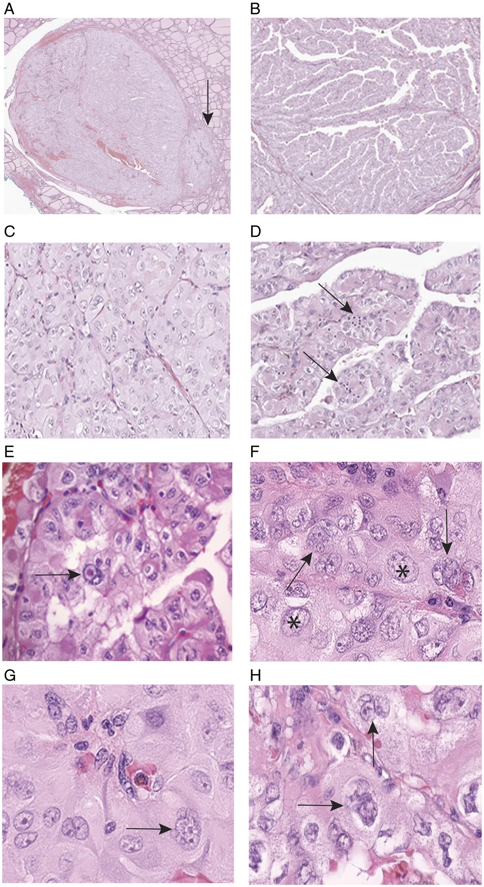
肿瘤呈弥漫乳头状(图 1B)与实性-乳头状(图 1C)架构,胞质斑片状透明与嗜酸性(图 1D–H);核不规则空泡状,染色质边集、核沟、偶见假包涵体(图 1E)。常见双核,以及特征性巨核(图 1F)与多核聚集(图 1F–H);可见少量瘤内淋巴细胞。
免疫组化与分子
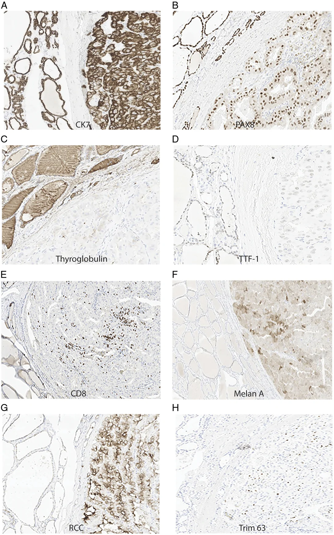
瘤细胞高表达 CK7、PAX8,甲状腺球蛋白极低、TTF-1 低;突触素、嗜铬粒素、降钙素阴性。瘤内淋巴细胞以 CD8+ T 细胞为主。Ki-67 约 2.5%;BRAF V600E 蛋白阴性;p53 呈野生型模式。相对正常甲状腺,肿瘤中等至高表达 RCC(有一定甲状腺交叉反应)与 Melan-A;Trim63 RNA 原位杂交呈特征性点状表达(图 2)。
异常形态、极低甲状腺球蛋白、低 TTF-1 与高 PAX8 最初强烈提示转移性肾细胞癌可能。TFE3/TFEB FISH 阴性。Thyroseq 与 Tempus 基于 RNA 的肿瘤来源评估分别以 87% 与 99% 概率支持甲状腺滤泡来源(含 NIS 等表达)。转录组见甲状腺球蛋白及黑色素细胞标志 MiTF、Melan-A、HMB45/PMEL。NGS 检出两处体细胞 TSC2 突变:一处缺失(c.2234del;VAF 11.7%)致 tuberin 结构域移码提前终止(p.K745R*fs25),另一处亦为截短突变(mid-protein 结构域)。无其他已知甲状腺癌致病驱动。图 3 示既往与本例 TSC2 突变在功能结构域上的分布。

讨论要点
作者将本病暂称为「乳头状 TSC2 甲状腺癌」:同时具滤泡与黑色素细胞分化、体细胞失活 TSC2 突变及独特形态。黑色素细胞分化既往偶见于甲状腺髓样癌,但据作者所知这是滤泡性甲状腺肿瘤中的首例(IHC Melan-A + 转录组 Melan-A/MiTF)。形态上巨核、多核聚集与瘤内 T 细胞等特征有助于与经典型 PTC 及其他 TSC2 突变甲状腺癌(多呈嗜酸细胞肾癌样)区分。主要局限为单例报告;期待更广泛、更准确的 TSC2 测序充实对该类肿瘤的认识。
- 47 F, 0.6 cm right-lobe thyroid tumour: thin capsule with focal invasion; diffuse papillary/solid-papillary architecture; clear/eosinophilic cytoplasm; irregular vesicular nuclei (margination, grooves, rare pseudo-inclusions); binucleation, macronuclei, multinuclear aggregates; intratumoral CD8+ T cells.
- IHC: high CK7/PAX8, very low thyroglobulin, low TTF-1; Melan-A and RCC+; Ki-67 ~2.5%; BRAF V600E−; wild-type p53 pattern.
- No TFE3/TFEB rearrangement; transcriptome confirms thyroid origin with Melan-A/MiTF; NGS: two somatic truncating TSC2 mutations (tuberin and mid-protein domains); no other known thyroid drivers.
- First demonstration of melanocytic differentiation in a thyroid follicular tumour; Melan-A may be a biomarker. Morphologic overlap with classic PTC and hard-to-sequence TSC2 suggest underdiagnosis. Consider TSC2-mutated thyroid carcinoma when morphology/IHC is unusual or known drivers are absent.
Scope: PMC open-access full text (CC BY-NC-ND 4.0) — abstract, key results, discussion highlights, and Figures 1–3 with legends; detailed methods, references and supplements not fully reproduced.
Abstract
Thyroid carcinomas with mutations in the TSC2 gene have been described rarely. Here, we report a unique thyroid carcinoma that has somatic mutations in TSC2, follicular and melanocytic differentiation and distinct morphologic features, supported by a comprehensive molecular-genetic analyses. No other known pathogenic driver mutations were identified. The TSC2 mutations are predicted to be truncating and result in constitutive activation of mammalian target of rapamycin (mTOR) signaling that was confirmed by transcriptome expression profiling. This is the first demonstration of a thyroid follicular tumor with melanocytic differentiation and corresponding Melan-A expression may be a biomarker for this thyroid cancer. Its diffuse papillary architecture, macronuclei, multinuclear aggregates and intratumoral T lymphocytes are morphologic features not yet described in other TSC2-mutated thyroid carcinomas. Morphologic overlap with classic thyroid papillary carcinoma and known technical difficulties in sequencing the large TSC2 gene make it likely that TSC2-mutated thyroid carcinomas have been underdiagnosed so far. We suggest that TSC2-mutated thyroid carcinoma should be considered in the differential diagnosis of any thyroid tumor that has unusual morphologic and immunohistochemical features or lacks known driver mutations in thyroid cancer.
Case and morphology
A 47-year-old woman presented with hyperparathyroidism, renal stones and hydronephrosis; TSH and free T4 were normal. Ultrasound showed a solid homogeneous hypoechoic nodule ~1.0×0.7×0.3 cm in the inferior right lobe/isthmus (ACR-TIRADS 4); preoperative FNA was atypia of uncertain significance (Bethesda). Parathyroidectomy identified a single adenoma; right hemithyroidectomy showed a well-demarcated 0.6 cm tumour with a thin capsule and infiltrative invasion along one edge (Fig. 1A). No extrathyroidal extension; margins negative; no regional nodes sampled.
The tumour showed diffuse papillary (Fig. 1B) and solid-papillary (Fig. 1C) architecture, patchy clear and eosinophilic cytoplasm (Fig. 1D–H), and irregular vesicular nuclei with chromatin margination, grooves and rare pseudo-inclusions (Fig. 1E). Binucleation, distinctive macronuclei (Fig. 1F) and multinuclear aggregates (Fig. 1F–H) were frequent; a small lymphocytic infiltrate was present.
Immunohistochemistry and molecular findings
Tumor cells expressed high CK7 and PAX8, very low thyroglobulin and low TTF-1; synaptophysin, chromogranin and calcitonin were negative. Intratumoral lymphocytes were predominantly CD8-positive T cells. Ki-67 was ~2.5%; BRAF V600E mutant protein was not expressed; p53 showed a wild-type pattern. Relative to normal thyroid, tumor cells expressed moderate-to-high RCC and Melan-A; Trim63 RNA ISH showed a characteristic dot-like pattern (Fig. 2).
Unusual morphology with very low thyroglobulin, low TTF-1 and high PAX8 initially strongly suggested metastatic renal cell carcinoma. TFE3/TFEB FISH was negative. Thyroseq and Tempus RNA-based assays supported thyroid follicular origin (87% and 99% probability), including NIS expression. Transcriptome profiling showed thyroglobulin and melanocytic markers MiTF, Melan-A and HMB45/PMEL. NGS identified two somatic TSC2 mutations: a deletion (c.2234del; VAF 11.7%) causing a frameshift/premature stop in the tuberin domain (p.K745R*fs25) and a second truncating mutation in the mid-protein domain. No other known pathogenic thyroid drivers were found. Figure 3 maps reported TSC2 mutations in thyroid carcinomas across functional domains.
Discussion highlights
The authors term this “papillary TSC2 thyroid carcinoma”: follicular plus melanocytic differentiation, somatic inactivating TSC2 mutations and distinct morphology. Melanocytic differentiation has been rare in medullary carcinoma; to their knowledge this is the first in a thyroid follicular tumour (IHC Melan-A plus transcriptome Melan-A/MiTF). Macronuclei, multinuclear aggregates and intratumoral T cells help separate it from classic PTC and from other TSC2-mutated thyroid carcinomas (often chromophobe RCC-like). Limitation: single case; broader, more accurate TSC2 sequencing should clarify the spectrum.