- 亚洲大洋洲神经病理学会(AOSNP)ADAPTR 工作组把室管膜瘤诊断按资源等级分为五级:RL I 仅组织学 → RL II 基础 IHC(GFAP、EMA 点状/环状、OLIG2、Ki-67,可选 SOX10)→ RL III 替代性 IHC → RL IV FISH/RT-PCR → RL V NGS 与甲基化谱;推荐分「可考虑」与「强烈推荐」。
- 替代 IHC 对照:ST 用 L1CAM/p65(ZFTA 融合多为双阳,联合敏感性 88%–100%、特异性 88%–92%);PF 用 H3 K27me3 缺失 + EZHIP 强核阳性(敏感性/特异性 99%/98%)确立 PFA;SP 用 MYCN(D4B2Y 克隆)提示 MYCN 扩增型;MPE 疑难时用 HOXB13。
- 哪些需要转诊或只能描述性诊断:ST 室管膜瘤在 RL III 及以下给「ST 室管膜瘤,NOS」;PFB 只能靠甲基化(RL V)确诊,H3 K27me3 保留(>80% 阳性)和/或 EZHIP 阴性时给「PF 室管膜瘤,NOS」;SE 和典型 MPE 在 RL I/II 即可达 WHO CNS5 诊断。
- 实用细节:OLIG2 阴性且 H3 K27me3 缺失的脊髓肿瘤须结合影像排除 PFA 下行转移;DMG 也可有 MYCN 扩增,RL III 应加做 H3 K27M/K27me3;脑干/中线 SE 样肿瘤可携 H3 K27M。表 2 列出 MN1、PLAGL1、PATZ1、BCOR 等室管膜瘤样实体的鉴别。
收录范围:PMC 开放获取全文(CC BY-NC-ND 4.0)——摘要、正文全部章节、表 1–2 与图 1–5 图注中英对照;流程图图片本身为英文原图。参考文献、补充表 S1–S6、作者贡献与图形摘要未收录。
摘要
室管膜瘤是少见的中枢神经系统(CNS)原发肿瘤,儿童与成人均可罹患。近年来,分子分析依据部位、CpG 岛甲基化模式及基因融合事件,识别出室管膜肿瘤的多个亚型。这些知识对于提升诊断精度、预后判断与有效治疗十分重要,但随之而来的副作用是:在许多低收入或中低收入国家,由于成本与技术障碍,最佳实践诊断往往无法获得。ADAPTR 小组(Adapting Diagnostic Approaches for Practical Taxonomy in Resource‐Restrained Regions,资源受限地区实用分类诊断方法适配)是亚洲大洋洲神经病理学会的一项倡议,旨在为不同程度病理资源受限的情况提供实用的 CNS 肿瘤诊断指南。本文依据 ADAPTR 资源等级,提出室管膜肿瘤的实用诊断方法。这些推荐是对世界卫生组织(WHO)中枢神经系统肿瘤分类指南的实用化适配,适合资源受限地区的常规诊断。
引言
实体瘤分类越来越依赖于将复杂分子信息与经典组织学评估相整合。这一要求在中枢神经系统(CNS)肿瘤中最为普遍、由来最久:部分肿瘤类型整合分子特征已被强制要求超过十年 [1]。室管膜瘤分级历来依赖组织学分级参数,可重复性和临床实用性均欠佳 [2]。自 2021 年 WHO CNS 肿瘤分类发布以来 [3],室管膜瘤的组织学亚型划分与分级因引入分子亚分类而得到改进,该方案以解剖部位、基因组改变和甲基化谱为依据 [4, 5]。在此方案中,部分室管膜肿瘤亚型的定义已转为完全基于复杂分子谱——例如,后颅窝 B 组(PFB)室管膜瘤现由其甲基化谱定义 [3]。诊断与预后精度的提升固然十分必要,但在低收入及中低收入国家(LICs 与 LMICs)开展所需分子检测,其成本令常规应用难以承受 [4, 6]。
AOSNP‐ADAPTR 倡议(亚洲大洋洲神经病理学会关于资源受限地区实用分类诊断方法适配的推荐)提出实用的、按资源分层的诊断路径:在 CNS 肿瘤诊断中强调形态学和免疫组化(IHC),并在必需或信息量很高时推荐靶向分子检测 [7, 8]。AOSNP‐ADAPTR 推荐是对 WHO 中枢神经系统肿瘤分类第 5 版 [3] 的实用化适配,旨在适应资源有限环境中的常规诊断实践,而不改变既定的诊断类别 [6, 7]。正如本组及其他研究者此前报道,LICs 与 LMICs 在实施许多(CNS)肿瘤诊断所需分子技术方面面临重大障碍 [6, 7, 8, 9]。
室管膜肿瘤的经典形态特征已有充分描述;按现行 WHO 中枢神经系统肿瘤分类(WHO CNS5),部分肿瘤类型仅凭形态即可诊断(如室管膜下瘤)[3]。然而,其他室管膜肿瘤的准确诊断需要高级分子检测。当遗传学检测不可行,或检测结果无法将肿瘤归入现行 WHO 分类时 [6],按现行 WHO 指南,诊断分别标注为 NOS(未特指)或 NEC(未另分类)[10]。
本文中,AOSNP‐ADAPTR 室管膜瘤工作组提出一种以组织学为导向、按资源等级划分的室管膜瘤诊断方法:利用替代性免疫组化标志物和临床—影像相关性,并在分子检测对 WHO CNS5 实体属必需且可能改变治疗时升级至分子检测。在特定情况下,建议或推荐转诊至资源更高的中心。
资源等级(RL)已在既往文献中定义 [6, 8],并转载于补充材料 S1。下文详述室管膜肿瘤的影像、形态及相关替代性免疫组化标志物,以及基于当地资源等级的 ADAPTR 推荐和诊断流程图。
资源受限环境中室管膜瘤诊断与治疗面临的挑战
室管膜瘤的诊断分类取决于其解剖部位,各部位具有不同的典型分子谱,需要不同的分子检测方法 [3]。例如,幕上(ST)室管膜瘤需要基因融合检测,而甲基化谱分析是 PFB 室管膜瘤的必要诊断标准 [3]。因此,很难对室管膜肿瘤作为一个整体统一规划分子检测,我们的 ADAPTR 推荐遵循了 WHO 分类按解剖部位逐一处理室管膜瘤的方式。
室管膜肿瘤的标准治疗仍以手术为主,大体全切(GTR)及及时的辅助放疗是重要的预后因素 [11, 12]。实现 GTR 所需的专业神经外科诊疗在提供上存在重大挑战:一项针对神经外科及小儿外科学会的国际调查估计,LICs/LMICs 每 360 万儿童仅有一名小儿神经外科医师 [13]。多伦多 Hospital for Sick Children 一项关于后颅窝室管膜瘤 GTR 率的研究显示,GTR 率由 1985 年的 35% 升至 2014 年的 77%,与无进展生存(PFS)和总生存(OS)的改善相关,同时伴有前期放疗 [14]。相比之下,LICs/LMICs 机构报道的 GTR 率分别为 17%(埃及)[15]、31%(伊朗)[16]、32%(秘鲁)[17] 和 38%(巴西)[18],这些研究均将较高死亡率归因于较低的 GTR 率 [19]。此外,在 LICs/LMICs,室管膜瘤放疗的公平可及性可能有限,交通费用与收入损失等社会经济因素影响治疗依从性 [16]。一项来自高收入及中高收入国家多个中心的多队列研究发现,相当比例的 PFB 室管膜瘤可仅行手术治疗 [20]。因此,分子分型虽然昂贵,但可能有助于识别可免于术后放疗的室管膜瘤亚型,为 LICs/LMICs 患者减轻辅助治疗的经济负担 [16, 20]。
室管膜瘤的影像特征
AOSNP‐ADAPTR 调查显示,LICs/LMICs 中超过一半的中心可进行增强磁共振成像(MRI),凸显了在此环境下影像资料对 CNS 肿瘤诊断的关键支持作用 [6]。沿用 ADAPTR 成人型弥漫性胶质瘤框架 [8],在所有资源等级中,影像特征都依据肿瘤部位(ST、后颅窝 [PF] 和脊髓 [SP])为室管膜肿瘤提供一线诊断背景。幕上肿瘤常为境界清楚的肿块,伴囊性变和钙化,实性部分呈明显但不均匀强化 [21]。后颅窝 A 组(PFA)室管膜瘤通常为伴钙化的外侧或中线肿瘤,强化不完全,侵入后颅窝诸孔(尤其是 Luschka 孔),导致明显脑积水,多见于幼儿 [22]。相反,PFB 室管膜瘤为侵袭性极低、无钙化的中线肿瘤,一般见于成人和青少年,起自第四脑室底,以囊性为主,实性部分完全均匀强化 [22]。基于 MRI 特征的机器学习算法也可能有助于区分 PFA 与 PFB 室管膜瘤 [23]。脊髓室管膜瘤通常位于髓内,好发于颈段—颈胸段;常伴脊髓空洞 [24, 25]。髓外延伸或软脑膜播散等侵袭性表现应提高对 SP 室管膜瘤,MYCN 扩增型的警惕 [26]。将影像特征纳入诊断流程,需要与当地(神经)影像专家密切协作。
室管膜瘤的形态特征
经典室管膜形态包括:瘤细胞核圆形至卵圆形,排列成血管周假菊形团、真性室管膜菊形团,以及模拟室管膜衬覆(管道)[1, 3, 27]。室管膜瘤通常与周围脑组织境界清楚 [1, 3, 27]。可能见到的其他组织学特征包括纤维性区域、钙化和囊性变 [1, 3, 27]。尽管 ADAPTR 推荐不要求超微结构检查,但所有室管膜瘤亚型均存在室管膜特征(如胞质内微绒毛、纤毛及复杂的细胞间拉链样连接)[28]。2016 年 WHO 分类 [27] 中描述的形态学模式(透明细胞型、伸长细胞型和乳头型)因缺乏临床病理实用价值,在 2021 年 WHO 分类 [3] 中不再列为亚型。这些形态学模式与特定解剖部位相关,因此在下文按解剖部位定义的亚型中加以描述。
现行分类仅依据组织学所见,为 ST、后颅窝和 SP 室管膜瘤类型赋予 CNS WHO 2 级或 3 级 [3]。然而,公认室管膜瘤的组织病理分级与患者预后并不一致 [2],且常与解剖部位相关 [29]。尽管在分子分类框架中组织学分级的意义可受质疑,但在资源有限的环境中,组织学分级可能是患者及其照护者唯一的预后指标,因此仍纳入本推荐。室管膜瘤的高级别特征包括活跃的核分裂和微血管增生 [2, 29],其预后价值高于核多形性或坏死等其他特征 [30]。
室管膜瘤的谱系及替代性免疫组化
资源等级 II(RL II)的室管膜瘤谱系标志物——ADAPTR 室管膜瘤工作组称之为「基础 IHC」——包括胶质纤维酸性蛋白(GFAP)和上皮膜抗原(EMA)(点状/环状腔面模式)的免疫组化表达 [31, 32, 33]。OLIG2 和 SOX10 阴性有助于将室管膜瘤与弥漫性胶质瘤以及神经鞘瘤、毛细胞型星形细胞瘤等类似病变相区分 [3, 34]。Ki‐67 指数未纳入 IHC 标志物,因为 WHO CNS5 室管膜瘤分级并不使用该指标 [3]。不过值得注意的是,一项 141 例的单中心研究发现,以 7% 为 Ki‐67 截断值,可作为所有解剖部位室管膜瘤分级的有用辅助 [35]。
资源等级 III(RL III)中可行的替代性 IHC 包括:(i)L1 细胞黏附分子(L1CAM)和 p65/RELA;(ii)H3 K27me3/H3 K27M;(iii)zeste 同源物增强子抑制蛋白(EZHIP);(iv)MYCN;(v)同源框 B13(HOXB13)。EBP50 表达于人上皮腔面,亦被提出作为潜在的 IHC 标志物 [36]。替代性 IHC 标志物在下文 ADAPTR 推荐中进一步阐述。与 cIMPACT‐NOW 第 11 次更新 [37] 保持一致,本文将热点 H3 p.K28 称为「H3 K27」。
ADAPTR 室管膜瘤评估推荐
与成人型弥漫性胶质瘤所用方法类似 [8],ADAPTR 室管膜瘤工作组在本文中给出 ADAPTR 的三个组成部分:(i)与室管膜肿瘤相关的诊断病理服务资源等级;(ii)各室管膜肿瘤类型在不同资源等级下的「以组织学为导向的整合诊断」格式推荐,包括在无法按 WHO CNS5 水平分级时给出的「ADAPTR 分级」;(iii)各室管膜肿瘤类型在不同资源等级下相应的诊断流程图。
诊断病理服务的资源等级
资源等级 I(RL I;组织学)强调识别经典室管膜形态,并在适用时给出 ADAPTR 组织学分级(II/III);当无法进一步检测时,允许仅凭形态进行治疗(表 1)。RL II 增加基础 IHC 以确认谱系。RL III 纳入替代性免疫标志物——ST 室管膜瘤用 L1CAM/p65,后颅窝室管膜瘤用 EZHIP/H3 K27me3,SP 室管膜瘤用 MYCN IHC——以近似判定 WHO CNS5 实体。资源等级 IV(RL IV)可进行靶向分子确认(如以 FISH 或逆转录聚合酶链反应 [RT‐PCR] 检测 ZFTA 或 YAP1 融合;以 FISH 检测 MYCN 扩增);资源等级 V(RL V)在前述各级结果不确定时或为进一步风险分层,使用综合平台(二代测序 [NGS] 和 DNA 甲基化谱)。虽然各资源等级都应开展多学科讨论,但在 RL I 和 RL II 作出诊断的病理医生尤其必须与治疗团队讨论其发现,以评估转诊至更高资源等级中心是否会影响治疗和预后判断。各部位的具体推荐详见下文。
| 资源等级 | 资源等级 I(RL I) | 资源等级 II(RL II) | 资源等级 III(RL III) | 资源等级 IV(RL IV) | 资源等级 V(RL V) | |||
|---|---|---|---|---|---|---|---|---|
| 可用资源 | 仅组织学 | + 基础诊断性 IHC 标志物 | + 替代性 IHC 分子标志物 | + 基础分子检测 | + 高级分子检测 | |||
| 病理服务 | 材料
技术
| 材料 (i) 和 (ii) 技术 1. 和 2. 3. 基础 IHC 标志物 | 材料 (i) 和 (ii) 技术 1. 至 3. 4. 替代性 IHC 分子标志物 | 材料 (i) 和 (ii) (iii) 新鲜组织 技术 1. 至 4. 5. FISH 6. RT‐PCR | 材料 (i)、(ii) 和 (iii) 技术 1. 至 6. 7. NGS DNA/RNA 测序 8. DNA 甲基化谱 | |||
| 基础 IHC | 无 | GFAP、EMA、OLIG2、Ki‐67;可选——SOX10 | 同 RL II | 同 RL II | 同 RL II | |||
| 替代性 IHC | 无 | 无 | 标志物 | 用于诊断: | 同 RL III | 同 RL III | ||
| L1CAM/p65 | ST 室管膜瘤,ZFTA 融合阳性 | |||||||
| EZHIP | PFA 与 PFB 室管膜瘤鉴别 | |||||||
| H3 K27M | 排除 H3 K27M 突变型弥漫性中线胶质瘤,H3 K27 变异型 | |||||||
| H3 K27me3 | PFA 与 PFB 室管膜瘤鉴别 中线部位弥漫性胶质瘤(OLIG2+)时,排除弥漫性中线胶质瘤,H3 K27 变异型 | |||||||
| MYCN | SP 室管膜瘤,MYCN 扩增型 | |||||||
| HOXB13 | MPE | |||||||
| 诊断性分子检测 | 无 | 无 | 无 | 5. FISH | 同 RL IV,另加 7. 用于诊断的 NGS DNA/RNA 测序 | |||
| ZFTA 融合 | ST 室管膜瘤,ZFTA 融合阳性 | ZFTA 融合 | ST 室管膜瘤,ZFTA 融合阳性 | |||||
| YAP1 融合 | ST 室管膜瘤,YAP1 融合阳性 | YAP1 融合 | ST 室管膜瘤,YAP1 融合阳性 | |||||
| MYCN 扩增 | SP 室管膜瘤,MYCN 扩增型 | MYCN 扩增 | SP 室管膜瘤,MYCN 扩增型 | |||||
| 6. RT‐PCR | 8. 用于甲基化诊断的 DNA 甲基化谱 | |||||||
| ZFTA 融合 | ST 室管膜瘤,ZFTA 融合阳性 | PFB 室管膜瘤 | ||||||
| YAP1 融合 | ST 室管膜瘤,YAP1 融合阳性 | 未定性病变 | ||||||
| 人员/病理医生 | 普通病理医生 | 普通病理医生 | 熟练判读 IHC 替代性分子标志物的病理医生 | 熟练判读分子检测并能给出整合诊断的神经病理医生 | 熟练判读分子检测并能给出整合诊断的神经病理医生 | |||
缩写:amp,扩增;EMA,上皮膜抗原;EZHIP,zeste 同源物增强子抑制蛋白;FISH,荧光原位杂交;FFPE,福尔马林固定石蜡包埋;GFAP,胶质纤维酸性蛋白;H&E,苏木精—伊红;HOXB13,同源框 B13;IHC,免疫组化;L1CAM,L1 细胞黏附分子;MPE,黏液乳头状室管膜瘤;NGS,二代测序;NF‐κB p65,核因子 NF‐κB p65 亚基;PFA,后颅窝 A 组(PFA)室管膜瘤;PFB,后颅窝 B 组(PFB)室管膜瘤;RT‐PCR,逆转录聚合酶链反应;SP 室管膜瘤,MYCN 扩增型:脊髓室管膜瘤,MYCN 扩增型;ST 室管膜瘤,YAP1 融合阳性:幕上室管膜瘤,YAP1 融合阳性;ST 室管膜瘤,ZFTA 融合阳性:幕上室管膜瘤,ZFTA 融合阳性。
不同资源等级下室管膜瘤「以组织学为导向的整合诊断」格式的 ADAPTR 推荐
ADAPTR 室管膜瘤工作组将「以组织学为导向的整合诊断格式」应用于各室管膜肿瘤类型(补充表 S1–S6 及图 1–5)。我们在推荐中纳入了 RL V,因为室管膜肿瘤可能需要 DNA 甲基化谱才能获得 WHO CNS5 诊断或解决不明确病例,这可能影响临床处理。虽然纳入了 RL V,但为确保推荐的可及性,我们的重点放在 RL I 至 IV。与其他 ADAPTR 推荐一致,我们将推荐分为「可考虑」与「强烈推荐」两类。
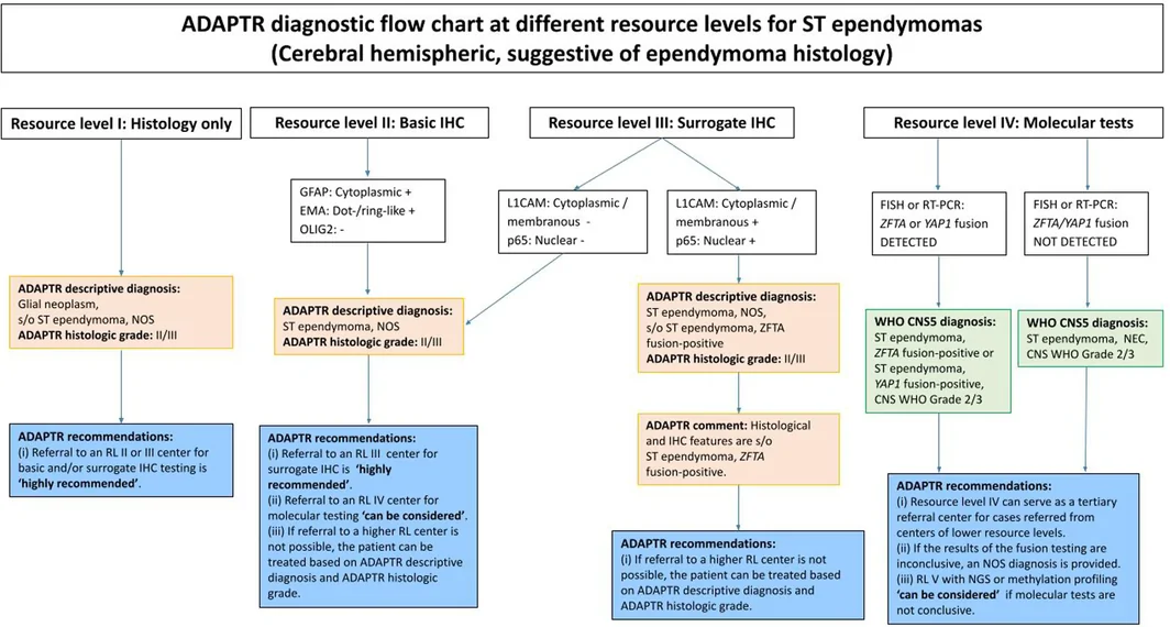
幕上室管膜瘤(ST 室管膜瘤)
WHO CNS5 将 ST 室管膜肿瘤分为三组:ST 室管膜瘤,ZFTA 融合阳性;ST 室管膜瘤,YAP1 融合阳性;ST 室管膜瘤,NOS。ST 室管膜瘤常见于儿童,有时也见于成人 [38],但流行病学特征部分取决于分子亚型 [5, 39]。影像上可见境界清楚的肿块,常伴囊性成分和钙化,实性部分强化明显但不均匀 [40]。尽管分子定义的 ST 室管膜瘤预后数据报道有限,ZFTA 融合阳性肿瘤预后最差,YAP1 融合阳性肿瘤的预后似乎较好 [4, 39],而 CDKN2A/B 纯合缺失的预后价值尚不确定 [41, 42]。不过,关于南亚人群中 ZFTA 融合与患者预后的关系,数据相互矛盾:印度 Vellore 的一项研究(n = 61)显示 ZFTA 融合对 PFS 或 OS 无显著影响 [43],而印度 Chandigarh 的另一项研究(n = 58)则显示 PFS 和 OS 显著较差 [44]。此类地区差异,尤其在罕见肿瘤中,需要更大队列和纵向研究才能准确判断预后。
除室管膜瘤的一般组织学特征外,ST 室管膜瘤还有若干适用于所有资源等级的组织学特征(图 1;补充表 S1)。ZFTA 融合阳性肿瘤常见分支状毛细血管网和透明细胞表型 [45],YAP1 融合阳性肿瘤常见 PAS 阳性嗜酸性颗粒小体 [46]。依据 WHO 标准 [3],ST 室管膜瘤可根据活跃核分裂和微血管增生等组织学所见 [30] 定为 CNS WHO 2 级或 3 级。转诊至 RL II 中心行基础 IHC 有助于确认室管膜来源,因此为「强烈推荐」。
ST 室管膜瘤分子改变的免疫组化替代标志物已可获得,包括 L1CAM(强而弥漫的膜染色)和 p65/RELA(核染色)[47, 48, 49, 50]。就这些标志物而言,ZFTA 融合阳性肿瘤(尤其是 ZFTA::RELA 融合)应为 L1CAM+/p65(NF‐κB)+,YAP1 融合阳性肿瘤或其他(NEC)为 L1CAM−/p65(NF‐κB)−。强而弥漫的胞质 L1CAM 染色检测 ZFTA 融合阳性室管膜瘤的敏感性高(92.3%–100%)[44, 51, 52]。然而它并非特异性标志物,在许多其他肿瘤类型中也有表达,此时并不提示存在 ZFTA 融合 [53]。p65/RELA 核染色对其主要融合伙伴基因 RELA 的敏感性不一(68%–100%),特异性高(92%–100%)[51]。联合 IHC(L1CAM/p65)作为 ZFTA 融合阳性肿瘤的替代指标,敏感性(88%–100%)和特异性(88%–92%)大幅提高 [44, 47, 51, 52]。为推进 ST 室管膜瘤的分子分型,转诊至 RL III 中心行替代性 IHC 同样为「强烈推荐」。
ST 室管膜瘤亚型的确定诊断依赖分子遗传学分析,以检出涉及 ZFTA(C11orf95)(即 ST 室管膜瘤,ZFTA 融合阳性)或 YAP1(即 ST 室管膜瘤,YAP1 融合阳性)的特征性融合基因 [4, 5, 39, 48, 49]。转诊至 RL IV 中心行 FISH 或 RT‐PCR 为「可考虑」,并需注意当地的治疗决策可能并不需要这一层级的信息。排除形态相似的肿瘤——包括 BCOR 变异肿瘤和 MN1 变异星形母细胞瘤(表 2)——也支持该诊断。因此,按 WHO CNS5 标准,仅凭替代标志物无法作出确定诊断;在 RL III 或更低的环境中,最恰当的诊断为 ADAPTR 描述性诊断「ST 室管膜瘤,NOS」。
| 肿瘤实体 | 区别于室管膜瘤的形态特征 | IHC 表型 | 分子所见 |
|---|---|---|---|
| 星形母细胞瘤,MN1 变异型 [54] | 星形母细胞性假菊形团 | D2‐40++ GFAP+ 至 +++ OLIG2+ 至 +++ EMA+ 至 +++ | MN1 融合(常见伙伴——BEND2) |
| 伴 PLAGL1 融合的室管膜瘤样神经上皮肿瘤 [55, 56] | 不同程度的 SE—室管膜瘤混合形态 | GFAP+++ OLIG2− SOX10− EMA+/− L1CAM− | PLAGL1 融合(常见伙伴——FOX01 [3′]、EWSR1 [5′]) |
| 伴 PATZ1 融合的神经上皮肿瘤 [57, 58] | 主要模式包括室管膜瘤样、伴多核细胞的胶质肿瘤、梭形细胞模式 | GATA2+/− GFAP + 至 +++ OLIG2+ 至 +++ CD34+ 至 +++ 突触素/嗜铬粒素+/− | PATZ1 融合(常见伙伴——EWSR1、MN1) |
| 伴 EP300::BCOR 或 CREBBP::BCORL1 融合的胶质瘤 [59, 60, 61] | 主要模式包括室管膜瘤样、少突胶质细胞瘤样、微囊性外观 | BCOR+/− GFAP+/− OLIG2+/− | EP300::BCOR 或 CREBBP::BCORL1 融合 |
缩写:GFAP,胶质纤维酸性蛋白;L1CAM,L1 细胞黏附分子;SE,室管膜下瘤。
后颅窝室管膜瘤(PF 室管膜瘤)
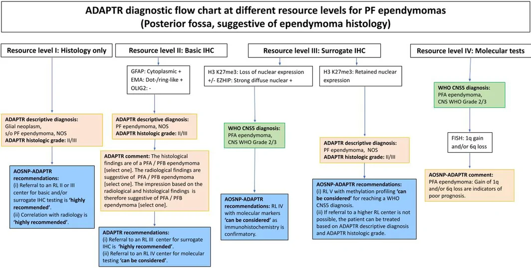
后颅窝室管膜肿瘤(图 2)目前分为三组:PFA 室管膜瘤(补充表 S2)、PFB 室管膜瘤(补充表 S3)和室管膜下瘤(SE)(见下文相应小节)[62]。PFA 和 PFB 室管膜瘤的诊断流程图合并于图 2。RL I 和 RL II 的描述对所有亚型通用,并为「强烈推荐」,需注明 ADAPTR 组织学分级及具体部位。
PFA 和 PFB 亚组主要由基因表达谱及全基因组 DNA 甲基化谱定义。两者发病年龄不同,更重要的是预后不同。PFA 室管膜瘤主要发生于婴幼儿,按现行 WHO 分类 [3],其特征为瘤细胞 H3 K27me3 表达缺失及 EZHIP 过表达;EZHIP 是一种参与抑制多梳抑制复合物 2(PRC2)的蛋白 [63]。这些分子改变驱动表观遗传失调,从而促成 PFA 室管膜瘤的侵袭性 [47]。
因此,转诊至 RL III 中心行替代性 IHC 为「强烈推荐」,因为在此资源等级即可获得 PFA 室管膜瘤的 WHO CNS5 诊断。就 PFA 诊断而言,按现行 WHO 分类 [3],H3 K27me3 核表达缺失是 PFA 室管膜瘤高度特异且敏感的标志物(混杂的非肿瘤细胞,包括内皮细胞和炎症细胞,保留 H3 K27me3 核表达,可作阳性内对照)[63]。需注意,cIMPACT‐NOW 第 11 次更新 [37] 已将术语由「整体减少」[64, 65] 明确为「缺失」。
EZHIP 是一种已有充分研究的 PRC2 活性抑制因子,作用类似 H3 K27M,在 PFA 室管膜瘤中诊断价值高 [66, 67],报道的敏感性和特异性分别高达 99% 和 98% [68]。在 cIMPACT‐NOW 第 11 次更新 [37] 中,EZHIP 强而广泛的核阳性目前被列为 PFA 室管膜瘤的理想(desirable)诊断标准。对 PFA 室管膜瘤,在 RL IV 以 FISH 证实 1q 获得和/或 6q 缺失提示预后较差,为「可考虑」[37, 69]。
相比之下,PFB 室管膜瘤通常保留 H3 K27me3 表达,见于年龄较大的儿童、青少年和成人,临床经过较为惰性 [62]。DNA 甲基化谱是 PFB 室管膜瘤确定性 WHO CNS5 诊断的必需条件 [3]。因此,未做甲基化谱、在 RL III 显示 H3 K27me3 核表达保留(建议截断值为 >80% 免疫阳性细胞)和/或 EZHIP 阴性的病例,给予 ADAPTR 诊断「PF 室管膜瘤,NOS」。RL IV 检测对 PFB 室管膜瘤的诊断不起作用。鉴于 LICs/LMICs 放疗可及性可能有限 [15, 16, 17, 18],若对治疗决策有影响,「可考虑」转诊至具备甲基化谱检测的 RL V 中心,以获得 PFB 室管膜瘤的 WHO CNS5 诊断。
近期又描述了具有新遗传学特征的后颅窝室管膜瘤,均至少需要 RL IV 至 RL V 检测。包括:对应后颅窝 SE 甲基化类别、伴 TERT 启动子突变和/或 6 号染色体缺失的 PF 室管膜瘤 [70];伴 ZFTA 融合、对应「ST 室管膜瘤,ZFTA 融合阳性」甲基化类别的 PF 室管膜瘤 [71];以及激活素受体 I 型(ACVR1)基因体细胞杂合编码区点突变且 H3 K27me3 表达保留者 [72]。
后颅窝的 SE 见于成人,与幕上和脊髓部位的 SE 相似,详见下文。
脊髓室管膜瘤(SP 室管膜瘤)
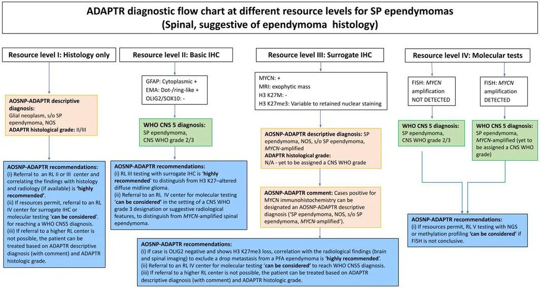
AOSNP‐ADAPTR 对 SP 室管膜瘤和 SP 室管膜瘤,MYCN 扩增型(图 3;补充表 S4)的推荐,大体沿用 ST 室管膜瘤一节所述。SP 室管膜瘤具有其他部位室管膜瘤的许多特征,包括境界清楚和细胞形态。在这一解剖部位,影像替代指标可能尤其有用,因为侵袭性影像表现——包括髓外延伸和软脑膜播散——是 SP 室管膜瘤‐MYCN 的特征 [73, 74, 75, 76]。
室管膜瘤的经典形态特征在 RL I 即适用于所有等级,因此可作出 ADAPTR 描述性诊断(提示 SP 室管膜瘤,NOS,ADAPTR 组织学分级 II/III)。应牢记伸长细胞(tanycytic)形态模式,因其在 SP 室管膜瘤中比例偏高 [3, 77]。在 RL II 即可作出 WHO CNS5 诊断「SP 室管膜瘤,CNS WHO 2 级或 3 级」,因为基础 IHC 结合组织学所见已满足 WHO CNS5 关于 SP 室管膜瘤的必要诊断标准 [3]。SOX10 有助于区分伸长细胞型室管膜瘤(SOX10 免疫阴性)与神经鞘瘤及毛细胞型星形细胞瘤(SOX10 免疫阳性)[3, 34]。OLIG2 有助于区分 SP 室管膜瘤(OLIG2 阴性)与其他胶质瘤(OLIG2 阳性)[3, 34]。尽管在 RL II 即可作出 WHO CNS5 诊断,但在高级别组织学和/或侵袭性影像表现的情况下,必须考虑 SP 室管膜瘤,MYCN 扩增型 [74, 75, 76]。此外,SP 室管膜瘤,MYCN 扩增型虽通常显示高级别组织学特征,但也有呈 CNS WHO 2 级组织学表现的罕见病例报道 [75]。
鉴于 SP 室管膜瘤,MYCN 扩增型未必总有高级别组织学,在 RL III「强烈推荐」MYCN IHC。MYCN IHC 与 MYCN 扩增分子检测一致性极好,因此被列为 RL III 的 MYCN 扩增替代性 IHC 标志物 [74, 75, 78]。所有研究均使用 Cell Signaling Technology(美国马萨诸塞州 Danvers)的 D4B2Y 克隆。有趣的是,Ghasemi 等注意到 MYCN 免疫阳性模式存在差异,组织学高级别区域表达更强 [75]。MYCN IHC 阳性病例若 MRI 显示外生性肿块,可给予 AOSNP‐ADAPTR 描述性诊断(提示 SP 室管膜瘤,MYCN 扩增型)[74, 75, 76, 78]。为与 WHO CNS5 对该实体的分级保持一致,未赋予 ADAPTR 组织学分级 [3]。
需注意,弥漫性中线胶质瘤,H3 K27 变异型也可伴 MYCN 扩增,因此在 RL III 还应进行 H3 K27M 和 K27me3 IHC [79]。值得注意的是,一项关于 SP 室管膜瘤,MYCN 扩增型的研究显示 H3 K27me3 核染色保留程度不一 [74]。RL II 的另一项重要推荐是:若病例 OLIG2 阴性且显示 H3 K27me3 缺失,「强烈推荐」进行影像对照,以排除 PFA 室管膜瘤的下行(drop)转移。
在 RL IV,以 FISH 检测 MYCN 扩增即足以分别满足 WHO 关于 SP 室管膜瘤,MYCN 扩增型的必要诊断标准和 SP 室管膜瘤的理想诊断标准 [75, 76, 78, 80]。因此,对可疑病例,在可行且 MYCN 状态可能影响治疗决策时,「可考虑」转诊至 RL IV 中心。近期有报道称,按 NF2 状态对 SP 室管膜瘤分层具有预后意义 [81],未来可能在 RL IV 检测中发挥作用。
黏液乳头状室管膜瘤
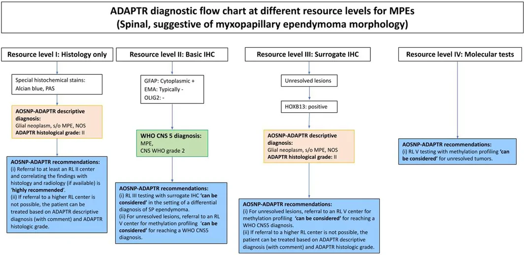
黏液乳头状室管膜瘤(MPE)(图 4;补充表 S5)形态独特,典型表现为立方至柱状胶质细胞围绕玻璃样变的纤维血管轴心排列,血管周有黏液样物质聚集 [3, 82]。MPE 也有特征性的解剖分布,绝大多数病例位于圆锥和终丝区域 [82, 83]。WHO CNS5 将 MPE 定为 CNS WHO 2 级 [3]。文献中记录有极少数「间变性」MPE,表现为细胞密度增加、黏液样物质减少及高级别组织学特征 [84]。
依据典型组织学和 GFAP 免疫阳性即可作出 MPE 的 WHO CNS5 诊断 [3]。在实践中,形态典型时,一些资深神经病理医生仅凭 H&E 染色即诊断此类肿瘤。因此,在 RL I 应可见 MPE 的经典特征,此时可作出 ADAPTR 描述性诊断「提示 MPE,NOS」。在 RL II,GFAP 染色阳性即可获得 WHO CNS5 诊断,因此为「强烈推荐」。RL III 及以上检测在形态不明确时才有意义,因此在此情形下为「可考虑」。HOXB13 被提议作为替代性 IHC 标志物(RL III),用于在形态重叠时区分 MPE 与 SP 室管膜瘤 [85, 86]。在近期一项 143 例脊髓肿瘤的单中心研究中,其诊断 MPE 的敏感性和特异性均为 100% [85, 86]。HOXB13 亦表达于马尾神经内分泌肿瘤,但该实体凭其神经内分泌特征和免疫表型很容易与 MPE 区分 [85, 86]。RL IV 的基础分子检测对 MPE 用处不大。RL V 的甲基化谱「可考虑」,对未定性病变可能提供有用信息,因为 MPE 具有独立的甲基化谱 [85, 86, 87]。呈室管膜瘤外观而甲基化谱符合 MPE 的病例,可能代表非经典形态的 MPE [85, 86, 87]。
室管膜下瘤
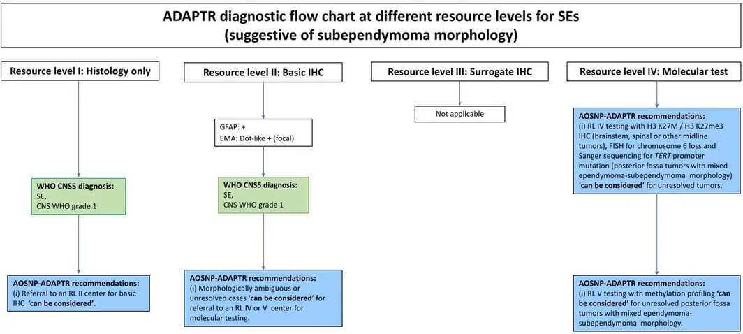
室管膜下瘤(SE,图 5;补充表 S6)具有众所周知、近乎特征性的形态外观,WHO CNS5 的必要诊断标准允许依据 H&E 染色所见的形态作出诊断(即 RL I)[3]。可进行基础 IHC(RL II)以确认其室管膜下胶质来源(弥漫 GFAP 免疫阳性及局灶 EMA 点状免疫反应)。然而,鉴于其形态独特,若病理医生有把握依据组织学诊断 SE,RL I 即满足 WHO CNS5 的必要诊断标准。
对具有 SE 型形态的肿瘤,可考虑 RL II 检测,但 ADAPTR 室管膜瘤工作组将其定为「可考虑」。因此,替代性 IHC 标志物的 RL III 检测被视为对 SE 诊断非必需。
后颅窝肿瘤呈室管膜瘤—SE 混合形态,或 SE 样肿瘤发生于脑干、脊髓或其他中线部位时,可对 SE 进行 RL IV 检测。在一个小样本 SE 系列(n = 24)中,延髓和脑桥具有 SE 型组织学的胶质瘤被发现携带 H3 p.K27M 变异(n = 4)[88],后续甲基化分析显示其形成一个既不同于 PF 室管膜瘤、也不同于弥漫性中线胶质瘤,H3 K27 变异型的独立聚类 [89]。中位随访 3.2 年,这些病例的临床结局与 CNS WHO 4 级的预后并不相符 [88]。因此,此类病例必须进行临床与影像对照。在脑干或中线部位,H3 K27M 和/或 H3 K27me3 IHC 因而可提供有用的附加信息 [88]。对于后颅窝室管膜瘤—室管膜下瘤混合形态这一特殊情形,分子检测(RL IV)可包括 TERT 启动子突变测序及 6 号染色体缺失评估,二者已显示与这种混合形态及较短 PFS 相关 [70]。
在 RL V,对未定性病变可考虑 DNA 甲基化谱,包括分类以及由全基因组 DNA 甲基化数据推导的染色体拷贝数变异判读。SE 按部位(ST‐SE、PF‐SE 和 SP‐SE)具有不同的甲基化谱,19 号染色体缺失见于所有部位 [90]。对于后颅窝室管膜瘤—室管膜下瘤混合性肿瘤,DNA 甲基化谱可能有助于分类和预后判断 [70]。室管膜瘤型组织学所占比例不一,目前尚不清楚室管膜瘤成分是否决定分级 [70, 91, 92]。
诊断流程图
本文为 ST 室管膜瘤(图 1)、PFA/PFB 室管膜瘤(图 2)、SP 室管膜瘤(图 3)、MPE(图 4)和 SE(图 5)制定了诊断流程图。这些流程图按资源等级为报告病理医生提供参考,并指明在特定资源等级何时适合给出 ADAPTR 描述性诊断、何时可获得 WHO CNS5 诊断。
ADAPTR 室管膜瘤工作组还考虑了具有室管膜瘤样形态的实体(表 2)。其中部分已纳入 WHO CNS5,包括星形母细胞瘤,MN1 变异型 [3]。cIMPACT‐NOW 第 10 次更新 [93] 给出了认定新的(暂定)肿瘤类型的最低标准建议。按照这些标准,伴 PATZ1 融合的神经上皮肿瘤 [57, 58] 和伴 PLAGL1 融合的室管膜瘤样神经上皮肿瘤 [55, 56] 符合暂定肿瘤类型标准,而伴 EP300::BCOR 或 CREBBP::BCORL1 融合的胶质瘤在该更新发表时尚未达到标准 [59]。这些实体之间 GFAP、EMA 和 OLIG2 染色程度存在显著重叠,许多肿瘤的室管膜瘤样外观只是其多形性形态谱的一部分(表 2)。这些新肿瘤类型的患者预后数据尚需进一步随访,但据称伴 PATZ1 融合的神经上皮肿瘤预后优于胶质母细胞瘤,IDH 野生型 [57, 58]。这些实体的准确诊断将依赖 RL V 检测,但未来或许可以借助 ADAPTR 描述性诊断,结合在 RL III 中心进行的相关替代性 IHC,来处理这些较新的诊断。
结论
AOSNP‐ADAPTR 室管膜瘤推荐提供了一个按资源分层、以组织学为导向的诊断框架,使神经病理医生即便在资源受限地区也能给出与 WHO CNS5 一致的诊断。这些指南与现有的 ADAPTR 成人型弥漫性胶质瘤框架互为补充,共同致力于协调资源受限地区的 CNS 肿瘤诊断。我们注意到,用于预测室管膜瘤分子分型的人工智能辅助数字病理工具正在出现,可能惠及 LICs/LMICs [94]。我们也承认,尽管 ADAPTR 描述性诊断和组织学分级将极大帮助 LICs/LMICs 的病理医生,但在亚洲—大洋洲地区及其他地方,推动更公平地获得分子检测——包括倡导在医疗卫生中增加病理投入——至关重要。
- The AOSNP ADAPTR working group stratifies ependymoma diagnosis into five resource levels: RL I histology only → RL II basic IHC (GFAP, dot/ring EMA, OLIG2, Ki-67, optional SOX10) → RL III surrogate IHC → RL IV FISH/RT-PCR → RL V NGS and methylation profiling; recommendations are graded “can be considered” or “highly recommended”.
- Surrogate IHC: L1CAM/p65 for ST (combined sensitivity 88%–100%, specificity 88%–92% for ZFTA fusion); H3 K27me3 loss plus strong nuclear EZHIP (99%/98%) establishes PFA; MYCN (clone D4B2Y) for SP ependymoma, MYCN-amplified; HOXB13 for ambiguous MPE.
- Referral vs descriptive diagnosis: ST ependymoma at RL III or lower → “ST ependymoma, NOS”; PFB requires methylation (RL V), otherwise H3 K27me3-retained (>80%) and/or EZHIP-negative → “PF ependymoma, NOS”; SE and classic MPE reach a WHO CNS5 diagnosis at RL I/II.
- Practical points: an OLIG2-negative, H3 K27me3-lost spinal tumour needs imaging to exclude a PFA drop metastasis; DMG can harbour MYCN amplification, so add H3 K27M/K27me3 at RL III; brainstem/midline SE-like tumours may carry H3 K27M. Table 2 lists ependymoma-like mimics (MN1, PLAGL1, PATZ1, BCOR).
Scope: PMC open-access full text (CC BY-NC-ND 4.0) — abstract, all main sections, Tables 1–2 and Figures 1–5 with captions. References, Supplementary Tables S1–S6, author contributions and the graphical abstract are not included.
Abstract
Ependymomas are uncommon primary tumors of the central nervous system (CNS) that affect both children and adults. In recent years, molecular analyses have identified multiple subtypes of ependymal tumors based on location, patterns of CpG island methylation, and gene fusion events. While this knowledge is highly important in progressing diagnostic precision, prognostication, and effective treatments, an unwanted side effect is that best practice diagnostics are often inaccessible because of cost and technical barriers for many low‐ or lower middle‐income countries. The ADAPTR group (Adapting Diagnostic Approaches for Practical Taxonomy in Resource‐Restrained Regions) is an initiative of the Asian Oceanian Society of Neuropathology and aims to provide practical guidelines for CNS tumor diagnostics at various levels of pathology resource restrictions. In this manuscript, we propose a practical approach to the diagnosis of ependymal tumors based on ADAPTR resource levels. These recommendations provide a practical adaptation of World Health Organisation Classification of Tumours of the Central Nervous System guidelines suited to routine diagnosis in resource‐restrained regions.
INTRODUCTION
Solid tumor classification increasingly relies on integration of complex molecular information with classical histological assessments. This requirement is most prevalent and longstanding in central nervous system (CNS) tumors, where integration of molecular features in some tumor types has been mandated for over a decade [1]. The grading of ependymoma has historically suffered from poor reproducibility and clinical utility, with a reliance on histological grading parameters [2]. Since the publication of the 2021 WHO Classification of CNS tumors [3], the histological subtyping and grading of ependymoma has been improved by the introduction of molecular subclassification, with a schema based on anatomical location, genomic alterations, and methylation profiling [4, 5]. Within this schema, the definition of some ependymal tumor subtypes has moved to one based solely on complex molecular profiles—for example, posterior fossa group B (PFB) ependymomas are now defined by their methylation profile [3]. While the improved precision in both diagnosis and prognosis is much needed, delivering the necessary molecular testing methodologies to low‐ and lower middle‐income countries (LICs and LMICs) incurs costs that are prohibitive for routine use [4, 6].
The AOSNP‐ADAPTR initiative (Asian Oceanian Society of Neuropathology recommendations for Adapting Diagnostic Approaches for Practical Taxonomy in Resource‐Restrained Regions) proposes practical, resource‐stratified pathways that emphasize morphology and immunohistochemistry (IHC) in CNS tumor diagnostics, with recommended targeted molecular tests when essential or highly informative [7, 8]. The AOSNP‐ADAPTR recommendations are a practical adaptation of the World Health Organization Classification of Tumors of the Central Nervous System 5th edition [3], intended to suit routine diagnostic practices in resource‐limited settings without altering established diagnostic categories [6, 7]. As previously reported by our group and others, LICs and LMICs face significant barriers to the implementation of molecular techniques required for the diagnosis of many (CNS) tumors [6, 7, 8, 9].
The classic morphologic features of ependymal tumors are well described, with the current World Health Organisation Classification of Tumours of the Central Nervous System (WHO CNS5) diagnosis of some tumor types possible on morphology alone (e.g., subependymoma) [3]. However, the accurate diagnosis of other ependymal tumors necessitates advanced molecular testing. When genetic testing is not feasible or does not categorize the tumor in the current WHO Classification [6], then the designation of NOS (not otherwise specified) or NEC (not elsewhere classified), respectively, is assigned to the diagnosis as per current WHO guidelines [10].
In this paper, the AOSNP‐ADAPTR Ependymoma Working Group outlines a histology‐oriented, resource‐level approach for ependymoma diagnosis that leverages surrogate immunohistochemical markers and clinical–radiological correlation, and escalates to molecular testing when it is essential for WHO CNS5 entities and may alter management. Referral pathways to higher‐resource centers are suggested or recommended in specific circumstances.
Resource levels (RLs) have been previously defined [6, 8] and are reproduced in Supporting Information S1. Imaging, morphology, and relevant surrogate immunohistochemical markers for ependymal tumors are detailed below, as well as ADAPTR recommendations based on local RLs with diagnostic flowcharts.
CHALLENGES IN DIAGNOSIS AND MANAGEMENT OF EPENDYMOMAS IN RESOURCE‐RESTRAINED SETTINGS
The diagnostic classification of an ependymoma is dictated by its anatomical location, with each site typified by different molecular profiles requiring different molecular testing methodologies [3]. For example, supratentorial (ST) ependymoma requires gene fusion testing, whereas methylation profiling is an essential diagnostic criterion for PFB ependymoma [3]. Therefore, it was difficult to synthesize molecular testing of ependymal tumors as a whole, and our ADAPTR recommendations have followed the WHO Classification's anatomical site‐by‐site approach for ependymoma.
The standard of care for ependymal tumors remains primarily surgical, with gross total resection (GTR) and timely adjuvant radiotherapy being an important prognostic factor [11, 12]. Significant challenges exist in the delivery of the expert neurosurgical care required for GTR, with an international survey of neurosurgical and pediatric surgical societies estimating one pediatric neurosurgeon for every 3.6 million children in LICs/LMICs [13]. In a study of GTR rates in posterior fossa ependymoma from Hospital for Sick Children, Toronto, GTR rates increased from 35% in 1985 to 77% in 2014, correlating with an improvement in both progression‐free (PFS) and overall survival (OS), alongside upfront radiotherapy [14]. By contrast, studies from institutions in LICs/LMICs report GTR rates of 17% (Egypt) [15], 31% (Iran) [16], 32% (Peru) [17], and 38% (Brazil) [18], all of which attribute higher mortality rates to lower rates of GTR [19]. Furthermore, equitable access to radiotherapy for the treatment of ependymomas may be limited in LICs/LMICs, with socio‐economic factors such as travel costs and loss of income affecting treatment adherence [16]. A multicohort study of centers from high‐income and upper‐middle income countries found that a considerable proportion of PFB ependymoma could be treated with surgical intervention alone [20]. Therefore, molecular profiling, while expensive, may play a role in identifying subtypes of ependymoma that can be spared post‐operative radiotherapy, saving patients in LICs/LMICs the financial burden of adjuvant treatment [16, 20].
Imaging features of ependymoma
The AOSNP‐ADAPTR survey showed that over half of centers in LICs/LMICs have access to magnetic resonance imaging (MRI) with contrast, highlighting the key supporting role of radiological data in CNS tumor diagnosis in this setting [6]. Following the ADAPTR adult‐type diffuse glioma framework [8], imaging features provide a first‐line diagnostic context for ependymal tumors at all RLs, based on tumor location (ST, posterior fossa [PF], and spinal [SP]). Supratentorial tumors are often well‐demarcated masses with cystic change and calcification, showing strong but heterogeneous enhancement in solid portions [21]. Posterior fossa group A (PFA) ependymoma are usually calcified lateral or median tumors, show incomplete enhancement, invasion into posterior fossa foramina (particularly the foramina of Luschka), resulting in significant hydrocephalus and are seen in young children [22]. In contrast, PFB ependymoma are minimally invasive and non‐calcified median tumors, generally seen in adults and adolescents, arising from the floor of the fourth ventricle, and predominantly cystic with complete homogeneous enhancement in the solid parts [22]. Machine learning algorithms based on MRI features may also assist in distinguishing PFA from PFB ependymomas [23]. Spinal ependymomas are usually intramedullary and frequently cervical–cervicothoracic; syringomyelia is common [24, 25]. Aggressive appearances such as extramedullary extension or leptomeningeal spread should raise suspicion for SP ependymoma, MYCN‐amplified [26]. Incorporating imaging features in diagnostic workflows requires close collaboration with local expert (neuro‐)radiologists.
Morphological features of ependymoma
Classic ependymal morphology includes tumor cells with round to ovoid nuclei, arranged in perivascular pseudorosettes, true ependymal rosettes and also as simulated ependymal lining (canals) [1, 3, 27]. Ependymomas are typically well‐demarcated from the surrounding brain [1, 3, 27]. Other histological features that may be encountered include fibrillary areas, calcification, and cystic degeneration [1, 3, 27]. Although ultrastructural findings are not required for ADAPTR recommendations, ependymal features (e.g., intracytoplasmic villi, cilia, and complex intercellular zipper‐like junctions) are present in all the ependymoma subtypes [28]. The morphological patterns previously described (clear cell, tanycytic, and papillary) in the 2016 WHO classification [27] are not listed as subtypes in the 2021 WHO classification [3], because of their lack of clinicopathological usefulness. These morphological patterns are associated with certain anatomical locations and are therefore described within the anatomically defined subtypes below.
The current classification assigns CNS WHO grades 2 or 3 to ST, posterior fossa, and SP ependymoma tumor types, based solely on histologic findings [3]. However, it is recognized that the histopathologic grading of ependymoma does not consistently relate to patient outcome [2] and is often related to anatomical site [29]. Though the relevance of histological grading in a molecular classification framework can be questioned, in a resource‐limited setting, histological grading may be the only indicator of prognosis for patients and their carers, and is therefore included in these recommendations. High‐grade features in ependymomas include brisk mitotic activity and microvascular proliferation [2, 29], which have more prognostic value than other features such as nuclear pleomorphism or necrosis [30].
Lineage and surrogate immunohistochemistry for ependymoma
Lineage markers for ependymoma at Resource Level II (RL II), termed “basic IHC” by the ADAPTR Ependymoma Working Group, include immunohistochemical expression of glial fibrillary acidic protein (GFAP) and epithelial membrane antigen (EMA) (dot‐like/ring‐like luminal patterns) [31, 32, 33]. OLIG2 and SOX10 negativity assist in distinguishing ependymomas from mimics such as diffuse gliomas, as well as schwannoma and pilocytic astrocytoma [3, 34]. Ki‐67 index has not been included as an IHC marker, as it is not used in the WHO CNS5 grading of ependymomas [3]. Nevertheless, it is noteworthy that in a single institution study of 141 cases, a Ki‐67 cut‐off of 7% was found to be a useful adjunct in grading ependymomas at all anatomical locations [35].
Surrogate IHC that is practical at Resource Level III (RL III) includes: (i) L1 cell adhesion molecule (L1CAM) and p65/RELA; (ii) H3 K27me3/H3 K27M; (iii) enhancer of zeste homolog inhibitory protein (EZHIP); (iv) MYCN; and (v) homeobox B13 (HOXB13). EBP50, expressed at the luminal surface of human epithelia, has also been proposed as a potential IHC marker [36]. Surrogate IHC markers are further described in the ADAPTR recommendations below. Aligning with cIMPACT‐NOW update 11 [37], we refer to the hotspot H3 p.K28 as “H3 K27” in this manuscript.
ADAPTR RECOMMENDATIONS FOR THE EVALUATION OF EPENDYMOMA
The ADAPTR Ependymoma Working Group presents the three ADAPTR components in this manuscript, similar to the approach used for adult‐type diffuse gliomas [8]: (i) RLs of diagnostic pathology services with relevance to ependymal tumors; (ii) recommendations for “Histology‐oriented integrated diagnosis” format for each ependymal tumor type at different RLs, including an “ADAPTR grade” where tumor grading cannot be performed at WHO CNS5 levels; (iii) corresponding diagnostic flow charts for each ependymal tumor type at different RLs.
Resource levels of diagnostic pathology services
Resource Level I (RL I; histology) emphasizes recognition of classic ependymal morphology and provides an ADAPTR histological grade (II/III) where applicable, with management permissible on morphology alone when further testing is not feasible (Table 1). RL II adds basic IHC to confirm lineage. RL III incorporates surrogate immunomarkers—L1CAM/p65 for ST ependymoma, EZHIP/H3 K27me3 for posterior fossa ependymoma, and MYCN IHC for SP ependymoma—to approximate WHO CNS5 entities. Resource Level IV (RL IV) enables targeted molecular confirmation (e.g., FISH or reverse transcription polymerase chain reaction [RT‐PCR] for ZFTA or YAP1 fusions; FISH for MYCN amplification), and Resource Level V (RL V) uses comprehensive platforms (next generation sequencing [NGS] and DNA methylation profiling) when prior levels are inconclusive or for further risk‐stratification. While multidisciplinary discussion should take place at all RLs, it is imperative that pathologists making diagnoses at RL I and RL II discuss their findings with the treating team, to assess if referral to a higher RL center will affect treatment and prognostication. Compartment‐specific recommendations are detailed below.
| Resource levels | Resource Level I (RL I) | Resource Level II (RL II) | Resource Level III (RL III) | Resource Level IV (RL IV) | Resource Level V (RL V) | |||
|---|---|---|---|---|---|---|---|---|
| Available resources | Histology only | + Basic diagnostic IHC markers | + Surrogate IHC molecular markers | + Basic molecular tests | + Advanced molecular tests | |||
| Pathology services | Material
Techniques
| Material (i) and (ii) Techniques 1. and 2. 3. Basic IHC markers | Material (i) and (ii) Techniques 1. to 3. 4. Surrogate IHC molecular markers | Material (i) and (ii) (iii) Fresh tissue Techniques 1. to 4. 5. FISH 6. RT‐PCR | Material (i), (ii) and (iii) Techniques 1. to 6. 7. DNA/RNA sequencing by NGS 8. DNA methylation profiling | |||
| Basic IHC | Nil | GFAP, EMA, OLIG2, Ki‐67 Optional—SOX10 | Same as RL II | Same as RL II | Same as RL II | |||
| Surrogate IHC | Nil | Nil | Markers | For diagnosis of: | Same as RL III | Same as RL III | ||
| L1CAM/p65 | ST ependymoma, ZFTA‐fusion positive | |||||||
| EZHIP | PFA versus PFB ependymoma | |||||||
| H3 K27M | To exclude H3 K27M‐mutant forms of diffuse midline glioma, H3 K27‐altered | |||||||
| H3 K27me3 | PFA versus PFB ependymoma To exclude diffuse midline glioma, H3 K27‐altered in the setting of diffuse gliomas (OLIG2+) in the midline | |||||||
| MYCN | SP ependymoma, MYCN‐amplified | |||||||
| HOXB13 | MPE | |||||||
| Diagnostic molecular assays | Nil | Nil | Nil | 5. FISH | Same as RL IV and 7. DNA/RNA sequencing by NGS for diagnosis | |||
| ZFTA fusion | ST ependymoma, ZFTA‐fusion positive | ZFTA fusion | ST ependymoma, ZFTA‐fusion positive | |||||
| YAP1 fusion | ST ependymoma, YAP1‐fusion positive | YAP1 fusion | ST ependymoma, YAP1‐fusion positive | |||||
| MYCN amp | SP ependymoma, MYCN‐amplified | MYCN amp | SP ependymoma, MYCN‐amplified | |||||
| 6. RT‐PCR | 8. DNA methylation profiling for methylation‐based diagnosis | |||||||
| ZFTA fusion | ST ependymoma, ZFTA‐fusion positive | PFB ependymoma | ||||||
| YAP1 fusion | ST ependymoma, YAP1‐fusion positive | Unresolved lesions | ||||||
| Personnel/pathologist | General pathologist | General pathologist | Pathologist skilled in interpreting the IHC surrogate molecular markers | Neuropathologist proficient in interpretation of molecular tests and providing an integrated diagnosis | Neuropathologist proficient in interpretation of molecular tests and providing an integrated diagnosis | |||
Abbreviations: amp, amplification; EMA, epithelial membrane antigen; EZHIP; enhancer of zeste homolog inhibitory protein; FISH, fluorescent in situ hybridization; FFPE, formalin fixed paraffin embedded; GFAP, glial fibrillary acidic protein; H&E, haematoxylin and eosin; HOXB13: homeobox B13; IHC, immunohistochemistry; L1CAM, L1 cell adhesion molecule; MPE, myxopapillary ependymoma; NGS, next generation sequencing; NF‐κB p65, nuclear factor NF‐kappa‐B p65 subunit; PFA, posterior fossa group A (PFA) ependymoma; PFB, posterior fossa group B (PFB) ependymoma; RT‐PCR, reverse transcription polymerase chain reaction; SP ependymoma, MYCN‐amplified: spinal ependymoma, MYCN‐amplified; ST ependymoma, YAP1‐fusion positive, supratentorial ependymoma, YAP1 fusion‐positive; ST ependymoma, ZFTA‐fusion positive, supratentorial ependymoma, ZFTA fusion‐positive.
ADAPTR recommendations for “Histology‐oriented integrated diagnosis” format for ependymomas at different resource levels
The ADAPTR Ependymoma Working Group applied the “Histology‐oriented integrated diagnosis format” to each ependymal tumor type (Tables S1–S6 and Figures 1, 2, 3, 4, 5). We have included RL V in these recommendations, as DNA methylation profiling may be required to achieve a WHO CNS5 diagnosis or resolve ambiguous cases in ependymal tumors, which may impact clinical management. While we have included RL V, our focus is on RLs I to IV to ensure the accessibility of these recommendations. In alignment with other ADAPTR recommendations, we categorize our recommendations as “can be considered” and “highly recommended.”
Supratentorial ependymoma (ST ependymoma)
The WHO CNS5 has classified ST ependymoma tumors into three groups: ST ependymoma, ZFTA fusion‐positive; ST ependymoma, YAP1 fusion‐positive; ST ependymoma, NOS. ST ependymoma often affects children and sometimes adults [38], although epidemiological features partially depend on molecular subtypes [5, 39]. Radiologically, a well‐demarcated mass, frequently with cystic components and calcification, is observed, associated with strong but heterogenous enhancement in solid parts [40]. Despite a limited number of reports on outcome data of molecularly‐defined ST ependymoma, ZFTA fusion‐positive tumors have the poorest outcome, and YAP1 fusion‐positive tumors appear to be prognostically more favorable [4, 39], with the prognostic value of homozygous CDKN2A/B deletion uncertain [41, 42]. However, there is conflicting data regarding the relationship of ZFTA fusion with patient outcome in South Asian populations. While one study from Vellore, India (n = 61) shows no significant effect of ZFTA fusion on PFS or OS [43], another from Chandigarh, India (n = 58) shows significantly inferior PFS and OS [44]. Such regional differences, particularly in rare tumors, will require larger cohorts and longitudinal studies for accurate prognostication.
There are several histologic features described for ST ependymoma suitable for all RLs, in addition to general histological features of ependymoma (Figure 1; Table S1). A branching network of capillaries and clear cell phenotypes is frequent in ZFTA fusion‐positive tumors [45], and PAS‐positive eosinophilic granular bodies are frequently observed in YAP1 fusion‐positive tumors [46]. ST ependymoma can be assigned CNS WHO grades 2 or 3 by histological findings including brisk mitoses and microvascular proliferation [30] as per WHO criteria [3]. Referral to an RL II center for basic IHC will aid in confirming ependymoma origin and is therefore “highly recommended.”
Immunohistochemical surrogate markers for the molecular alterations of ST ependymoma are available, including L1CAM (strong diffuse membranous staining) and p65/RELA (nuclear staining) [47, 48, 49, 50]. For these markers, ZFTA fusion‐positive tumors (especially with a ZFTA::RELA fusion) should be L1CAM+/p65 (NF‐κB)+, and YAP1 fusion‐positive tumors or others (NEC) L1CAM−/p65 (NF‐κB)−. Strong and diffuse cytoplasmic L1CAM staining has high sensitivity (92.3%–100%) for detecting ZFTA fusion‐positive ependymomas [44, 51, 52]. However, it is not a specific marker, and demonstrates expression in many other tumor types, where it does not indicate an underlying ZFTA fusion [53]. Nuclear p65/RELA staining has variable sensitivity (68%–100%) and high specificity (92%‐100%) for its main fusion gene partner, RELA [51]. Combinatorial IHC approaches (L1CAM/p65) result in greatly improved sensitivity (88%–100%) and specificity (88%–92%) [44, 47, 51, 52] as a surrogate for ZFTA fusion‐positive tumors. Referral to an RL III center for surrogate IHC is also “highly recommended” to progress toward molecular subtyping of ST ependymoma.
The definitive diagnosis of ST ependymoma subtype relies on molecular genetic analyses to detect pathognomonic fusion genes involving ZFTA (C11orf95) (i.e., ST ependymoma, ZFTA fusion‐positive) or YAP1 (i.e., ST ependymoma, YAP1 fusion‐positive) [4, 5, 39, 48, 49]. Referral to an RL IV center for FISH or RT‐PCR “can be considered,” noting that local treatment decisions may not require this level of information. The diagnosis is also supported by the exclusion of morphologically similar tumors including BCOR‐altered tumors and MN1‐altered astroblastoma (Table 2). Therefore, definite diagnoses cannot be made by surrogate markers alone based on WHO CNS5 criteria, rendering an ADAPTR descriptive diagnosis of ST ependymoma, NOS, as the most appropriate diagnosis in RL III or lower settings.
| Tumor entity | Distinguishing morphological features from ependymoma | IHC profile | Molecular findings |
|---|---|---|---|
| Astroblastoma, MN1‐altered [54] | Astroblastic pseudorosette | D2‐40++ GFAP+ to +++ OLIG2+ to +++ EMA+ to +++ | MN1 fusion (common partner—BEND2) |
| Ependymoma‐like neuroepithelial tumor with PLAGL1 fusions [55, 56] | Variable mixed SE‐ependymoma morphology | GFAP+++ OLIG2− SOX10− EMA+/− L1CAM− | PLAGL1 fusion (common partners—FOX01 [3′], EWSR1 [5′]) |
| Neuroepithelial tumor with PATZ1 fusion [57, 58] | Major patterns include ependymoma‐like, glial tumor with multinucleated cells, spindle cell pattern | GATA2+/− GFAP + to +++ OLIG2+ to +++ CD34+ to +++ Synaptophysin/chromogranin+/− | PATZ1 fusion (common partners—EWSR1, MN1) |
| Gliomas with EP300::BCOR or CREBBP::BCORL1 fusions [59, 60, 61] | Major patterns include ependymoma‐like, oligodendroglioma‐like, microcystic appearances | BCOR+/− GFAP+/− OLIG2+/− | EP300::BCOR or CREBBP::BCORL1 fusions |
Abbreviations: GFAP, glial fibrillary acidic protein; L1CAM, L1 cell adhesion molecule; SE, subependymoma.
Posterior fossa ependymoma (PF ependymoma)
Ependymal tumors of the posterior fossa (Figure 2) are currently classified into three groups: PFA ependymoma (Table S2), PFB ependymoma (Table S3), and subependymoma (SE) (see section below) [62]. The diagnostic flow charts for PFA and PFB ependymomas are combined in Figure 2. The RL I and RL II description is common to all subtypes and is “highly recommended,” with ADAPTR histological grade and specific location mentioned.
PFA and PFB subgroups are predominantly defined by gene expression and whole‐genome DNA methylation profiles. They differ in age of onset and, more importantly, in prognosis. PFA ependymomas predominantly occur in infants and young children, and are characterized in the current WHO Classification [3] by loss of H3 K27me3 expression in tumor cells and overexpression of EZHIP, a protein implicated in inhibition of the Polycomb Repressive Complex 2 (PRC2) [63]. These molecular changes drive epigenetic dysregulation, thus contributing to the aggressive nature of PFA ependymomas [47].
Referral to RL III centers for surrogate IHC is therefore “highly recommended,” as a WHO CNS5 diagnosis of PFA ependymoma can be achieved at this RL. For a diagnosis of PFA, loss of nuclear expression of H3 K27me3 in the current WHO Classification [3] is a highly specific and sensitive marker for PFA ependymoma (admixed non‐neoplastic cells, including endothelial and inflammatory cells, retain nuclear H3 K27me3 and serve as positive internal controls) [63]. It should be noted that in cIMPACT‐NOW update 11 [37], the terminology has now been clarified from “global reduction” [64, 65] to “loss.”
EZHIP, a well‐characterized inhibitor of PRC2 activity analogous to H3 K27M, demonstrates high diagnostic utility in PFA ependymoma [66, 67], with high sensitivity and specificity reported (99% and 98%, respectively) [68]. Demonstration of strong global nuclear positivity for EZHIP is currently listed as a desirable diagnostic criterion for PFA ependymoma in cIMPACT‐NOW Update 11 [37]. For PFA ependymoma, at RL IV, demonstration of 1q gain and/or 6q loss by FISH is indicative of a poorer prognosis and “can be considered” [37, 69].
In contrast, PFB ependymomas typically retain H3 K27me3 expression, are seen in older children, adolescents and adults, and exhibit a more indolent clinical course [62]. DNA methylation profiling is mandatory for a definitive WHO CNS5 diagnosis of PFB ependymoma [3]. Hence, cases that do not have methylation profiling, exhibit retention of nuclear H3 K27me3 (with a proposed cut‐off of >80% immunopositive cells) and/or are negative for EZHIP at RL III would be given an ADAPTR diagnosis of “PF ependymoma, NOS.” RL IV testing does not play a role in the diagnosis of PFB ependymoma. Given that access to radiotherapy can be limited in LICs/LMICs [15, 16, 17, 18], referral to an RL V centre with methylation profiling “can be considered” in reaching a WHO CNS5 diagnosis of PFB ependymoma if it will impact upon treatment decisions.
More recently, ependymomas of the posterior fossa with novel genetic signatures have been described, all of which would require at least RL IV to RL V testing. These include PF ependymomas that correspond to the methylation class of posterior fossa SE with TERT promoter mutation and/or chromosome 6 loss [70]; PF ependymomas with ZFTA fusion that correspond to the methylation class of ST ependymoma, ZFTA‐fusion positive [71]; and those with somatic heterozygous coding point mutations in the activin receptor type I (ACVR1) gene and retained H3 K27me3 expression [72].
SEs in the posterior fossa are seen in adults and are similar to those in the supratentorial and spinal compartments. They are described in detail below.
Spinal ependymoma (SP ependymoma)
The AOSNP‐ADAPTR recommendations for SP ependymomas and SP ependymoma, MYCN‐amplified (Figure 3; Table S4) largely follow those delineated in the ST ependymoma section. SP ependymomas share many of the features of ependymomas at other locations, including their circumscription and cytomorphology. Radiological surrogates may prove particularly helpful in this anatomical compartment, as aggressive radiological appearances are a feature of SP ependymoma‐MYCN, including extramedullary extension and leptomeningeal dissemination [73, 74, 75, 76].
The classic morphological features of an ependymoma are common to all RLs at RL I, where an ADAPTR descriptive diagnosis (suggestive of SP ependymoma, NOS, ADAPTR histological grade II/III) can therefore be made. The tanycytic morphological pattern should be kept in mind, because of its overrepresentation in SP ependymoma [3, 77]. A WHO CNS5 diagnosis of “SP ependymoma, CNS WHO grades 2 or 3” can be rendered at RL II, as basic IHC combined with histological findings meet the WHO CNS5 essential diagnostic criteria for SP ependymoma [3]. SOX10 can aid in distinguishing tanycytic ependymoma (SOX10 immunonegative) from schwannoma and pilocytic astrocytoma (SOX10 immunopositive) [3, 34]. OLIG2 can assist in distinguishing SP ependymoma (OLIG2 negative) from other gliomas (OLIG2 positive) [3, 34]. Although a WHO CNS5 diagnosis can be made at RL II, it is important to consider SP ependymoma, MYCN‐amplified in the setting of high‐grade histology and/or aggressive radiology [74, 75, 76]. Additionally, although SP ependymoma, MYCN‐amplified typically demonstrates high‐grade histological features, rare cases with CNS WHO grade 2 histological appearances have been reported [75].
Given that high‐grade histology may not always be present in SP ependymoma, MYCN‐amplified, MYCN IHC is “highly recommended” at RL III. MYCN IHC has excellent concordance with molecular testing for MYCN amplification and is therefore listed as a surrogate IHC marker of MYCN amplification at RL III [74, 75, 78]. All studies used the D4B2Y clone, Cell Signaling Technology (Danvers, Massachusetts, USA). Interestingly, Ghasemi and colleagues noted differential patterns of MYCN immunopositivity, with stronger expression observed in histologically high‐grade areas of tumor [75]. Cases positive for MYCN IHC can be designated an AOSNP‐ADAPTR descriptive diagnosis (suggestive of SP ependymoma, MYCN‐amplified) in the presence of an exophytic mass on MRI [74, 75, 76, 78]. An ADAPTR histological grade has not been assigned to align with WHO CNS5 grading of this entity [3].
It should be noted that diffuse midline glioma, H3 K27‐altered can also harbor MYCN amplification, and therefore IHC for H3 K27M and K27me3 should also be undertaken at RL III [79]. Notably, variable retention of H3 K27me3 nuclear staining has been demonstrated in one study of SP ependymoma, MYCN‐amplified [74]. Another important recommendation at RL II includes if the case is OLIG2 negative and shows H3 K27me3 loss, radiological correlation is “highly recommended” to exclude a drop metastasis from PFA ependymoma.
At RL IV, FISH testing for MYCN amplification would be sufficient to fulfil the WHO essential and desirable diagnostic criteria for SP ependymoma, MYCN‐amplified and SP ependymoma, respectively [75, 76, 78, 80]. Therefore, referral to an RL IV center “can be considered” in suspicious cases where feasible, and where knowledge of the MYCN status is likely to influence treatment decisions. More recently, stratification of SP ependymoma by NF2 status has been reported to have prognostic significance [81], and may play a role in RL IV testing in the future.
Myxopapillary ependymoma
Myxopapillary ependymoma (MPEs) (Figure 4; Table S5) have a distinct morphological appearance, typified by cuboidal to columnar glial cells arranged around hyalinized fibrovascular cores, with perivascular aggregation of myxoid material [3, 82]. MPE also has a characteristic anatomical distribution, with the vast majority of cases found in the region of the conus and filum terminale [82, 83]. The WHO CNS5 designates MPE as CNS WHO grade 2 [3]. Exceptional examples of “anaplastic” MPE have been documented in the literature, displaying increased cellularity, reduced myxoid material, and high‐grade histological features [84].
A WHO CNS5 diagnosis of MPE can be made on the basis of typical histology and GFAP immunopositivity [3]. In practice, some senior neuropathologists diagnose these tumors on H&E staining alone when the morphology is typical. Therefore, at RL I, the classic features of MPE should be seen, in which case an ADAPTR descriptive diagnosis of “suggestive of MPE, NOS” can be made. At RL II, a WHO CNS5 diagnosis can be achieved with positive GFAP staining and is therefore “highly recommended.” RL III level testing and higher becomes relevant where there is morphological ambiguity, and therefore, “can be considered” in this setting. HOXB13 is proposed as a surrogate IHC marker (RL III) to distinguish between MPE and SP ependymoma in instances of morphological overlap [85, 86]. In a recent single institutional study of 143 spinal tumors, it has shown 100% sensitivity and specificity for the diagnosis of MPE [85, 86]. HOXB13 is also expressed in cauda equina neuroendocrine tumor, an entity readily discernible from MPE by its neuroendocrine features and immunoprofile [85, 86]. Basic molecular testing at RL IV does not have much utility in MPE. Methylation profiling at RL V “can be considered” and may provide useful information in cases of unresolved lesions, as MPE has a discrete methylation profile [85, 86, 87]. Cases with ependymoma appearances that align with the MPE methylation profile likely represent MPE with non‐classic morphology [85, 86, 87].
Subependymoma
Subependymoma (SE, Figure 5; Table S6) has a well‐known, near pathognomonic morphological appearance, with WHO CNS5 essential diagnostic criteria enabling a diagnosis on morphology as seen on H&E staining (i.e., RL I) [3]. Basic IHC (RL II) can be performed to confirm their subependymal glial origin (diffuse GFAP immunopositivity and focal EMA dot‐like immunoreactivity). However, given the distinct morphological appearance, if the pathologist is confident in making a diagnosis of SE on histology, RL I fulfills the essential diagnostic criteria for WHO CNS5.
Tumors with SE‐type morphology may warrant consideration of RL II testing, although this is designated “can be considered” by the ADAPTR ependymoma Working Group. RL III testing with surrogate IHC markers was therefore considered unnecessary for a diagnosis of SE.
RL IV testing for SE could be undertaken in situations of mixed ependymoma‐SE morphology in the posterior fossa tumors, or where the SE‐like tumor occurs in the brainstem, spine, or other midline locations. In a small series of SEs (n = 24), gliomas with SE‐type histology in the medulla and pons were found to harbor H3 p.K27M variants (n = 4) [88], and form a cluster distinct from both PF‐ependymoma and diffuse midline glioma, H3 K27‐altered on subsequent methylation analysis [89]. With a median follow‐up of 3.2 years, the clinical outcome of these cases did not align with a CNS WHO grade 4 prognosis [88]. Clinical and radiological correlation in such cases is therefore imperative. IHC for H3 K27M and/or H3 K27me3 may therefore provide useful additional information in the setting of a brainstem or midline location [88]. Molecular testing (RL IV) in the unique situation of mixed ependymoma‐subependymoma morphology in the posterior fossa can include sequencing for TERT promoter mutation and assessment of chromosome 6 loss, which have shown an association with this mixed morphological appearance and shorter PFS [70].
At RL V, DNA methylation profiling, including classification and chromosomal copy number variant calls derived from whole‐genome DNA methylation data, could be considered for unresolved lesions. SEs have distinct methylation profiles as per their locations (ST‐SE, PF‐SE, and SP‐SE), with chromosome 19 loss common to all locations [90]. In the setting of mixed ependymoma‐subependymoma tumors of the posterior fossa, DNA‐methylation profiling may be helpful in classification and prognostication [70]. The proportion of ependymoma‐type histology can be variable, and it is unclear at present as to whether the ependymoma component dictates the grade [70, 91, 92].
Diagnostic flow charts
Diagnostic flow charts were generated for ST ependymoma (Figure 1), PFA/PFB ependymomas (Figure 2), SP ependymoma (Figure 3), MPE (Figure 4), and SE (Figure 5). The diagnostic flow charts provide a reference for reporting pathologists based on RL and indicate when an ADAPTR descriptive diagnosis is suitable or a WHO CNS5 diagnosis is attainable at a particular RL.
The ADAPTR Ependymoma Working Group also considered entities with ependymoma‐like morphology (Table 2). Some of these are already included in WHO CNS5, including astroblastoma, MN1‐altered [3]. cIMPACT‐NOW update 10 [93] gives recommendations for minimum criteria for recognizing new (provisional) tumor types as such. Using those criteria, neuroepithelial tumor with PATZ1 fusion [57, 58] and ependymoma‐like neuroepithelial tumor with PLAGL1 fusions [55, 56] meet the criteria for provisional tumor types, while gliomas with EP300::BCOR or CREBBP::BCORL1 fusions did not at the time this update was published [59]. There is significant overlap in the degrees of GFAP, EMA, and OLIG2 staining between these entities, with many harboring ependymoma‐like appearances as part of a polymorphous morphological spectrum (Table 2). Patient outcome data for these new tumor types requires further follow‐up, although neuroepithelial tumor with PATZ1 fusion are said to have a better prognosis compared to glioblastoma, IDH‐wildtype [57, 58]. The accurate diagnoses of these entities will rely on RL V testing, although it is possible that an ADAPTR descriptive diagnosis, along with relevant surrogate IHC performed at an RL III center, could address these newer diagnoses in the future.
CONCLUSION
The AOSNP‐ADAPTR ependymoma recommendations provide a resource‐stratified, histology‐oriented diagnostic framework that enables neuropathologists to deliver WHO CNS5‐aligned diagnoses even in resource‐restrained regions. These guidelines complement existing ADAPTR frameworks for adult‐type diffuse gliomas and collectively aim to harmonize CNS tumor diagnostics in resource‐restrained regions. We note the emergence of artificial intelligence‐assisted digital pathology tools for the prediction of molecular subtyping in ependymoma and its potential benefit in LICs/LMICs [94]. We acknowledge that while ADAPTR descriptive diagnoses and histological grading will be of great assistance to pathologists in LICs/LMICs, working toward more equitable access to molecular testing, including advocacy for pathology spending in healthcare, is vital in the Asian‐Oceanian region and elsewhere.