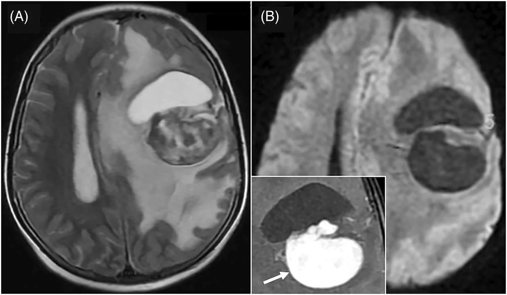
- 11 岁女孩,双眼视物模糊、右臂进行性无力 2 周;MRI 左侧大脑半球 53 × 57 mm 囊实性肿块,明显强化、无弥散受限,术中完全位于脑实质内、基底浸润运动区白质。
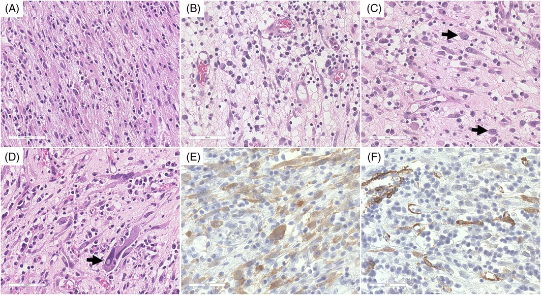
- 镜下:成纤维细胞/肌纤维母细胞样梭形细胞,密度不均,空泡核、小核仁;散在节细胞样与多形性细胞,核分裂罕见;间质巨噬细胞、淋巴细胞、浆细胞浸润伴局灶黏液样变。
- IHC 排除思路:GFAP、OLIG2、EMA、PR、STAT6 阴性(排除胶质瘤、脑膜瘤、SFT);CD68+ 但 Langerin、CD1a、S100 阴性(排除组织细胞增生症);阳性仅 CD99、ALK1(弥漫胞质、局灶核)与 actin,Ki-67 5%。RNA 测序检出 ALK 融合(病例描述处原文拼作 TMP4,讨论部分为 TPM4)。
- 要点:IMT 未列入 WHO CNS5,但颅内可发生;节细胞样细胞、多形性和浆细胞为主的浸润易误判为胶质瘤、富于淋巴浆细胞脑膜瘤、IPT 或组织细胞病变——遇脑内梭形细胞+炎性背景,应加做 ALK 并必要时行融合检测(ALK 阴性时考虑 ROS1、NTRK3)。附 ISN 虚拟玻片链接。
收录范围:PMC 开放获取全文(CC BY 4.0)——临床病史、检查所见、最终诊断、讨论、结论及图 1–2 与图注中英对照;患者知情同意声明与参考文献未收录。
临床病史
11 岁女孩,既往无重要病史,因双眼视物模糊及右臂进行性无力两周,就诊于越南国家癌症医院。脑部磁共振成像(MRI)显示左侧顶叶半球一境界清楚的实性—囊性病变,大小 53 × 57 mm,占位效应明显(图 1)。全身影像未见其他部位病变。术中见肿瘤完全位于脑实质内,基底部浸润运动区白质。切除实性部分;囊性部分因与重要传导束粘连而予以保留。
检查所见
组织病理学显示成纤维细胞样及肌纤维母细胞样细胞增生,细胞密度不均(图 2A、B),核呈空泡状,核仁小,胞质淡嗜酸性(框 1)。可见散在节细胞样细胞和多形性细胞(图 2C、D),核分裂罕见。间质含巨噬细胞、淋巴细胞和浆细胞,伴局灶黏液样变。GFAP、OLIG2、EMA、PR 和 STAT6 阴性,据此排除胶质瘤、脑膜瘤和孤立性纤维性肿瘤。ATRX 和 INI1 表达保留。CD68 阳性,而 Langerin、CD1a 和 S100 阴性,排除组织细胞增生症。其余阳性的免疫染色仅有 CD99、ALK1 和 actin(图 2E、F),更倾向间叶性肿瘤一类。增殖指数 KI67 为 5%。RNA 测序检出 TMP4::ALK 融合。DNA 测序(Ion AmpliSeq Cancer Hotspot Panel,Life Technologies)未检出其他突变。
框 1 虚拟玻片。访问地址:https://isn‐slidearchive.org/?col=ISN&fol=Archive&file=BPA‐6927662.svs
最终诊断
炎性肌纤维母细胞性肿瘤。
讨论
IMT 是一种中间型(交界性)恶性的梭形细胞肿瘤,长期被视为炎性假瘤(IPT),在 2020 年 WHO 软组织肿瘤分类中被确认并描述为独立实体 [1]。颅内 IMT 极为罕见:文献报道的 0–24 岁患者中枢神经系统(CNS)IMT/IPT 25 例中,颅内占 21 例 [2]。由于在 CNS 罕见,IMT 未被纳入 2021 年 WHO CNS 肿瘤分类,该分类转而列出了组织学特征与之重叠的新实体 [3]。
IMT 的临床和 MRI 特征均无特异性,诊断依赖组织病理和免疫组化。组织学上,IMT 表现为梭形细胞增生伴密集淋巴浆细胞浸润,有三种可并存的主要模式:(1)结节性筋膜炎样,(2)纤维瘤病/纤维组织细胞样,(3)硬纤维瘤样。坏死、束状或鲱鱼骨样结构、多核、多形性、节细胞样细胞及非典型核分裂提示更具侵袭性的亚型 [1]。
梭形细胞不同程度表达平滑肌 actin 和 desmin 等肌源性标志物,但通常不表达胶质标志物、S100、CD117 和 DOG1。40%–60% 的病例可检出 ALK1 表达,与 ALK 融合相关,既是诊断标志物,也是 ALK 靶向酪氨酸激酶抑制剂治疗的预测标志物 [1, 2]。其亚细胞着色模式随融合伙伴而异 [1]。TPM4 是儿童 IMT 常见的融合伙伴,通常产生弥漫胞质着色,正如本例所见 [1]。在 ALK 阴性 IMT 中,ROS1 和 NTRK3 重排最常见 [1, 2]。
IMT 的组织学特征并非特征性的,在 CNS 中可能类似其他实体。颅内伴 FET:CREB 融合的间叶性肿瘤和 CIC 重排肉瘤可有重叠特征 [3]。节细胞样细胞、细胞多形性及以浆细胞为主的浸润,需要与胶质瘤、富于淋巴浆细胞的脑膜瘤、IPT、组织细胞增生症或自身免疫病等其他实体相鉴别。仔细评估组织学模式和包括 ALK 在内的免疫组化表型,并结合分子检测,是将 IMT 与这些类似病变区分开的关键。
手术切除是 CNS‐IMT 的主要治疗。无法手术或复发/转移病例可使用糖皮质激素、化疗、放疗和 ALK 抑制剂,其中 ALK 抑制剂作为二线治疗显示出良好效果 [2]。ALK 重排通常以免疫组化或 FISH 检测。若结果阴性,现代补充分子检测(NGS panel、RNA‐seq)可发现假阴性结果或其他改变 [1]。因此,全面分子谱分析对作出最佳决策至关重要。
结论
颅内 IMT 是罕见肿瘤,其发生机制知之甚少,临床和影像特征无特异性。病理医生应知晓 CNS 中存在 IMT,进行 ALK 免疫染色,必要时行分子分析以识别基因融合。正确识别该实体及其分子特征,对诊断和治疗均至关重要。
- 11-year-old girl with 2 weeks of bilateral blurred vision and progressive right arm weakness; MRI showed a 53 × 57 mm solid-cystic left hemispheric mass with intense enhancement and no diffusion restriction, entirely intraparenchymal with its base infiltrating motor white matter.
- Histology: fibroblast-/myofibroblast-like spindle cells of variable cellularity, vesicular nuclei, small nucleoli; scattered ganglion-like and pleomorphic cells, rare mitoses; macrophages, lymphocytes and plasma cells with focal myxoid change.
- IHC work-up: GFAP, OLIG2, EMA, PR, STAT6 negative (excluding glioma, meningioma, SFT); CD68+ but Langerin, CD1a, S100 negative (excluding histiocytosis); only CD99, ALK1 (diffuse cytoplasmic, focal nuclear) and actin positive; Ki-67 5%. RNA sequencing found an ALK fusion (spelled “TMP4” in Findings; TPM4 in Discussion).
- Take-home: IMT is not listed in WHO CNS5 but occurs intracranially; ganglion-like cells, pleomorphism and plasma-cell-rich infiltrates can mimic glioma, lymphoplasmacyte-rich meningioma, IPT or histiocytosis — stain ALK and test for fusions when needed (ROS1, NTRK3 if ALK-negative). ISN virtual slide linked.
Scope: PMC open-access full text (CC BY 4.0) — clinical history, findings, final diagnosis, discussion, conclusion and Figures 1–2 with captions. Patient consent statement and references not included.
CLINICAL HISTORY
An 11‐year‐old girl, with no significant medical history, presented to the Vietnam National Cancer Hospital with blurred vision in both eyes and progressive weakness in the right arm over a two‐week period. Brain magnetic resonance imaging (MRI) revealed a well‐defined, solid and cystic lesion in the left parietal hemisphere, measuring 53 × 57 mm, causing a significant mass effect (Figure 1). Whole‐body imaging showed no evidence of lesions elsewhere. Intraoperatively, the tumor was entirely intraparenchymal, with its base infiltrating the motor white matter. The solid portion was resected, and the cystic part preserved due to adherence to critical tracts.
FINDINGS
Histopathology showed a proliferation of fibroblast‐ and myofibroblast‐like cells with heterogeneous cellularity (Figure 2A,B), vesicular nuclei, small nucleoli, and pale eosinophilic cytoplasm (Box 1). Scattered ganglion‐like and pleomorphic cells were observed (Figure 2C,D), while mitoses were rare. The stroma contained macrophages, lymphocytes, and plasma cells, with focal myxoid changes. Diagnoses of glioma, meningioma, and solitary fibrous tumor were ruled out, based on the negativity of GFAP, OLIG2, EMA, PR, and STAT6. ATRX and INI1 were maintained. CD68 was positive, but Langerin, CD1a, and S100 were negative, excluding histiocytosis. The only other positive immunostainings were CD99, ALK1, and actin (Figure 2E,F), which rather pointed toward the group of mesenchymal tumors. Proliferation index KI67 was 5%. RNA sequencing identified a TMP4::ALK fusion. No additional mutations were detected by DNA sequencing (Ion AmpliSeq Cancer Hotspot Panel, Life Technologies).
BOX 1 Virtual glass slide. Access at https://isn‐slidearchive.org/?col=ISN&fol=Archive&file=BPA‐6927662.svs
FINAL DIAGNOSIS
Inflammatory myofibroblastic tumor.
DISCUSSION
IMT, a spindle cell neoplasm of intermediate malignancy, long considered an inflammatory pseudotumor (IPT), has been recognized and described as a distinct entity in the 2020 WHO classification of soft tissue tumors [1]. Intracranial IMT is extremely rare, accounting for 21 out of 25 reported cases in the literature of central nervous system (CNS) IMT/IPT in patients aged 0–24 years [2]. Due to its rarity in the CNS, IMT is not included in the 2021 WHO Classification of CNS Tumors, which instead lists new entities with overlapping histological features [3].
Clinical and MRI features of IMTs are nonspecific, and diagnosis relies on histopathology and immunohistochemistry. Histologically, IMTs show spindle cell proliferation with dense lymphoplasmacytic infiltrate and three main patterns that may coexist: (1) nodular fasciitis‐like, (2) fibromatosis/fibrohistiocytic, and (3) desmoid‐like. Necrosis, fascicular or herringbone architecture, multinucleation, pleomorphism, ganglion‐like cells, and atypical mitoses indicate more aggressive variants [1].
Spindle cells variably express myoid markers such as smooth muscle actin and desmin, but are typically negative for glial markers, S100, CD117, and DOG1. ALK1 expression is detected in 40%–60% of cases, correlates with ALK fusion, and serves as both a diagnostic and predictive marker for ALK‐targeted tyrosine kinase inhibitor therapy [1, 2]. Its subcellular staining pattern varies with fusion partners [1]. TPM4 is a frequent fusion partner in pediatric IMTs, typically producing diffuse cytoplasmic staining, as observed in our case [1]. In ALK‐negative IMTs, ROS1 and NTRK3 rearrangements are most common [1, 2].
Histological features of IMT are not pathognomonic and may mimic other entities in the CNS. Intracranial mesenchymal tumor with FET:CREB fusion and CIC‐rearranged sarcoma may exhibit overlapping features [3]. The presence of ganglion‐like cells, cellular pleomorphism, and a predominant plasmacytic infiltrate requires distinction from other entities such as gliomas, lymphoplasmacytic‐rich meningioma, IPT, histiocytosis, or autoimmune diseases. Careful evaluation of histological pattern and immunohistochemical profile, including ALK, as well as molecular assessments, are essential to differentiate IMT from these mimics.
Surgical resection is the primary treatment for CNS‐IMT. When not feasible or in recurrent/metastatic cases, corticosteroids, chemotherapy, radiotherapy, and ALK inhibitors may be used, with ALK inhibitors showing promising second‐line results [2]. ALK rearrangements are usually detected by immunohistochemistry or FISH. If negative, modern additional molecular testing (NGS panels, RNA‐seq) can detect false‐negative results or other alterations [1]. Comprehensive molecular profiling is therefore crucial for optimal decisions.
CONCLUSION
Intracranial IMTs are rare neoplasms with poorly understood tumorigenesis and nonspecific clinical and radiological features. Pathologists should be aware of the existence of IMT in the CNS and perform ALK immunostaining and, if necessary, molecular analysis to identify gene fusions. Proper identification of this entity and its molecular characteristics is crucial for diagnosis and therapeutic purposes.