- 设计:西班牙一家神经组织库对截至 2024-01-01 的 212 例神经病理确诊 CJD(排除遗传型)做盲法 CTE 筛查——仅用 AT8 p-tau 染额叶皮质与海马;阳性或可疑者按第二版 NINDS/NIBIB 共识扩大取材,并用 McKee 分期与 Bieniek 严重度评估。
- 结果:3/212(1.4%)确诊 CTE,均为男性、均为高严重度(McKee III、I、III),2 例前足球运动员(其中 1 例同时打五人制足球)为 MM/MV1 型,1 例前英式橄榄球运动员为 VV2 型;另 8 例可疑者经扩大取材排除。队列女性占 55%,但阳性者全为有接触性运动史的男性。
- 诊断要点:CJD 本身可伴广泛 tau 病变(PART、ARTAG 样神经元/胶质 tau),单有神经元或胶质 tau 并不特异;确立 CTE 仍须找到特征性病变——皮质沟底血管周围神经元 p-tau 聚集(可伴棘状星形胶质细胞),且不局限于软膜下/浅层。
- 合并病理多见:病例 1 同时有中度 AD-NC(A3B2C2)与 LATE-NC 3 期,病例 3 有 A1B2C2 与 LATE-NC 2 期。表 1–2 并列本组与 Nemani 等既往 3 例的临床、PrP 染色模式、CTE 病变分布与最终神经病理诊断。
收录范围:PMC 开放获取全文(CC BY 4.0)——摘要、引言、材料与方法、结果(含 3 例详述)、讨论、结论,表 1–2 与图 1 图注中英对照;作者贡献、资助、利益冲突、伦理声明与参考文献未收录。
摘要
慢性创伤性脑病(CTE)是一种与反复头部撞击相关的 tau 蛋白病,反复头部撞击最常见于接触性运动;然而,其与朊病毒病的关系仍不明确。Hospital Clínic‐IDIBAPS 神经组织库在对患者临床病史不知情的情况下,对经神经病理学确诊的克雅病(CJD)患者的海马和额叶皮质样本进行磷酸化 tau 染色,以开展死后 CTE 筛查。对于阳性病例及具有提示性特征的病例,依据 NINDS/NIBIB 的 CTE 共识标准扩大分析范围,并回顾临床与神经病理学资料。在 212 例 CJD 病例中检出 3 例 CTE(1.4%),均有重度 CTE 病理改变:2 例为退役足球运动员(其中一例为精英运动员),1 例为退役英式橄榄球运动员。所有患者均在出生后第一个至第二个十年间开始参与运动,并在第五个十年间结束。CJD 的临床表现具有异质性,各病例的初始症状不同。神经病理学检查显示,足球运动员的组织学类型为 MM1/MV1,英式橄榄球运动员为 VV2,分别为 CJD 最常见和第二常见的变异型。这三例病例展示了 CTE 与 CJD 可在不同的临床病理表现下共存。仍需进一步研究,以确定共同机制是否可能促成这两种病理改变的共存。
引言
接触性运动与神经退行性疾病风险增加相关 [1, 2, 3]。在这些疾病中,慢性创伤性脑病(CTE)已成为与反复头部撞击关联最为一致的疾病 [4]。
CTE 于 1928 年首次在拳击手中被描述为“拳击醉酒综合征”,后来称为“拳击性痴呆” [5, 6];目前在神经病理学上被认定为一种 tau 蛋白病,其特征为皮质脑沟深部血管周围神经元内过度磷酸化 tau 的积聚 [7, 8]。根据修订后的美国国家神经疾病与卒中研究所(NINDS)和美国国家生物医学成像与生物工程研究所(NIBIB)共识标准(2021),单个特征性病变即足以确诊,疾病严重程度则根据 p‐tau 病理改变的分布进行分期 [8]。
普通人群中 CTE 的患病率尚不清楚。在将脑捐献给脑库的接触性运动运动员尸检研究中,患病率较高,介于 54% 至 99% [9, 10, 11]。相比之下,已报道的频率从欧洲社区老龄化队列中的 0% 到常规神经病理学病例系列中的 2.2% 不等 [12, 13]。
CTE 常与其他神经退行性疾病或病理改变共存,例如阿尔茨海默病、路易体病理或肌萎缩侧索硬化 [8, 14]。这种重叠使临床病理关联更加复杂,并突显了探索共病的重要性。
克雅病(CJD)是一种快速进展的致死性神经退行性疾病,其神经病理学特征为脑内海绵状改变和朊蛋白的细胞外沉积。最常见的组织学类型为 MM1 和 VV2,分别对应于 PRNP 基因第 129 位密码子为甲硫氨酸纯合型(MM1)或缬氨酸纯合型(VV2)的患者,各自具有不同的分子、神经病理学及临床特征 [15]。
尽管关于 CTE 及其共病的文献日益增多,其与朊病毒病的关系仍基本未经探索。迄今仅有一项既往研究报道了三例 CTE 与 CJD 并存的病例 [16]。
本研究调查了一个经神经病理学确诊的 CJD 患者系列中 CTE 的频率,并探索 CTE 阳性病例的临床与神经病理学特征。
材料与方法
研究设计
在 NTB‐Hospital Clínic‐IDIBAPS,对截至 2024 年 1 月 1 日可获得的经神经病理学确诊的 CJD 患者开展回顾性横断面研究。对所有疑似可能由朊病毒引起的快速进展性痴呆患者,均系统性地提出脑捐献建议。排除遗传性类型。此外,还纳入了病程较长、最初未怀疑 CJD 但随后经神经病理学确诊的非典型病例。
CTE 筛查按照第二次 NINDS/NIBIB 共识标准的简化版本进行,筛查时对神经病理学诊断及临床资料不知情 [7]。对脑库队列中所有受试者的海马和额叶皮质进行磷酸化 tau 免疫组织化学染色评估(p‐tau;AT8,美国赛默飞世尔科技公司)。
评估重点为 CTE 的特征性病变,其定义为位于皮质脑沟深部的血管周围神经元内过度磷酸化 tau 聚集,可伴有或不伴有棘状星形胶质细胞,且不局限于软脑膜下或浅表区域 [7, 8]。
对于表现出提示性特征的病例(如脑沟深部 tau 病理改变,或不伴 β‐淀粉样蛋白的皮质浅层神经原纤维缠结),在有石蜡包埋组织块可用时,将分析范围扩展至共识标准建议的其他脑区:额中回、颞上回及颞中回、顶下小叶、海马、杏仁核、基底节、丘脑、中脑(包括黑质)、脑桥(包括蓝斑)、延髓以及小脑皮质 [7]。还检查了包括枕叶皮质在内的其他皮质区域。对于符合 CTE 诊断标准的病例,依据第二次 NINDS/NIBIB 共识标准确定 CTE 严重程度,并额外采用 McKee 分期系统评估特征性 CTE 病理改变的范围 [17]。评估包括在杏仁核、丘脑、乳头体及小脑齿状核追加 p‐tau 染色 [7]。记录各受检区域是否存在特征性 CTE 病变,以及所检查的切片数量。
临床病理特征分析
回顾所有 CTE 筛查阳性病例的临床及神经病理学资料。临床变量包括性别、发病年龄、死亡年龄、反复头部损伤史、接触性运动暴露及参与时长,以及临床表现。
依据神经组织库的标准流程进行神经病理特征分析 [18],并在必要时根据现行诊断标准更新分类。评估内容包括 CJD 的有无及其亚型 [15, 19],依据美国国家老龄化研究所–阿尔茨海默病协会(NIA‐AA)/NINDS 标准评定 AD 神经病理改变的 ABC 评分 [20],tau 病理改变的 Braak 分期(I–VI)[21],Aβ 淀粉样蛋白沉积的 Thal 分期(0–5)[22],以及阿尔茨海默病登记联合体(CERAD)的神经炎性斑块评分(0–3)[23]。根据 McKeith 标准评估 α‐突触核蛋白病理改变 [24],并采用边缘系统为主的年龄相关 TAR DNA 结合蛋白 43(TDP‐43)脑病神经病理改变(LATE‐NC)分类评估 TDP‐43 病理改变 [25]。
结果
CJD 病例中 CTE 的筛查与频率
样本包括 212 名确诊 CJD 的个体,其中 55%(n = 117)为女性。通过额叶皮质和海马筛查,发现三名男性具有特征性 CTE 病变。八例具有提示性特征的病例经扩大取材后排除了 CTE [7]。三例确诊病例均表现出高级别 CTE 病理改变。下文将这些确诊病例称为病例 1、病例 2 和病例 3。这些病例与既往报道病例的临床及神经病理学特征汇总见表 1 和表 2。
| 特征 | 病例 1 | 病例 2 | 病例 3 | 病例 4a | 病例 5a | 病例 6a |
|---|---|---|---|---|---|---|
| 性别 | 男 | 男 | 男 | 男 | 男 | 男 |
| 发病年龄 | 60 多岁后期 | 60 多岁中期 | 80 多岁初期 | 79 岁 | 64 岁 | 48 岁 |
| 死亡年龄 | 70 多岁初期 | 60 多岁中期 | 80 多岁初期 | 84 岁 | 68 岁 | 48 岁 |
| RHI 暴露的主要来源 | 足球 | 足球和五人制足球 | 英式橄榄球 | 美式橄榄球和服兵役 | 美式橄榄球 | 多次头部创伤、斗殴及机动车事故 |
| 运动史与暴露时长 | 自儿童时期起踢足球 >50 年,其中精英级别 >20 年 | 自儿童时期至 30 多岁踢足球,随后以守门员身份参与五人制足球至 40 多岁中期;参与运动 >30 年 | 自青春期后期至成年中期参与英式橄榄球;参与运动 28 年,包括国际级别比赛 | 从高中到 NFL 参加美式橄榄球运动;共参与 9 年 | 从高中到 NFL 参加美式橄榄球运动;共参与 18 年 | 非职业运动员;多次 TBI 暴露 |
| 症状 | 进行性认知下降(6 年)、时间及空间定向障碍、癫痫发作、肌阵挛、无动性缄默(3 个月) | 右上肢麻木、震颤、异己手现象、失语(3 个月) | 步态不稳、定向障碍、幻视、肌阵挛(3 个月) | 愤怒、记忆障碍、运动症状、无动性缄默 | 健忘、失语、偏执、步态障碍、认知功能迅速下降 | 眼肌麻痹、意识混乱、强直、幻觉、神经功能迅速恶化 |
注:病例 1–3 来自本研究。
缩写:CJD,克雅病;CTE,慢性创伤性脑病;NFL,美国国家美式橄榄球联盟;RHI,反复头部撞击;TBI,创伤性脑损伤。
病例 4–6 此前由 Nemani 等报道 [16]。
| 特征 | 病例 1 | 病例 2 | 病例 3 | 病例 4a | 病例 5a | 病例 6a |
|---|---|---|---|---|---|---|
| 死亡年龄 | 70 多岁初期 | 60 多岁中期 | 80 多岁初期 | 84 岁 | 68 岁 | 48 岁 |
| 脑重(未固定) | 1200 g | 1635 g | 1210 g | 未报告 | 未报告 | 未报告 |
| 大体病理 | 弥漫性萎缩,以海马尤为明显 | 轻度全脑水肿;无明显萎缩 | 轻至中度额顶叶萎缩;杏仁核、海马及小脑萎缩极轻微 | 皮质及小脑萎缩 | 广泛重度皮质萎缩 | 枕叶及小脑萎缩最严重,重于额叶、顶叶及颞叶皮质 |
| PrP 免疫组织化学染色 | 突触型模式,主要累及小脑及枕叶皮质 | 弥漫性突触型模式,以枕叶及小脑皮质最强,呈“斑片状空泡周围”模式 | 广泛的突触型及神经元周围 PrP 沉积,主要位于小脑。 | 小脑皮质呈突触型模式,颞叶皮质呈粗颗粒型模式 | 粗细颗粒型模式,伴成熟斑块及斑块样结构形成 | 新皮质区域呈突触型模式,小脑呈“笔触状”模式 |
| McKee CTE 分期 | III | I | III | IV | II | II |
| Bieniek CTE 严重程度 | 重度 | 重度 | 重度 | 重度 | 轻度 | 轻度 |
| Braak 分期 | III | II | III | III–IV | I–II | I–II |
| 特征性 CTE 病变 | 2/2 额叶 | 1/2 颞叶 + 1/1 梭状回 | 2/4 额叶 +1/2 颞叶 | 运动皮质、颞叶及顶叶皮质 | 脑沟深部偶见血管周围簇状聚集;未注明区域 | 额叶、顶叶及颞叶皮质 |
| ABC 评分 | A3B2C2 | A1B1C0 | A1B2C2 | A2B2C0 | A2B1C0 | A1B1C0 |
| 最终神经病理学诊断 | sCJD(MM/MV1) + 高级别 CTE + 中等级别 AD‐NC + LATE | sCJD(MM/MV1‐MM2C)+ 高级别 CTE | sCJD(VV2) + 高级别 CTE + 中等级别 AD‐NC | sCJD(MV1‐2C) + 高级别 CTE + 中等级别 AD‐NC + LATE + α‐突触核蛋白(嗅球) | sCJD(MV2K‐C) + 低级别 CTE | sCJD(MM1/MV1) + 低级别 CTE |
注:病例 1–3 来自本研究。对于病例 1–3,特征性 CTE 病变按各解剖区域中显示该病变的切片数/所检查的切片数报告。检查了来自顶叶和枕叶皮质的各至少一张切片、额叶皮质的两张切片,以及颞叶皮质和海马/梭状回区域的各至少一张切片,以确定是否存在特征性 CTE 病变。对于病例 4–6,由于未报告各区域的受检切片数,故特征性 CTE 病变的解剖分布按 Nemani 等的描述报告 [16]。病例 4–6 的 Bieniek CTE 严重程度由作者依据所报告的 McKee CTE 分期回顾性判定。
缩写:ABC 评分(A:淀粉样蛋白,B:Braak,C:CERAD);AD‐NC,阿尔茨海默病神经病理改变;CJD,克雅病;CTE,慢性创伤性脑病;LATE,边缘系统为主的年龄相关 TDP‐43 脑病;PrP,朊蛋白;sCJD,散发性克雅病。
病例 4–6 此前由 Nemani 等(2018)报道 [16]。
伴有 CTE 的 CJD 病例的临床及神经病理学特征
病例 1
一名 60 多岁后期的男性出现进行性认知障碍,最初表现为记忆减退,随后发展为定向障碍和语言功能缺损。他曾是一名职业足球运动员,自儿童时期起参与足球运动超过五十年,其中以前锋身份参加精英级别比赛超过 20 年。
神经心理学评估显示记忆、语言、定向力和注意力受损。脑 MRI 显示重度海马萎缩。
症状出现六年后,患者神经功能迅速恶化,表现为步态不稳、肌阵挛和无动性缄默。脑电图显示周期性癫痫样放电,脑脊液 14–3–3 检测阳性。患者于 3 个月后死亡,临床诊断为可能的阿尔茨海默病及 CJD。
神经病理学检查显示脑重 1235 g,存在弥漫性脑萎缩,以海马受累为主。组织学分析显示朊病毒病(散发性克雅病,MM/MV1 亚型)与符合高分期 CTE 的广泛 tau 蛋白病共存。
朊病毒病理改变的特征为海绵状改变、神经元丢失及胶质增生,PrP 免疫染色以突触型模式为主,主要累及小脑及枕叶皮质。
AT8 免疫染色显示广泛的 tau 蛋白病,以星形胶质细胞受累为主,并伴有中度神经原纤维病理改变,主要累及额叶皮质、边缘系统区域、基底节及脑干头端。新皮质所见包括颗粒状绒毛状星形胶质细胞、棘状星形胶质细胞,以及皮质脑沟深部血管周围神经元内的 tau 聚集,符合 CTE 的特征性病变。所检查的 2 张额叶皮质切片中有 2 张发现特征性 CTE 病变(表 2)。Tau 病理改变扩展至海马、杏仁核、丘脑、下丘脑、黑质及脑干,且包含 3R 和 4R 两种亚型,其中星形胶质细胞病理改变中的 4R 相对占优势。其他 tau 病理改变包括额叶和颞叶白质内的 p‐tau 阳性星形胶质细胞、盘绕小体和球状胶质细胞包涵体,具有年龄相关 tau 星形胶质细胞病的特征。
其他所见包括中等级别的 AD 神经病理改变(A3B2C2)、轻度脑淀粉样血管病,以及边缘系统 TDP‐43 病理改变(LATE‐NC 第 3 期)。未见 α‐突触核蛋白病理改变。
病例 2
一名 60 多岁的男性,有高血压、2 型糖尿病病史,自儿童时期至 40 多岁中期参与业余足球运动,并自 30 多岁起以守门员身份参与五人制足球,因持续 2 个月的进行性神经系统症状就诊;最初为感觉障碍,随后出现右上肢肌阵挛和辨距不良。
脑 MRI 显示左侧顶叶、岛叶及额叶皮质轻度弥散异常。脑电图显示左侧大脑半球周期性偏侧癫痫样放电。脑脊液(CSF)分析显示 14–3–3 蛋白阴性。
疾病进展迅速。在 1 个月内,患者出现异己手现象、不对称性运动功能障碍(以右侧为主)、表达性失语及凝视障碍,随后进展为嗜睡和言语输出极少。生前最终临床诊断为 CJD。
大体检查显示脑重 1635 g,存在轻度全脑水肿,伴轻微小脑蚓部萎缩。
组织病理学检查显示朊病毒病(散发性克雅病,MM/MV1 亚型,伴 M2C 病灶)与高级别 CTE 共存。可见广泛的神经元丢失、星形胶质细胞增生及小胶质细胞活化,伴轻至中度皮质海绵状改变,其特征为贯穿各皮质层的小至中等大小空泡,以枕叶皮质最为显著,同时可见孤立的大型融合空泡灶,支持存在 M2C 特征。基底节及小脑亦受累,小脑分子层海绵状改变明显。脑干核团基本未受累。
PrP 免疫组织化学染色显示弥漫性突触型沉积,以枕叶皮质和小脑最为强烈,伴局灶性空泡周围染色。蛋白质印迹检测证实为 PrP 1 型。
AT8 免疫染色显示以星形胶质细胞为主的 tau 病理改变,伴有神经原纤维改变,包括神经原纤维缠结和神经网丝。颞下皮质可见皮质脑沟深部血管周围神经元内 tau 聚集,符合 CTE 的特征性病变。在所检查的两张颞叶皮质切片中的一张以及一张梭状回切片中的一张发现特征性 CTE 病变。其他 tau 病理改变累及海马及海马旁区域,Meynert 基底核及蓝斑神经元仅轻微受累。这些所见符合高分期 CTE 的诊断。
存在低级别的 AD 神经病理改变(A1B1C0)。未检测到 TDP‐43 或 α‐突触核蛋白病理改变。
病例 3
一名 80 多岁初期的男性,曾是精英英式橄榄球运动员,从十几岁后期至 40 多岁中期从事该项运动,在腰椎手术后 3 周出现步态不稳,随后在 2 个月内神经功能迅速恶化。既往病史包括血脂异常、心律失常、肥厚型心肌病及腰椎管狭窄。
入院时,患者嗜睡,定向力部分保留,伴计算障碍、运用能力受损、言语流畅性降低、步态障碍及后倾。脑 MRI 显示右侧尾状核弥散高信号、脑室周围白质 T2/FLAIR 高信号、中度皮质及皮质下萎缩,以及左侧小脑萎缩。CSF 感染相关检测、肿瘤相关神经抗体及 14–3–3 蛋白均为阴性,脑电图显示弥漫性慢波。
住院期间,患者在 72 h 内迅速恶化,出现四肢瘫痪、言语不连贯、幻视、舌肌阵挛及麻痹性肠梗阻。患者于症状出现后 3 个月死亡。
大体检查显示脑重 1210 g,存在轻至中度额顶叶萎缩,杏仁核、海马及小脑仅轻微受累。组织学显示朊病毒病(散发性克雅病,VV2 亚型)与高级别 CTE 共存。海绵状脑病主要累及皮质下及小脑区域,小脑皮质、海马 CA1 区、下托、杏仁核、丘脑、基底节及脑干中至重度受累,而新皮质改变较轻。海绵状改变以小空泡为主,偶见中等大小空泡。可见明显的神经元丢失和胶质增生,符合海马硬化。
免疫组织化学染色显示广泛的突触型及神经元周围 PrP 沉积,主要累及皮质下及小脑区域,符合 VV2 亚型。
Tau 病理改变累及神经元及胶质细胞,包括额叶、颞叶及岛叶脑沟深部血管周围神经元内的 tau 聚集,符合 CTE 的特征性病变。在所检查的四张额叶皮质切片中的两张及两张颞叶皮质切片中的一张发现特征性 CTE 病变。星形胶质细胞成分的特征为受累新皮质区域内的棘状星形胶质细胞和颗粒状绒毛状星形胶质细胞。颞叶外侧皮质浅层可见轻度神经原纤维病理改变。乳头体、丘脑及顶叶皮质亦受累,黑质、中脑被盖及 Meynert 基底核偶见神经原纤维缠结,壳核及齿状核受累较少,符合高分期 CTE 的诊断。存在中度 AD 神经病理改变(A1B2C2),伴边缘系统 TDP‐43 病理改变(LATE‐NC 第 2 期)。Meynert 基底核中偶见 α‐突触核蛋白阳性神经突。
代表性的神经病理学所见,包括海绵状改变、PrP 沉积及 CTE 相关 tau 病理改变,见图 1。
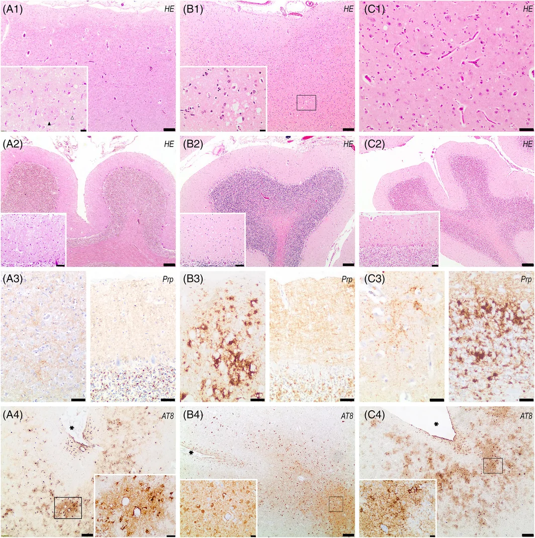
在这三例病例中,多个皮质区域均发现特征性 CTE 病变,但其分布和范围因病例而异(表 2)。病例 1–3 的 McKee 分期分别为 III、I 和 III,而按 Bieniek 标准,三例均被归类为重度。
讨论
据我们所知,这是首次在经神经病理学确诊的 CJD 病例中开展系统性 CTE 筛查。这些病例也是西班牙首次报道的经神经病理学确诊的 CTE 病例。在本研究之前,仅有一项西班牙研究根据创伤性脑损伤(TBI)病史描述了四例临床上提示 CTE 的病例,但均未经神经病理学证实 [26]。
我们筛查了 212 名经神经病理学确诊的 CJD 受试者,发现三例高分期 CTE,频率为 1.4%。一项既往研究报道了三例 CTE 与 CJD 共存的病例,包括在 UNITE 脑库中发现的两例(2/55,3.6%),以及在美国国家朊病毒病病理监测中心偶然发现的一例 [16]。尽管自 Nemani 等发表研究以来,波士顿大学 CTE 中心的规模已显著扩大,并进行了常规 PrP 免疫组织化学筛查,但仍未发现更多合并 CJD 的病例。已知或疑似朊病毒病患者通常被转诊至美国国家朊病毒病病理监测中心,这可能限制了在波士顿大学(BU)CTE 队列中发现 CJD 的机会。我们的结果首次提供了通过对明确界定的 CJD 队列进行系统性筛查而得出的 CTE 出现频率估计。
对这一频率的解释存在固有局限。西班牙普通人群中尚无 CTE 患病率可供参照,现有基于人群的研究来自接触性运动暴露模式不同的地区,因而限制了直接比较。未经选择的队列中,已报道的患病率从欧洲社区老龄化队列中的 0%,到美国、巴西和澳大利亚社区队列及尸检队列中的约 0.6–0.8% 不等,支持社区人群中偶然发现的 CTE 罕见这一观点 [13, 27, 28, 29]。在此背景下,我们观察到的频率较高;然而,鉴于研究设计与人群特征的差异,对这一差异的解释应持谨慎态度。
我们这三例病例的 CJD 临床表现具有异质性,各患者初始症状不同。神经病理学检查显示,足球运动员为 MM1/MV1 组织学类型,英式橄榄球运动员为 VV2 组织学类型,即散发性克雅病最常见的两种变异型。这种异质性与 Nemani 等的研究一致;该研究报道 CTE + CJD 病例的疾病相关朊蛋白性质及组织病理学表型与仅有 CJD 的对照病例无差异,提示 CTE 和 CJD 可能作为独立的病理过程共存 [16]。所有已报道病例,包括本系列的三例及既往发表病例,其临床特征与神经病理学所见的详细比较见表 1 和表 2。
多种接触性运动的运动员中均已广泛报道 CTE。我们的病例包括首次报道的足球及英式橄榄球运动员朊病毒病诊断,尤其还包括首例有记录的五人制足球运动员 CTE 病例。人们通常认为,五人制足球因场地较小且在室内进行,头部撞击较少;但近期证据显示,五人制足球的头球发生率与传统足球相当(每 1000 比赛小时的发生率〔IR〕分别为 32.9 和 36.9)[30]。此外,五人制足球中需要医疗处置的潜在头部损伤发生率实际上更高(每 1000 比赛小时分别为 122.3 和 76.4),非故意球击头部所占比例也更高(16.7% 对 6%)[31]。该患病个体在整个运动生涯中均参与五人制足球及传统足球,增加了反复头部撞击(RHI)的累积暴露。这些结果提示,五人制足球运动员的 RHI 暴露可能与传统足球运动员相当,甚至更高,因此应将其纳入 CTE 监测项目。
我们的发现引出的一个根本问题是,反复创伤是否可能成为散发性克雅病发生的危险因素。早期流行病学研究提示身体创伤与 CJD 之间存在关联 [32]。然而,这些观察结果未在更严格的现代研究中得到证实,而且很可能反映的是未被识别的医源性传播,而非创伤相关机制 [33]。值得注意的是,我们队列中的绝大多数 CJD 病例均无 CTE 证据。
在 CJD 背景下解释 tau 病理改变同样需要谨慎。Kovacs 等报道,散发性克雅病中的 tau 病理改变范围广泛,包括符合原发性年龄相关 tau 蛋白病和年龄相关 tau 星形胶质细胞病的神经元及胶质细胞 tau 病理改变 [34]。因此,仅存在神经元或胶质细胞 tau 病理改变并不对 CTE 具有特异性,识别 CTE 的特征性病变仍是确立诊断的必要条件。
从性别角度看,CTE 的报道以男性为主,这既反映了男性参与接触性运动的比例较高,也反映了既往研究可能存在选择偏倚,对家庭暴力等其他 RHI 来源的代表性可能不足 [14, 35]。我们采用了无偏倚的筛查方法;尽管女性占 CJD 队列的 55%,但三例 CTE 阳性病例均为男性,且均有明确记录的接触性运动参与史。这些结果提示,在我们的研究背景下,运动相关 RHI 暴露是促成 CTE 的主要因素,与历史上这些活动以男性为主的情况一致。
本研究存在若干局限。由于采用回顾性设计,详细的临床及运动暴露资料有限,且未能系统性获取 CTE 阴性病例的接触性运动史。初始筛查仅限于单侧大脑半球的额叶皮质和海马,可能降低了对早期病理改变的敏感性,尽管对于具有提示性特征的病例进行了扩大评估。
尽管存在上述局限,本研究的优势在于采用现行 CTE 诊断标准,以及不受性别或运动暴露影响的无偏倚筛查方法,对一个大型、经神经病理学确诊的 CJD 队列进行了系统评估。检出三例高级别 CTE 病例,表明该方法有效识别了具有临床意义的病例。
结论
本研究首次对经神经病理学确诊的 CJD 队列进行系统性 CTE 评估,发现三例高分期 CTE 与 CJD 共存的病例(1.4%)。这些病例也是西班牙首次报道的经神经病理学确诊的 CTE 病例。三例均发生于退役男性接触性运动运动员,其临床及神经病理学表现具有异质性。这些结果突显了在神经系统及神经病理学评估中系统记录反复头部撞击暴露的重要性。CTE 与 CJD 的共存提示可能存在重叠的神经退行性机制,值得进一步研究。
- Design: blinded CTE screening of 212 neuropathologically confirmed CJD cases (genetic forms excluded) from a Spanish Neurological Tissue Bank, using AT8 p-tau on frontal cortex and hippocampus only; positive/suggestive cases were expanded per the second NINDS/NIBIB consensus and staged by McKee and Bieniek.
- Results: 3/212 (1.4%) had CTE, all men, all high severity (McKee III, I, III) — two former soccer players (one also a futsal goalkeeper) with MM/MV1 and one former rugby player with VV2; eight suggestive cases were excluded after expanded sampling. Women made up 55% of the cohort, yet all positives were men with contact-sport histories.
- Diagnostic point: sporadic CJD can carry a broad spectrum of tau pathology (PART- and ARTAG-like), so neuronal or glial tau alone is not specific; the pathognomonic lesion — perivascular neuronal p-tau at the depth of a cortical sulcus, not restricted to subpial/superficial regions — remains essential.
- Co-pathologies were common: Case 1 had intermediate AD-NC (A3B2C2) and LATE-NC stage 3; Case 3 had A1B2C2 and LATE-NC stage 2. Tables 1–2 compare these three cases with the three previously reported by Nemani et al.
Scope: PMC open-access full text (CC BY 4.0) — abstract, introduction, methods, results (including the three case descriptions), discussion and conclusion, Tables 1–2 and Figure 1 with caption. Author contributions, funding, conflicts, ethics statements and references are not included.
Abstract
Chronic traumatic encephalopathy (CTE) is a tauopathy associated with repetitive head impacts, experienced most commonly in contact sports; however, its relationship with prion diseases remains unclear. Postmortem CTE screening using phosphorylated tau staining was conducted at the Neurological Tissue Bank of Hospital Clínic‐IDIBAPS on hippocampal and frontal cortex samples from patients with neuropathologically confirmed Creutzfeldt–Jakob disease (CJD), blinded to patients' clinical histories. In positive and suggestive cases, the analysis was expanded in accordance with the NINDS/NIBIB consensus criteria for CTE, and clinical and neuropathological information was reviewed. CTE was detected in 3 out of 212 CJD cases (1.4%), all with severe CTE pathology: 2 former soccer players (one of them an elite player) and 1 former rugby player. All had started playing sports between the first and second decades of life and ended in the fifth. The clinical presentation of CJD was heterogeneous, with different initial symptoms in each case. Neuropathological examination revealed an MM1/MV1 histotype in the soccer players and a VV2 histotype in the rugby player, the first‐ and second‐most common variants in CJD, respectively. These three cases illustrate the coexistence of CTE and CJD across heterogeneous clinicopathological presentations. Further studies are needed to determine whether shared mechanisms may contribute to the coexistence of both pathologies.
INTRODUCTION
Contact sports are associated with an increased risk of neurodegenerative diseases [1, 2, 3]. Among these, chronic traumatic encephalopathy (CTE) has emerged as the condition most consistently linked to repetitive head impacts [4].
First described in 1928 in boxers as “punch‐drunk syndrome,” later termed “pugilistic dementia” [5, 6], CTE is currently recognized neuropathologically as a tauopathy characterized by the perivascular accumulation of hyperphosphorylated tau in neurons at the depths of cortical sulci [7, 8]. According to the revised National Institute of Neurological Disorders and Stroke (NINDS) and National Institute of Biomedical Imaging and Bioengineering (NIBIB) consensus criteria (2021), a single pathognomonic lesion is sufficient for diagnosis, and disease severity is staged by the distribution of the p‐tau pathology [8].
The prevalence of CTE in the general population is unknown. In autopsy studies of contact sport athletes who donated their brains to brain banks, the prevalence is high, ranging from 54% to 99% [9, 10, 11]. In contrast, reported frequencies range from 0% in a European community‐based aging cohort to 2.2% in a routine neuropathology series [12, 13].
CTE often coexists with other neurodegenerative conditions, such as Alzheimer's disease, Lewy body pathology, or amyotrophic lateral sclerosis [8, 14]. This overlap complicates clinicopathological correlations and highlights the importance of exploring comorbidities.
Creutzfeldt–Jakob disease (CJD) is a rapidly progressive and fatal neurodegenerative disorder characterized neuropathologically by spongiform changes and extracellular deposition of prion protein in the brain. The most common histotypes are MM1 and VV2, corresponding to patients homozygous for methionine (MM1) or valine (VV2) at codon 129 of the PRNP gene, each associated with distinct molecular, neuropathological, and clinical features [15].
Despite the growing literature on CTE and its comorbidities, its relationship with prion diseases remains largely unexplored. To date, only one previous study reported three cases of concomitant CTE and CJD [16].
In this study, we investigated the frequency of CTE in a series of patients with neuropathologically confirmed CJD and to explore the clinical and neuropathological characteristics of CTE‐positive cases.
MATERIALS AND METHODS
Study design
A retrospective cross‐sectional study was conducted in patients with neuropathologically confirmed CJD available up to January 1, 2024, at NTB‐Hospital Clínic‐IDIBAPS. Brain donation was systematically proposed to all patients with suspected rapidly progressive dementia of possible prion origin. Genetic forms were excluded. In addition, atypical cases with prolonged disease duration, in which CJD had not been initially suspected but were subsequently confirmed neuropathologically, were also incorporated.
CTE screening was performed blinded to the neuropathological diagnosis and clinical information, following a reduced version of the second NINDS/NIBIB consensus criteria [7]. Immunohistochemical staining for phosphorylated tau (p‐tau; AT8, Thermo Fisher Scientific, USA) was evaluated in the hippocampus and frontal cortex of all subjects in the brain bank cohort.
The assessment focused on the pathognomonic lesion of CTE, defined as perivascular aggregates of hyperphosphorylated tau in neurons, with or without thorn‐shaped astrocytes, located at the depth of a cortical sulcus and not restricted to the subpial or superficial regions [7, 8].
In cases showing suggestive features (e.g., tau pathology at the sulcal depths or superficial cortical neurofibrillary tangles without amyloid‐β), the analysis was expanded to include additional brain regions recommended by the consensus criteria: middle frontal gyrus, superior and middle temporal gyri, inferior parietal lobule, hippocampus, amygdala, basal ganglia, thalamus, midbrain (including substantia nigra), pons (including locus coeruleus), medulla oblongata, and cerebellar cortex when paraffin‐embedded tissue blocks were available [7]. Additional cortical regions, including the occipital cortex, were also examined. In cases fulfilling diagnostic criteria for CTE, CTE severity was determined according to the second NINDS/NIBIB consensus criteria, and the extent of pathognomonic CTE pathology was additionally assessed using the McKee staging system [17]. The assessment included additional p‐tau staining in the amygdala, thalamus, mammillary bodies, and dentate nucleus of the cerebellum [7]. The presence of pathognomonic CTE lesions was recorded for each examined region, together with the number of sections examined.
Clinicopathological characterization
The clinical and neuropathologic data from all cases with positive CTE screening were reviewed. Clinical variables included sex, age at disease onset, age at death, history of repetitive head injury, exposure to contact sports and duration of practice, and clinical presentation.
Neuropathological characterization was performed according to the standard protocols of the Neurological Tissue Bank [18], and classifications were updated when necessary according to current diagnostic criteria. The assessment included the presence and subtype of CJD [15, 19], the ABC score for AD neuropathologic change according to National Institute on Aging–Alzheimer’s Association (NIA‐AA)/NINDS criteria [20], Braak staging of tau pathology (I–VI) [21], Thal phase for Aβ amyloid deposition (0–5) [22], and Consortium to Establish a Registry for Alzheimer's Disease (CERAD) score for neuritic plaques (0–3) [23]. α‐Synuclein pathology was evaluated according to the McKeith criteria [24], and TDP‐43 pathology was assessed with Limbic‐predominant age‐related TAR DNA‐binding protein 43 (TDP‐43) encephalopathy neuropathological change (LATE‐NC) classification [25].
RESULTS
Screening and frequency of CTE in CJD cases
The sample consisted of 212 individuals diagnosed with CJD, of whom 55% (n = 117) were women. Screening of the frontal cortex and hippocampus identified three men with pathognomonic CTE lesions. Eight cases with suggestive features were ruled out for CTE with expanded sampling [7]. The three confirmed cases exhibited high‐grade CTE pathology. From this point forward, the confirmed cases will be referred to as Case 1, Case 2, and Case 3. A summary of the clinical and neuropathological characteristics of these cases, together with previously reported cases, is provided in Tables 1 and 2.
| Feature | Case 1 | Case 2 | Case 3 | Case 4a | Case 5a | Case 6a |
|---|---|---|---|---|---|---|
| Gender | Male | Male | Male | Male | Male | Male |
| Age at disease onset | Late 60s | Mid 60s | Early 80s | 79 years | 64 years | 48 years |
| Age at death | Early 70s | Mid 60s | Early 80s | 84 years | 68 years | 48 years |
| Primary source of RHI exposure | Soccer | Soccer and futsal | Rugby | Football and military service | Football | Multiple head traumas, fistfights, and motor vehicle accidents |
| Sports history and duration of exposure | Soccer from childhood for >50 years, including >20 years at elite level | Soccer from childhood to his 30s, followed by futsal as goalkeeper until the mid‐40s; >30 years of practice | Rugby from late adolescence to mid‐adulthood; 28 years of practice, including international‐level competition | Football from high school to NFL; a total of 9 years of practice | Football from high school to NFL; a total of 18 years of practice | Not a professional athlete; multiple TBI exposures |
| Symptoms | Progressive cognitive decline (6 years), spatiotemporal disorientation, seizures, myoclonus, akinetic mutism (3 months) | Right upper limb numbness, tremor, alien hand phenomenon, aphasia (3 months) | Gait instability, disorientation, visual hallucinations, myoclonus (3 months) | Anger, memory impairment, motor symptoms, akinetic mutism | Forgetfulness, aphasia, paranoia, gait disturbance, rapid cognitive decline | Ophthalmoplegia, confusion, rigidity, hallucinations, rapid neurological deterioration |
Note: Cases 1–3 correspond to the present study.
Abbreviations: CJD, Creutzfeldt–Jakob disease; CTE, chronic traumatic encephalopathy; NFL, National Football League; RHI, repetitive head impacts; TBI, traumatic brain injury.
Cases 4–6 were previously reported by Nemani et al. [16].
| Feature | Case 1 | Case 2 | Case 3 | Case 4a | Case 5a | Case 6a |
|---|---|---|---|---|---|---|
| Age at death | Early 70s | Mid 60s | Early 80s | 84 years | 68 years | 48 years |
| Brain weight (unfixed) | 1200 g | 1635 g | 1210 g | Not reported | Not reported | Not reported |
| Macroscopic pathology | Diffuse atrophy, especially in the hippocampus | Mild generalized edema; no significant atrophy | Mild–moderate frontoparietal atrophy; minimal in amygdala, hippocampus and cerebellum | Cortical and cerebellar atrophy | Widespread severe cortical atrophy | Most severe atrophy in occipital and cerebellum than in the frontal, parietal and temporal cortices |
| PrP immunohistochemistry | Synaptic pattern, predominantly affecting cerebellar and occipital cortices | Diffuse synaptic pattern, most intense in occipital and cerebellar cortex with a “patchy perivacuolar” pattern | Widespread synaptic and perineuronal PrP deposits, mainly in cerebellum. | Synaptic pattern in cerebellar cortex, and coarse pattern in temporal cortex | Coarse fine granular pattern, with mature plaques and plaque‐like formations | Synaptic pattern in neocortical regions with “brush‐stroke” cerebellar pattern |
| McKee CTE stage | III | I | III | IV | II | II |
| Bieniek CTE severity | High | High | High | High | Low | Low |
| Braak stage | III | II | III | III–IV | I–II | I–II |
| Pathognomonic CTE lesions | 2/2 frontal | 1/2 temporal + 1/1 fusiform | 2/4 frontal +1/2 temporal | Motor, temporal, and parietal cortices | Occasional perivascular clusters at sulcal depths; region not specified | Frontal, parietal, and temporal cortices |
| ABC score | A3B2C2 | A1B1C0 | A1B2C2 | A2B2C0 | A2B1C0 | A1B1C0 |
| Final neuropathological diagnosis | sCJD (MM/MV1) + high‐grade CTE + intermediate AD‐NC + LATE | sCJD (MM/MV1‐MM2C) + high‐grade CTE | sCJD (VV2) + high‐grade CTE + intermediate AD‐NC | sCJD (MV1‐2C) + high‐grade CTE + intermediate AD‐NC + LATE + α‐syn (olfactory bulbs) | sCJD (MV2K‐C) + low‐grade CTE | sCJD (MM1/MV1) + low‐grade CTE |
Note: Cases 1–3 correspond to the present study. For Cases 1–3, pathognomonic CTE lesions are reported as the number of sections showing the lesion/number of sections examined for each anatomical region. At least one section from the parietal and occipital cortices, two sections from the frontal cortex, and at least one section from the temporal cortex and hippocampal/fusiform region were examined for the presence of pathognomonic CTE lesions. For Cases 4–6, the anatomical distribution of pathognomonic CTE lesions is reported as described by Nemani et al. [16], as the number of sections examined for each region was not reported. Bieniek CTE severity for Cases 4–6 was retrospectively assigned by the authors based on the reported McKee CTE stage.
Abbreviations: ABC score (A: amyloid, B: Braak, C: CERAD); AD‐NC, Alzheimer disease neuropathologic changes; CJD, Creuzfeld–Jakob disease; CTE, chronic traumatic encephalopathy; LATE, limbic‐predominant age‐related TDP‐43 encephalopathy; PrP, prion protein; sCJD, sproadic Creuzfeld–Jakob disease.
Cases 4–6 were previously reported by Nemani et al. (2018) [16].
Clinical and neuropathological features of CJD cases with CTE
Case 1
A man in his late 60s presented with progressive cognitive impairment initially characterized by memory loss, later evolving to include disorientation and language deficits. He was a former professional soccer player who had over five decades of soccer participation beginning in childhood, including more than 20 years of elite‐level competition as a striker.
Neuropsychological assessment revealed deficits in memory, language, orientation, and attention. Brain MRI showed severe hippocampal atrophy.
Six years after symptom onset, the patient experienced rapid neurological deterioration, including gait instability, myoclonus, and akinetic mutism. EEG showed periodic epileptiform discharges, and cerebrospinal fluid 14–3–3 was positive. He died 3 months later with a clinical diagnosis of possible Alzheimer's disease and CJD.
Neuropathological examination revealed a brain weighing 1235 g, with diffuse brain atrophy, predominantly affecting the hippocampus. Histological analysis demonstrated coexistent prion disease (sporadic CJD, MM/MV1 subtype) and extensive tauopathy consistent with high‐stage CTE.
Prion pathology was characterized by spongiform change, neuronal loss, and gliosis, with a predominantly synaptic PrP immunostaining pattern affecting mainly cerebellar and occipital cortices.
AT8 immunostaining revealed extensive tauopathy with predominant astroglial involvement and moderate neurofibrillary pathology, affecting mainly the frontal cortex, limbic regions, basal ganglia, and rostral brainstem. Neocortical findings included granular fuzzy astrocytes, thorn‐shaped astrocytes, as well as perivascular neuronal tau aggregates at the depths of cortical sulci, consistent with the pathognomonic lesion of CTE. Pathognomonic CTE lesions were identified in 2 of 2 frontal cortex sections examined (Table 2). Tau pathology extended to the hippocampus, amygdala, thalamus, hypothalamus, substantia nigra, and brainstem, and included both 3R and 4R isoforms, with a relative predominance of 4R in astroglial pathology. Other tau pathology included p‐tau‐positive astrocytes, coiled bodies, and globular glial inclusions in frontal and temporal white matter characteristic of aging‐related tau astrogliopathy.
Additional findings included intermediate AD neuropathologic changes (A3B2C2), mild cerebral amyloid angiopathy, and limbic TDP‐43 pathology (LATE‐NC Stage 3). α‐Synuclein pathology was absent.
Case 2
A man in his 60s, with hypertension, Type 2 diabetes mellitus, and a history of amateur soccer playing from childhood to his mid‐40s, with participation in futsal as a goalkeeper from his 30s onward, presented with a 2‐month history of progressive neurological symptoms, initially sensory disturbances followed by right upper limb myoclonus and dysmetria.
Brain MRI revealed mild diffusion abnormalities in the left parietal, insular, and frontal cortices. EEG showed periodic lateralized epileptiform discharges in the left hemisphere. Cerebrospinal fluid (CSF) analysis was negative for 14–3–3 protein.
The disease course was rapidly progressive. Within 1 month, he developed alien hand phenomenon, asymmetric motor impairment (right‐predominant), expressive aphasia, and impaired gaze, progressing to somnolence and minimal verbal output. The final antemortem clinical diagnosis was CJD.
On gross examination, the brain weighed 1635 g and exhibited mild generalized edema with discrete vermian cerebellar atrophy.
Histopathological examination demonstrated coexistent prion disease (sporadic CJD, MM/MV1 subtype with M2C foci) and high‐grade CTE. There was widespread neuronal loss, astrogliosis, and microglial activation, with mild‐to‐moderate cortical spongiform change characterized by small‐ to medium‐sized vacuoles across cortical layers, most prominent in the occipital cortex, along with isolated foci of large confluent vacuoles supporting the presence of M2C features. The basal ganglia and cerebellum were also involved, with prominent spongiform changes in the cerebellar molecular layer. Brainstem nuclei were largely spared.
PrP immunohistochemistry showed diffuse synaptic deposits, most intense in the occipital cortex and cerebellum, with focal perivacuolar staining. Western blot confirmed PrP Type 1.
AT8 immunostaining revealed predominantly astroglial tau pathology with associated neurofibrillary changes, including neurofibrillary tangles and neuropil threads. Perivascular neuronal tau aggregates at the depths of cortical sulci were identified in the inferior temporal cortex, consistent with the pathognomonic lesion of CTE. Pathognomonic CTE lesions were identified in one of two temporal cortex sections and one of one fusiform gyrus sections examined. Additional tau pathology involved hippocampal and parahippocampal regions, with minimal involvement of the basal nucleus of Meynert and neurons of the locus coeruleus. Findings were diagnostic of high‐stage CTE.
Low AD neuropathologic changes were present (A1B1C0). No TDP‐43 or α‐synuclein pathology was detected.
Case 3
A man in his early 80s, a former elite rugby player active from his late teens to mid‐40s, presented with gait instability 3 weeks after lumbar surgery, followed by rapid neurological deterioration over 2 months. His medical history included dyslipidemia, cardiac arrhythmia, hypertrophic cardiomyopathy, and lumbar spinal stenosis.
On admission, he was somnolent and partially oriented, with dyscalculia, impaired praxis, hypofluent speech, gait impairment, and retropulsion. Brain MRI showed diffusion hyperintensity in the right caudate nucleus, T2/FLAIR periventricular white matter hyperintensities, moderate cortico‐subcortical atrophy, and left cerebellar atrophy. CSF was negative for infection, onconeural antibodies, and 14–3–3 protein, and EEG showed diffuse slowing.
During hospitalization, he deteriorated rapidly over 72 h, developing quadriplegia, incoherent speech, visual hallucinations, lingual myoclonus, and paralytic ileus. He died 3 months after symptom onset.
On gross examination, the brain weighed 1210 g and showed mild–moderate frontoparietal atrophy, with minimal involvement of the amygdala, hippocampus, and cerebellum. Histology revealed coexistent prion disease (sporadic CJD, VV2 subtype) and high‐grade CTE. Spongiform encephalopathy predominantly affected subcortical and cerebellar regions, with moderate‐to‐severe involvement of the cerebellar cortex, hippocampal CA1, subiculum, amygdala, thalamus, basal ganglia, and brainstem, while neocortical changes were mild. Spongiform changes were characterized by predominantly small vacuoles with occasional medium‐sized forms. Marked neuronal loss and gliosis consistent with hippocampal sclerosis were present.
Immunohistochemistry showed widespread synaptic and perineuronal PrP deposits, predominantly affecting subcortical and cerebellar regions, consistent with the VV2 subtype.
Tau pathology showed neuronal and glial involvement, including perivascular neuronal tau aggregates at the depths of frontal, temporal, and insular sulci, consistent with the pathognomonic lesion of CTE. Pathognomonic CTE lesions were identified in two of four frontal cortex sections and one of two temporal cortex sections examined. The astroglial component was characterized by thorn‐shaped and granular fuzzy astrocytes in affected neocortical regions. Mild superficial neurofibrillary pathology was observed in the lateral temporal cortex. Additional involvement of mammillary bodies, thalamus, and parietal cortex was present, with occasional neurofibrillary tangles in the substantia nigra, midbrain tegmentum, and basal nucleus of Meynert, and less frequent involvement of the putamen and dentate nucleus, diagnostic for high‐stage CTE. Moderate AD neuropathologic changes were present (A1B2C2), along with limbic TDP‐43 pathology (LATE‐NC stage 2). Occasional α‐synuclein‐positive neurites were observed in the basal nucleus of Meynert.
Representative neuropathological findings, including spongiform changes, PrP deposition, and CTE‐related tau pathology, are shown in Figure 1.
Across the three cases, pathognomonic CTE lesions were identified in multiple cortical regions, although their distribution and extent varied between cases (Table 2). McKee staging was III, I, and III for Cases 1–3, respectively, whereas all three cases were classified as high severity according to the Bieniek criteria.
DISCUSSION
To the best of our knowledge, this is the first systematic screening for CTE in neuropathologically confirmed CJD cases. These cases also represent the first neuropathologically confirmed diagnoses of CTE reported in Spain. Prior to this work, only one Spanish study had described four clinically suggestive CTE cases based on a history of traumatic brain injury (TBI), but none were neuropathologically confirmed [26].
We screened 212 subjects with neuropathologically confirmed CJD and identified three cases with high‐stage CTE, representing a frequency of 1.4%. A previous study reported three cases with coexisting CTE and CJD, including two identified within the UNITE Brain Bank (2/55, 3.6%) and one incidentally found at the National Prion Disease Pathology Surveillance Center [16]. Although the Boston University CTE Center has substantially expanded since the publication of Nemani et al., no additional cases of concomitant CJD have been identified despite routine PrP immunohistochemical screening. Patients with known or suspected prion disease are generally referred to the National Prion Disease Pathology Surveillance Center, which may limit the opportunity to identify CJD within the BU CTE cohort. Our findings provide the first estimate of CTE occurrence derived from systematic screening in a defined CJD cohort.
The interpretation of this frequency presents inherent limitations. There is no reference prevalence of CTE in the general Spanish population, and available population‐based studies derive from regions with different exposure patterns to contact sports, limiting direct comparisons. Reported prevalences in unselected cohorts range from 0% in a European community‐based aging cohort to approximately 0.6–0.8% in community‐based and autopsy‐based cohorts from the United States, Brazil, and Australia, supporting the notion that incidental CTE is rare in community populations [13, 27, 28, 29]. In this context, our observed frequency is higher; however, this difference should be interpreted with caution given differences in study design and population characteristics.
The clinical presentation of CJD was heterogeneous among our three cases, with different initial symptoms in each patient. Neuropathological examination revealed MM1/MV1 histotype in the soccer players and VV2 histotype in the rugby player, the two most common sporadic CJD variants. This heterogeneity is consistent with Nemani et al., who reported that disease‐related prion protein properties and histopathological phenotype in CTE + CJD cases did not differ from CJD‐only controls, suggesting that CTE and CJD may coexist as independent pathological processes [16]. A detailed comparison of clinical characteristics and neuropathological findings across all reported cases, including those three from the present series and previously published cases, is provided in Tables 1 and 2.
CTE has been widely reported in athletes from various contact sports. Our cases include the first reported diagnoses of prion disease in soccer and rugby players, and notably, the first documented case of CTE in a futsal player. Contrary to the common assumption that futsal involves fewer head impacts due to its smaller field and indoor setting, recent evidence demonstrates that the incidence rate of headers in futsal is comparable to that of traditional soccer (IR 32.9 vs. 36.9 per 1000 match hours) [30]. Moreover, the rate of potential head injuries requiring medical attention is actually higher in futsal (122.3 vs. 76.4 per 1000 match hours), with unintentional ball‐to‐head impacts being proportionally more frequent (16.7% vs. 6%) [31]. The affected individual played both futsal and traditional soccer throughout his career, increasing cumulative repetitive head impacts (RHI) exposure. These findings suggest that futsal players may have comparable or even greater RHI exposure than traditional soccer players, warranting their inclusion in CTE surveillance programs.
A fundamental question arising from our findings is whether repetitive trauma could act as a risk factor for the development of sporadic CJD. Early epidemiological studies suggested an association between physical trauma and CJD [32]. However, these observations have not been confirmed in more rigorous contemporary investigations, and they likely reflected unrecognized iatrogenic transmission rather than trauma‐related mechanisms [33]. Notably, the vast majority of CJD cases in our cohort showed no evidence of CTE.
The interpretation of tau pathology in the setting of CJD also requires caution. Kovacs et al. reported a broad spectrum of tau pathology in sporadic CJD, including neuronal and glial tau pathology compatible with primary age‐related tauopathy and ageing‐related tau astrogliopathy [34]. Therefore, the presence of neuronal or glial tau pathology alone is not specific for CTE, and identification of the pathognomonic CTE lesion remains essential for establishing the diagnosis.
From a gender perspective, CTE has predominantly been reported in men, reflecting both higher male participation in contact sports and potential selection bias in prior studies, which may have underrepresented other sources of RHI such as domestic violence [14, 35]. We applied an unbiased screening approach; although women comprised 55% of our CJD cohort, all three CTE‐positive cases were male with a documented history of contact sports participation. These findings suggest that, in our setting, sport‐related RHI exposure is the primary contributor to CTE, consistent with the historical male predominance in these activities.
This study has several limitations. Due to its retrospective design, detailed clinical and sport exposure information was limited, and contact sports history was not systematically available for CTE‐negative cases. The initial screening was limited to the frontal cortex and hippocampus of a single hemisphere, potentially reducing sensitivity to early pathology, although cases with suggestive features underwent expanded assessment.
Despite these limitations, the study benefits from the systematic evaluation of a large neuropathologically confirmed CJD cohort using current CTE diagnostic criteria and an unbiased screening approach independent of sex or sports exposure. The identification of three high‐grade CTE cases indicates that the methodology effectively detected clinically relevant cases.
CONCLUSION
This study provides the first systematic assessment of CTE in a neuropathologically confirmed CJD cohort and identified three cases of coexisting high‐stage CTE and CJD (1.4%). These cases also represent the first neuropathologically confirmed diagnoses of CTE reported in Spain. All three occurred in male former contact‐sport athletes with heterogeneous clinical and neuropathological profiles. These findings highlight the importance of systematic documentation of repetitive head impact exposure in neurological and neuropathological evaluations. The coexistence of CTE and CJD raises the possibility of overlapping neurodegenerative mechanisms that warrant further investigation.