- 58 岁女性,5 年来反复头晕、意识受损发作,发作前有强烈恐惧感,曾被当作先兆偏头痛;MRI 示右侧海马与杏仁核 2.5 cm 边界模糊的多结节病变,T2-FLAIR 高信号,无水肿、无强化、无弥散受限,波谱有胆碱峰。
- 镜下:低倍见苍白、略融合的结节;结节内细胞核大偏位、核仁明显、胞质丰富细颗粒嗜酸,似神经元,埋于疏松纤维基质,胞质内/细胞周围空泡变常见;无核分裂,Ki-67 约 1%。
- IHC 组合是关键:OLIG2 阳性、突触素弱阳性,而 NeuN、嗜铬粒蛋白、神经丝蛋白阴性;GFAP 仅基质阳性,CD34 限于血管,NSE 在低倍更清楚地勾勒结节模式。
- TSO500 检出 MAP2K1 2 号外显子框内缺失 p.Gln56_Val60del(VAF 2.5%)及 ATR 失活变异(VAF 4%),肿瘤细胞含量 30%–40%——低 VAF 也可支持诊断。讨论回顾 MVNT「别管我」病变的手术指征与 MAP2K1/BRAF 非 V600E/FGFR2 融合驱动谱。附 ISN 虚拟玻片。
收录范围:PMC 开放获取全文(CC BY-NC-ND 4.0)——病例介绍、检查所见、最终诊断、讨论及图 1–2 与图注中英对照;作者贡献、资助、利益冲突声明与参考文献未收录。
病例介绍
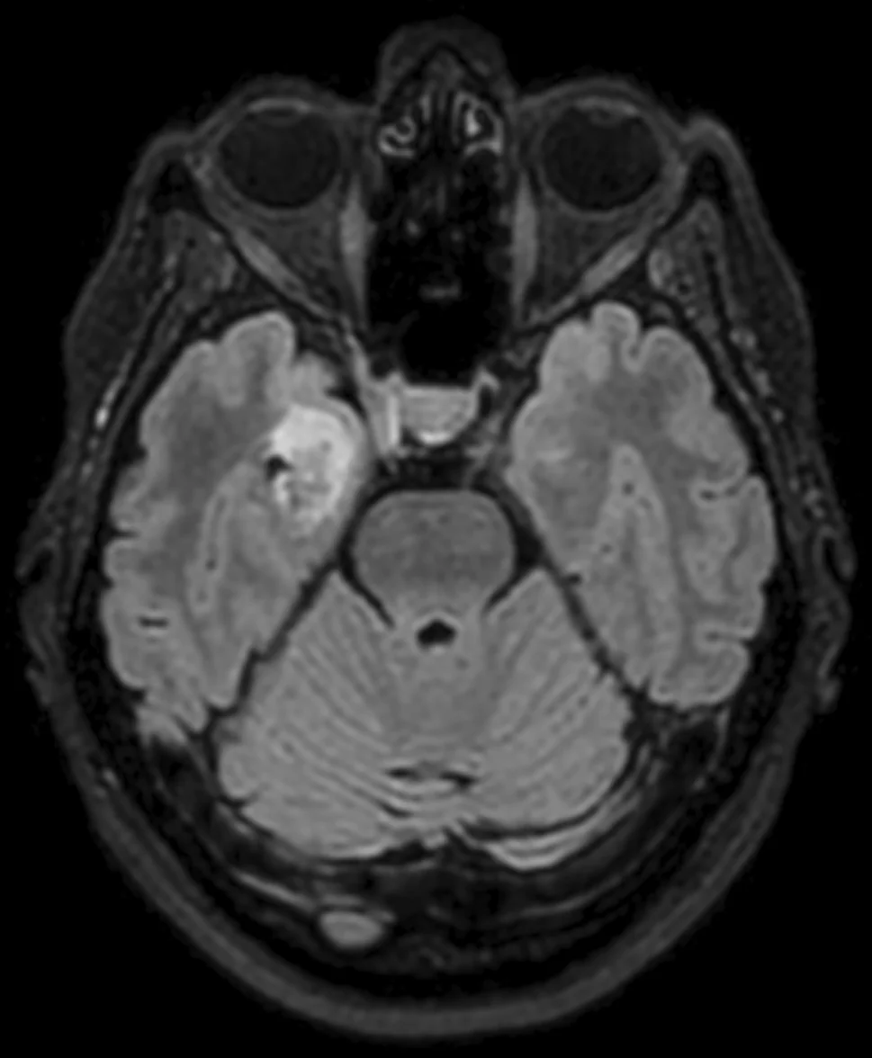
58 岁女性,反复出现头晕和对周围环境意识受损的发作,发作前有强烈的恐惧感。病史始于 5 年前,当时经脑部 CT 排除血管性疾病后,这些发作被解释为可能的先兆偏头痛。由于发作频率随时间逐渐增加,遂行脑部 MRI,显示右侧海马和杏仁核区一 2.5 cm、边界模糊的多结节状病变。病变在 T1 加权序列上呈轻度低信号,在 T2-FLAIR 序列上呈高信号,无瘤周水肿、无强化,DWI 无弥散受限。MRI 波谱显示胆碱峰,不伴肌醇峰。影像学所见总体提示低级别肿瘤(图 1)。鉴于症状持续,患者接受开颅手术,病变大体全切。术后患者出现轻度感受性和表达性失语,目前正在逐渐恢复。随后随访期间未记录到癫痫发作。
检查所见
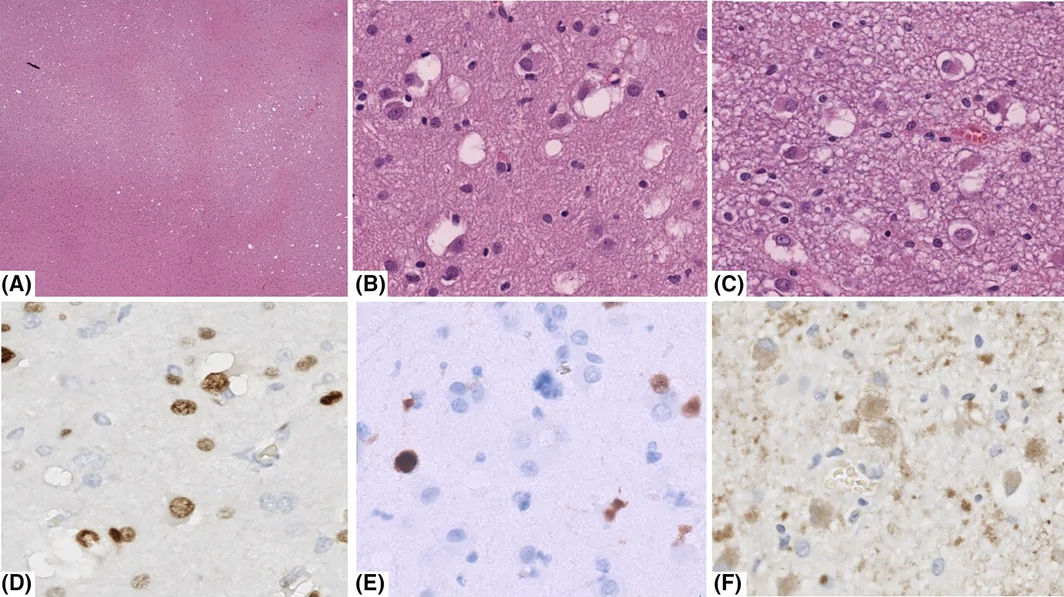
组织病理学检查见海马皮质和白质碎片。低倍镜下,由于存在苍白、略相互融合的结节状区域,实质显得不均匀(图 2A,框 1)。中倍镜下,苍白结节细胞密度轻度增加,细胞核大、偏位,含一个或多个核仁,胞质丰富、细颗粒状、嗜酸性,类似神经元成分。这些细胞埋于疏松的纤维状基质中,常见胞质内或细胞周围空泡变(图 2B、C)。未见核分裂。Ki-67 增殖指数约 1%。免疫组化显示,肿瘤细胞 OLIG2 阳性(图 2D)、突触素弱阳性(图 2F),而嗜铬粒蛋白、NeuN(图 2E)和神经丝蛋白阴性;GFAP 在基质中阳性,而肿瘤细胞阴性;CD34 仅限于血管;NSE 在细胞中阴性,但在低倍镜下更清晰地勾勒出结节状模式。临床病理特征总体提示多结节空泡状神经元肿瘤(MVNT)的诊断。检出 MAP2K1 基因 2 号外显子的致病性突变(p.Gln56_Val60del)(TruSight Oncology 500,Illumina),进一步支持该诊断。值得注意的是,病变还携带 ATR 基因 10 号外显子的另一致病性变异(p.Ile774Tyrfs*5)。在肿瘤细胞含量为 30%–40% 的标本中,两种突变的变异等位基因频率均较低(分别为 2.5% 和 4%)。
框 1 虚拟玻片。访问地址:https://isn‐slidearchive.org/?col=ISN&fol=Archive&file=BPA‐3743922.tif。
最终诊断
多结节空泡状神经元肿瘤(MVNT),CNS WHO 1 级。
讨论
MVNT 是一种罕见肿瘤,迄今英文文献报道的病理证实病例不足 60 例 [1],其中仅少数提供了分子特征。MVNT 可发生于很宽的年龄范围,发病高峰在中年,女性略多。临床表现通常包括癫痫发作、头痛、头晕或长期癫痫,但 MVNT 也可无症状,在 MRI 检查中偶然发现,而 CT 扫描在多数病例中无法发现病变。MVNT 即使在全切或次全切后也不进展,常被称为「别管我」(“leave-me-alone”)病变。是否手术可根据是否缺乏特征性影像学表现(如气泡样信号改变)和/或是否存在症状来考虑。越来越多的证据表明,编码丝裂原活化蛋白激酶(MAPK)激酶 1(MEK1)的 MAP2K1 突变是 MVNT 的致癌驱动因素,个别病例携带 BRAF 非 V600E 突变或 FGFR2 融合 [2, 3]。
本例肿瘤位于海马区,属典型部位。尽管影像学特征本身并不特异,但根据 2021 年 WHO 中枢神经系统肿瘤分类,病理所见(即多结节模式,以及存在共表达 OLIG2 和突触素、而 NeuN 缺失的伴空泡变的神经元成分)可诊断 MVNT。重要的是,本例携带 MAP2K1 框内致病性缺失(p.Gln56_Val60del),进一步支持诊断。该变异此前已在两例已发表的 MVNT 病例中报道 [3]。作为新发现,我们记录到 ATR 基因的失活突变,该基因编码一种在 DNA 损伤应答中起关键作用的丝氨酸/苏氨酸激酶。MAP2K1 与 ATR 变异之间可能存在的相互作用有待进一步探索。
- 58-year-old woman with 5 years of dizzy spells and impaired awareness preceded by intense fear, initially read as migraine with aura; MRI showed a 2.5-cm blurred multinodular lesion in the right hippocampus/amygdala, T2-FLAIR hyperintense, no edema, enhancement or diffusion restriction, with a choline peak.
- Histology: pale, somewhat confluent nodules of neuron-like cells with large eccentric nuclei, nucleoli and abundant granular eosinophilic cytoplasm in a loose fibrillar matrix, with frequent cytoplasmic/pericellular vacuoles; no mitoses, Ki-67 ~1%.
- IHC: OLIG2+, synaptophysin weakly +, NeuN/chromogranin/neurofilament −; GFAP only in the matrix, CD34 restricted to vessels, NSE outlining the nodular pattern at low power.
- TSO500 found MAP2K1 exon 2 in-frame deletion p.Gln56_Val60del (VAF 2.5%) plus an inactivating ATR variant (VAF 4%) at 30%–40% tumour cellularity. Discussion covers the “leave-me-alone” lesion, surgical indications and the MAP2K1 / BRAF non-V600E / FGFR2 driver spectrum. ISN virtual slide linked.
Scope: PMC open-access full text (CC BY-NC-ND 4.0) — case presentation, findings, final diagnosis, discussion and Figures 1–2 with captions. Author contributions, funding, conflict statements and references are not included.
CASE PRESENTATION
A 58‐year‐old woman experienced episodes of dizziness and impaired awareness of her surroundings, preceded by an intense feeling of fear. Her history had started 5 years earlier when, after the exclusion of a vascular disorder by a brain CT scan, these episodes were interpreted as possible migraines with aura. Due to their gradual increase in frequency over time, a brain MRI was performed, which revealed a 2.5‐cm blurred multinodular lesion localized in the right hippocampus and amygdala. The lesion showed mild hypointensity in T1‐weighted sequences and hyperintensity in T2‐FLAIR sequences, without perilesional edema, contrast enhancement or diffusion restriction on DWI. An MRI spectroscopy documented a choline peak without a myo‐inositol peak association. The radiological findings overall suggested a low‐grade neoplasm (Figure 1). Given the persistence of the symptoms, the patient underwent craniotomy with gross total resection of the lesion. After the surgery, the patient experienced mild receptive and expressive aphasia, which is gradually resolving. No seizures have been recorded during subsequent follow‐up.
FINDINGS
Histopathological examination showed fragments of hippocampal cortex and white matter. At low power, the parenchyma appeared uneven due to the presence of pale somewhat confluent nodular areas (Figure 2A, Box 1). At medium magnification, the pale nodules displayed a mildly increased cellularity with cells featuring large, eccentric, nuclei with one or more nucleoli and abundant, finely granular and eosinophilic cytoplasm, resembling neuronal elements. The cells were embedded in a loose fibrillar matrix and showed frequent cytoplasmic or pericellular vacuolar changes (Figure 2B,C). No mitoses were observed. Ki‐67 proliferative index was about 1%. By immunohistochemistry, the neoplastic cells were positive for OLIG2 (Figure 2D) and weakly positive for synaptophysin (Figure 2F), while negative for chromogranin, NeuN (Figure 2E) and neurofilaments; GFAP was positive in the matrix but not in the neoplastic cells; CD34 was restricted to vessels; NSE was negative in the cells but highlighted more clearly the nodular pattern at low magnification. The clinico‐pathological features overall suggested the diagnosis of multinodular and vacuolating neuronal tumor (MVNT). A pathogenic mutation (p.Gln56_Val60del) in exon 2 of the MAP2K1 gene was found (TruSight Oncology 500, Illumina), further supporting the diagnosis. Interestingly, the lesion carried an additional pathogenic variant (p.Ile774Tyrfs*5) in exon 10 of the ATR gene. Both mutations were present at a low variant allele frequency (i.e., 2.5% and 4%, respectively) in a specimen with 30%–40% tumor cellularity.
BOX 1 Virtual glass slide. Access at https://isn‐slidearchive.org/?col=ISN&fol=Archive&file=BPA‐3743922.tif.
FINAL DIAGNOSIS
Multinodular and vacuolating neuronal tumor (MVNT), CNS WHO grade 1.
DISCUSSION
MVNT is a rare neoplasm, with fewer than 60 pathology‐proven cases reported in the English literature to date [1], only a minority providing molecular characterization. MVNT may arise in a wide age range, peaking in the middle age, with a slight female predominance. The clinical presentation commonly includes seizures, headache, dizziness or long‐standing epilepsy, but MVNT may also be asymptomatic and be discovered incidentally on MRI study, while CT scan fails to identify the lesion in most cases. MVNT does not progress even after total or subtotal resection and is often referred to as a “leave‐me‐alone” lesion. Surgery can be considered depending on the absence of the pathognomonic radiological features (e.g., bubbly signal alterations) and/or on the presence of symptoms. Increasing evidence indicates that mutations in MAP2K1 encoding Mitogen‐Activated Protein Kinase (MAPK) Kinase 1 (MEK1) are the oncogenic driver of MVNT, with isolated cases harboring BRAF non‐V600E mutations or FGFR2 fusions [2, 3].
In our case, the tumor was located in the hippocampal region, a typical location. Although the radiological features were not specific per se, the pathological findings (i.e., the multinodular pattern and the presence of neuronal elements with vacuolar changes co‐expressing OLIG2 and synaptophysin in absence of NeuN) were diagnostic of MVNT, according to the 2021 WHO Classification of the CNS tumors. Importantly, our case harbored an in‐frame pathogenic deletion in MAP2K1 (p.Gln56_Val60del), further supporting the diagnosis. This variant has been previously reported in two published MVNT cases [3]. As an element of novelty, we documented the presence of an inactivating mutation in the ATR gene, encoding a serine/threonine kinase with a pivotal role in the DNA damage response. A possible interplay between MAP2K1 and ATR variants needs to be further explored.