- 男婴 2 月龄起头围一个月内由 36 cm 增至 44 cm,影像示左侧幕上 11.8 × 8.9 × 7.4 cm 不均质肿块伴中线移位与梗阻性脑积水,急诊半颅骨切除部分减瘤。
- 镜下为高级别弥漫浸润性胶质瘤:细胞密度高、核多形、病理性核分裂、微血管增生、假栅栏状及地图样坏死;GFAP、SOX2 弥漫阳性,ATRX 保留,Ki-67 达 30%–40%,IDH1 R132H 与 pan-TRK 阴性。
- 分层检测路径:ALK(D5F3)弥漫强胞质阳性(不同于上皮样纤维组织细胞瘤中同一融合的胞质点状着色)→ ALK 分离探针 FISH 84.5% 核重排 → 锚定多重 PCR RNA 融合 panel 定出 PRKAR2A(外显子 2)::ALK(外显子 20)→ 复发灶 586 基因 DNA panel 再证实,无其他致病改变,TMB 1.14/Mb、MSS。
- 卡铂 + 依托泊苷化疗后进展、再次减瘤;1 岁起口服劳拉替尼单药(每日 25 mg),至今情况良好。作者强调常规化疗对 IHG 疗效有限,可考虑 1 岁前即启用 ALK 抑制剂。
收录范围:CAP TODAY 网站公开全文——AMP 合作说明、作者、正文(病例介绍、讨论)、图 1–3 与图注及作者简介中英对照;参考文献(12 条)未收录,正文上标为原文引文编号。
CAP TODAY 与分子病理学会(AMP)合作,为 CAP TODAY 读者带来分子病例报告。AMP 会员利用自身实践中的临床病例撰写这些报告,展示分子检测在诊断、预后和治疗中的重要作用。以下报告来自哥伦比亚大学内外科医学院。如欲投稿病例报告,请发送电子邮件至 AMP:amp@amp.org。有关 AMP 及所有已发表病例报告的更多信息,请访问 www.amp.org。
Mine Yilmaz, MD; Sidra Zaheer, MD; Craig Soderquist, MD; Mahesh Mansukhani, MD; Susan Hsiao, MD, PhD; Marie Smithgall, MD
2026 年 10 月——婴儿型大脑半球胶质瘤(IHG)是一种由分子定义的儿童高级别胶质瘤,首次在 2021 年世界卫生组织中枢神经系统肿瘤分类中得到认定。1这类肿瘤主要发生于出生后第一年内,特征为大脑半球巨大肿块,常见受体酪氨酸激酶(RTK)基因融合,最常涉及 ALK、NTRK1/2/3、ROS1 或 MET。2与其他高级别胶质瘤不同,IHG 通常基因组复杂性低,其肿瘤发生由单一的主导融合事件驱动。
IHG 的准确分类有赖于整合组织病理所见与分子检测,尤其是融合检测。识别特定的 RTK 融合具有诊断、预后及潜在的治疗意义。本文报告一例携带新型 PRKAR2A::ALK 融合的 IHG,该融合通过互补的分子方法得以鉴定。全转录组测序证实了融合转录本的表达,以及包括 TYK2、TNK2、EGFL7、MAPK11 和 MAPK12 在内的多种信号蛋白的异常激活。
病例介绍。男婴,9 个月,最初于 2 月龄时因头围迅速增大就诊,一个月内由 36 cm 增至 44 cm。神经影像显示一个巨大的不均质肿块,大小 11.8 × 8.9 × 7.4 cm,累及左侧幕上大脑半球,伴明显占位效应、中线移位和梗阻性脑积水。患儿接受了急诊左侧半颅骨切除术,行肿瘤部分减瘤并放置脑室-腹腔分流管。
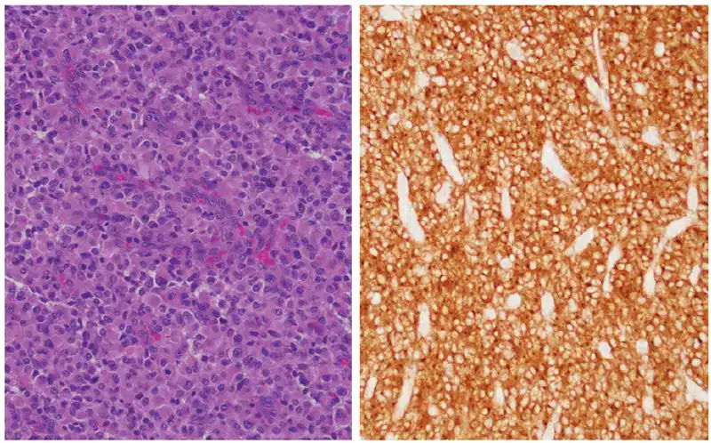
组织学检查显示弥漫浸润性胶质肿瘤,细胞密度高,核多形性明显,核分裂象多见(包括病理性核分裂),伴微血管增生、局灶假栅栏状坏死和广泛地图样坏死。肿瘤细胞 GFAP 和 SOX2 弥漫阳性,支持胶质分化。ATRX 表达保留。Ki-67 增殖指数最高达 30%–40%。IDH1 R132H 和 pan-TRK 免疫染色阴性。ALK(克隆 D5F3)免疫组化显示弥漫强阳性胞质着色(图 1)。值得注意的是,这一着色模式不同于已报道的携带 PRKAR2A::ALK 融合的上皮样纤维组织细胞瘤——后者 ALK 表达特征性地呈胞质点状模式,3而本例为弥漫胞质着色。
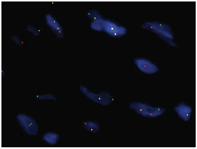
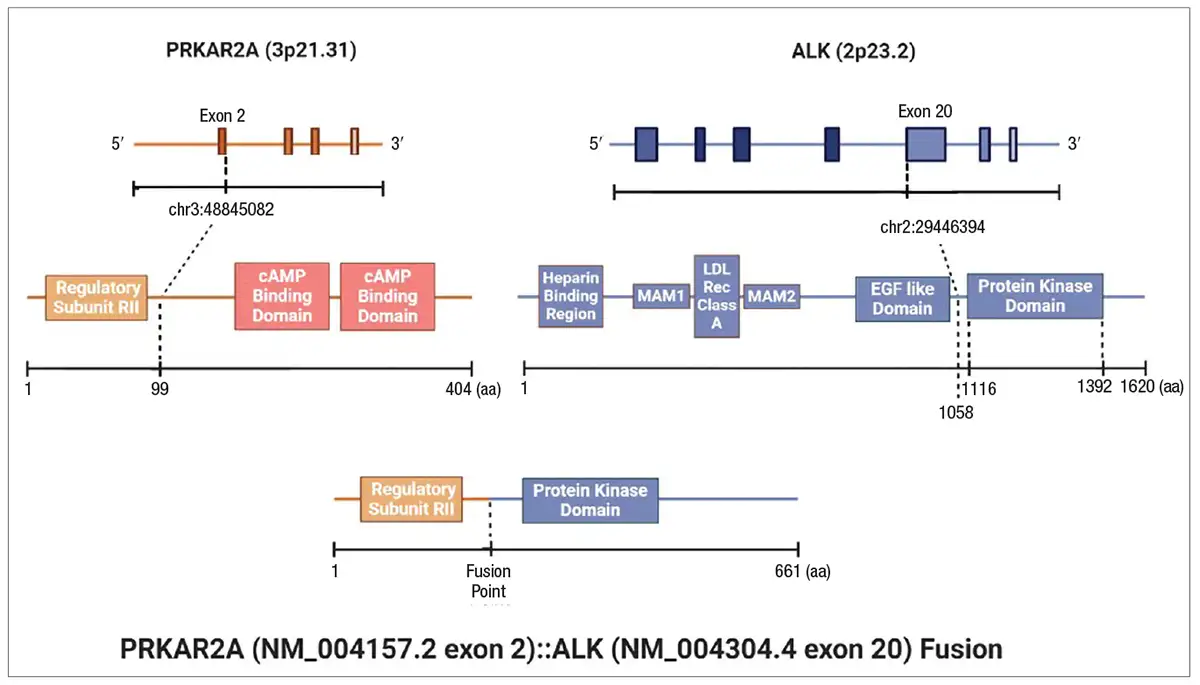
鉴于 ALK 弥漫免疫反应性,遂使用 ALK(2p23)分离探针进行荧光原位杂交,在 84.5% 的受检肿瘤细胞核中显示重排(图 2)。为进一步明确该重排,采用锚定多重 PCR 融合 panel 进行了基于 RNA 的二代测序。该分析鉴定出一个预测为框内的 PRKAR2A(2 号外显子)::ALK(20 号外显子)融合(图 3)。
根据组织学特征及 RTK 融合的鉴定,该肿瘤被归类为婴儿型大脑半球胶质瘤。患儿接受了卡铂和依托泊苷化疗;然而,复查影像显示肿瘤进展。再次行手术减瘤,并对复发肿瘤进行了额外的分子检测。
采用 586 基因 panel 的全面 DNA 二代测序再次检出 PRKAR2A::ALK 融合,未检出其他致病性单核苷酸变异、插入或缺失、拷贝数改变或结构重排。肿瘤突变负荷低(1.14 个突变/Mb),肿瘤为微卫星稳定。全转录组测序证实了融合转录本的表达,并显示包括 TYK2、TNK2、EGFL7、MAPK11 和 MAPK12 在内的多种信号蛋白异常激活。这些基因在多种恶性肿瘤中与免疫调节、肿瘤进展、血管生成及致癌信号通路相关。4,5特别是,MAPK11 和 MAPK12 属于 p38 MAPK 家族,是更广泛的 MAPK 信号网络的组成部分;该网络在癌症中常失调,并已成为若干肿瘤的治疗靶点。6尽管这些转录改变在婴儿型大脑半球胶质瘤中的临床意义尚不确定,但其激活提示可能存在下游 MAPK 通路的参与,并为今后探索靶向治疗和免疫调节方法提供了潜在方向。总之,这些发现进一步支持 PRKAR2A::ALK 融合是该肿瘤的主要致癌驱动因素。
讨论。IHG 的定义是存在 RTK 融合,后者在一个基因组本身稳定的肿瘤中充当主导致癌事件。其中,ALK 重排是一个已明确的亚组;然而,IHG 中 ALK 融合伙伴的多样性仍在不断扩展。PRKAR2A::ALK 融合是一种罕见的 ALK 重排,此前曾见于上皮样纤维组织细胞瘤,7但据我们所知,尚未在婴儿型大脑半球胶质瘤中报道。
该融合由编码蛋白激酶 A 调节亚基的 PRKAR2A 的 1–2 号外显子,与编码酪氨酸激酶结构域的 ALK 的 20 号外显子框内融合而成。尽管这一特定融合的功能后果尚未经实验证实,但其结构与其他激活性 ALK 融合类似,预计会导致激酶的组成性激活。8在原发与复发肿瘤中均检出同一融合、且无其他致病性改变,支持其作为早期驱动事件的作用。
本例凸显了多模态分子诊断方法的价值。ALK 弥漫免疫反应性作为初筛工具,FISH 证实基因重排,基于 RNA 的二代测序则精确鉴定出融合伙伴和断点。随后的 DNA 水平和转录组水平分析提供了正交验证,并显示了 IHG 的标志性特征——基因组复杂性低。这种分层检测在儿童中枢神经系统肿瘤中尤为重要,因为准确的分子分类直接关系到诊断和治疗考量。
从治疗角度看,ALK 融合的鉴定使靶向激酶抑制成为可能。然而,ALK 抑制剂在婴儿中的临床经验仍然有限,与年龄相关的监管限制可能延迟治疗的开始。在 IHG 中,常规化疗似乎基本无效,且常有复发报道。9-11因此,为改善预后,可能有必要在 1 岁前考虑 ALK 抑制剂治疗。
本例患儿于 1 岁时开始口服单药劳拉替尼(一种 ALK 抑制剂)(每日 25 mg),12自开始用药以来情况良好。尽管如此,对可干预融合的分子鉴定对于指导临床试验入组和为未来治疗决策提供依据仍然至关重要。
总之,这例携带新型 ALK 融合伙伴的 IHG 扩展了该病已知的分子谱。转录组分析显示五种参与细胞通讯的信号蛋白异常激活,其中四种在细胞内信号转导中起关键作用。从管理角度看,鉴于常规化疗疗效有限,本例凸显了考虑早期 ALK 抑制剂治疗的重要性。它也强调了需要登记数据和前瞻性研究,以更好地明确婴儿患者的临床结局并优化管理策略。
Yilmaz 医生和 Zaheer 医生撰写本报告时为分子遗传病理学专科培训医师;Soderquist 医生为病理与细胞生物学副教授;Mansukhani 医生为病理与细胞生物学教授;Hsiao 医生为副教授,Smithgall 医生为病理与细胞生物学助理教授——均任职于哥伦比亚大学内外科医学院病理与细胞生物学系。Yilmaz 医生现为西奈山伊坎医学院病理学助理教授。Zaheer 医生现为阿肯色大学医学科学院血液病理学及分子遗传病理学分部助理教授。
- A boy whose head circumference grew from 36 to 44 cm in one month from age 2 months; imaging showed an 11.8 × 8.9 × 7.4 cm heterogeneous left supratentorial mass with midline shift and obstructive hydrocephalus; emergent hemicraniectomy with partial debulking.
- Histology: high-grade diffusely infiltrating glioma with high cellularity, pleomorphism, atypical mitoses, microvascular proliferation, pseudopalisading and geographic necrosis; GFAP/SOX2 diffuse, ATRX retained, Ki-67 up to 30–40%, IDH1 R132H and pan-TRK negative.
- Layered work-up: diffuse strong cytoplasmic ALK (D5F3) — unlike the dot-like pattern of the same fusion in epithelioid fibrous histiocytoma → ALK break-apart FISH in 84.5% of nuclei → anchored multiplex PCR RNA panel: PRKAR2A (exon 2)::ALK (exon 20) → 586-gene DNA panel on the recurrence: no other pathogenic alterations, TMB 1.14/Mb, MSS.
- Progression after carboplatin/etoposide and repeat debulking; single-agent lorlatinib (25 mg daily) from age one, doing well. The authors argue for considering ALK inhibition before one year of age given limited chemotherapy efficacy.
Scope: full public text from the CAP TODAY website — AMP series note, authors, body (case presentation, discussion), Figures 1–3 with captions and author affiliations. The 12 references are not included; superscripts are the original citation numbers.
CAP TODAY and the Association for Molecular Pathology have teamed up to bring molecular case reports to CAP TODAY readers. AMP members write the reports using clinical cases from their own practices that show molecular testing’s important role in diagnosis, prognosis, and treatment. The following report comes from Columbia University College of Physicians and Surgeons. If you would like to submit a case report, please send an email to the AMP at amp@amp.org. For more information about the AMP and all previously published case reports, visit www.amp.org.
Mine Yilmaz, MD; Sidra Zaheer, MD; Craig Soderquist, MD; Mahesh Mansukhani, MD; Susan Hsiao, MD, PhD; Marie Smithgall, MD
October 2026—Infant-type hemispheric glioma (IHG) is a molecularly defined pediatric high-grade glioma first recognized in the 2021 World Health Organization classification of tumors of the central nervous system.1 These tumors arise predominantly within the first year of life and are characterized by large hemispheric masses and frequent receptor tyrosine kinase (RTK) gene fusions, most commonly involving ALK, NTRK1/2/3, ROS1, or MET.2 Unlike other high-grade gliomas, IHGs typically demonstrate low genomic complexity, with oncogenesis driven by a single dominant fusion event.
Accurate classification of IHG relies on the integration of histopathologic findings with molecular testing, particularly fusion detection. Identification of specific RTK fusions has diagnostic, prognostic, and potential therapeutic implications. Here, we report a case of IHG harboring a novel PRKAR2A::ALK fusion, identified using complementary molecular approaches. Whole transcriptome sequencing confirmed expression of the fusion transcript as well as aberrant activation of multiple signaling proteins, including TYK2, TNK2, EGFL7, MAPK11, and MAPK12.
Case presentation. A nine-month-old boy initially presented at two months of age with a rapidly increasing head circumference, enlarging from 36 cm to 44 cm over one month. Neuroimaging revealed a large heterogeneous mass measuring 11.8 × 8.9 × 7.4 cm involving the left supratentorial cerebral hemisphere, with marked mass effect, midline shift, and obstructive hydrocephalus. The patient underwent emergent left hemicraniectomy with partial tumor debulking and ventriculoperitoneal shunt placement.
Histologic examination demonstrated a diffusely infiltrating glial neoplasm with high cellularity, marked nuclear pleomorphism, frequent mitotic figures including atypical forms, microvascular proliferation, focal pseudopalisading necrosis, and extensive geographic necrosis. Tumor cells showed diffuse positivity for GFAP and SOX2, supporting glial differentiation. ATRX expression was retained. The Ki-67 proliferation index reached up to 30 to 40 percent. IDH1 R132H and pan-TRK immunostains were negative. ALK (clone D5F3) immunohistochemistry demonstrated diffuse strong cytoplasmic positivity (Fig. 1). Notably, this staining pattern differs from that reported in epithelioid fibrous histiocytoma harboring PRKAR2A::ALK fusion, in which ALK expression characteristically shows a dot-like cytoplasmic pattern,3 whereas our case exhibited diffuse cytoplasmic staining.
Given the diffuse ALK immunoreactivity, fluorescence in situ hybridization using an ALK (2p23) break-apart probe was performed and demonstrated rearrangement in 84.5 percent of analyzed tumor nuclei (Fig. 2). To further characterize the rearrangement, RNA-based next-generation sequencing was performed using an anchored multiplex PCR fusion panel. This analysis identified a predicted in-frame PRKAR2A (exon 2)::ALK (exon 20) fusion (Fig. 3).
Based on the histologic features and identification of an RTK fusion, the tumor was classified as infant-type hemispheric glioma. The patient received carboplatin and etoposide chemotherapy; however, interval imaging demonstrated tumor progression. Repeat surgical debulking was performed, and additional molecular studies were undertaken on the recurrent tumor.
Comprehensive DNA-based NGS using a 586-gene panel again identified the PRKAR2A::ALK fusion, with no additional pathogenic single nucleotide variants, insertions or deletions, copy number alterations, or structural rearrangements detected. Tumor mutation burden was low (1.14 mutations/Mb), and the tumor was microsatellite stable. Whole transcriptome sequencing confirmed expression of the fusion transcript and demonstrated aberrant activation of multiple signaling proteins, including TYK2, TNK2, EGFL7, MAPK11, and MAPK12. These genes have been implicated in immune regulation, tumor progression, angiogenesis, and oncogenic signaling pathways across a variety of malignancies.4,5 In particular, MAPK11 and MAPK12 belong to the p38 MAPK family, a component of the broader MAPK signaling network that is frequently dysregulated in cancer and has emerged as a therapeutic target in several neoplasms.6 While the clinical significance of these transcriptional alterations in infant-type hemispheric glioma remains uncertain, their activation raises the possibility of downstream MAPK pathway involvement and suggests potential avenues for future investigation into targeted therapies and immunomodulatory approaches. Overall, these findings further support the role of the PRKAR2A::ALK fusion as the primary oncogenic driver in this tumor.
Discussion. IHG is defined by the presence of RTK fusions that serve as dominant oncogenic events in an otherwise genomically stable tumor. Among these, ALK rearrangements represent a well-established subset; however, the diversity of ALK fusion partners in IHG continues to expand. The PRKAR2A::ALK fusion is a rare ALK rearrangement previously described in epithelioid fibrous histiocytoma,7 but, to our knowledge, it has not been reported in infant-type hemispheric glioma.
The fusion involves exons 1–2 of PRKAR2A, encoding a regulatory subunit of protein kinase A, fused in-frame to exon 20 of ALK, which encodes the tyrosine kinase domain. Although the functional consequences of this specific fusion have not been experimentally characterized, its structure is analogous to other activating ALK fusions and is predicted to result in constitutive kinase activation.8 Detection of the same fusion in both the primary and recurrent tumor, in the absence of additional pathogenic alterations, supports its role as an early driver event.
This case highlights the value of a multimodal molecular diagnostic approach. Diffuse ALK immunoreactivity served as an initial screening tool, while FISH confirmed gene rearrangement and the RNA-based NGS precisely identified the fusion partner and breakpoint. Subsequent DNA-based and transcriptome-level analyses provided orthogonal confirmation and demonstrated low genomic complexity, a hallmark of IHG. Such layered testing is particularly important in pediatric CNS tumors, where accurate molecular classification directly informs diagnosis and therapeutic consideration.
From a therapeutic standpoint, identification of ALK fusions raises the possibility of targeted kinase inhibition. However, clinical experience with ALK inhibitors in infants remains limited, and age-related regulatory constraints may delay treatment initiation. In IHGs, conventional chemotherapy appears largely ineffective with frequent recurrence reported.9-11 Therefore, consideration of ALK inhibitor therapy prior to one year of age may be warranted to improve outcomes.
In this case, the patient was started on single-agent oral lorlatinib (an ALK inhibitor) (25 mg daily)12 at age one year and has been doing well since initiation. Nevertheless, molecular identification of actionable fusions remains essential for guiding clinical trial enrollment and informing future therapeutic decision-making.
In conclusion, this case of IHG with a novel ALK fusion partner expands the known molecular spectrum of the disease. Transcriptome analysis demonstrated aberrant activation of five signaling proteins involved in cellular communication, four of which play key roles in intracellular signal transduction. From a management perspective, this case highlights the importance of considering early ALK inhibitor therapy given the limited efficacy of conventional chemotherapy. It also underscores the need for registry data and prospective studies to better define clinical outcomes and optimize management strategies in infants.
Dr. Yilmaz and Dr. Zaheer were fellows in molecular genetic pathology at the time of the writing of this report; Dr. Soderquist is associate professor of pathology and cell biology; Dr. Mansukhani is professor of pathology and cell biology; and Dr. Hsiao is associate professor and Dr. Smithgall is assistant professor of pathology and cell biology—all in the Department of Pathology and Cell Biology, Columbia University College of Physicians and Surgeons. Dr. Yilmaz is now assistant professor of pathology, Icahn School of Medicine at Mount Sinai. Dr. Zaheer is assistant professor in the hematopathology and molecular genetic pathology divisions, University of Arkansas for Medical Sciences.