- CAP 联合 ASCP 与 ASCO,于 2026 年 10 月 21 日在 Arch Pathol Lab Med 提前在线发布 2016 版胃食管腺癌(GEA)HER2 指南的更新版:3 条强推荐 + 4 条重申的良好实践声明;2016 版仍然有效。
- 推荐 1:晚期 GEA 初诊时在质量最佳的标本(原发或转移)上检测 HER2,病理医生选取形态级别最低区域所在蜡块,形态异质时可选多个蜡块。推荐 2:以 IHC(必要时 ISH)为主,基因组检测(液体/组织)可同时或随后用于临床决策。推荐 3:HER2 阳性患者进展后拟后续治疗时,可在复发/再发灶复检 HER2,首选组织。
- 判读:Rüschoff-Hofmann 评分;手术标本以 ≥10% 肿瘤细胞为阈值,活检以 ≥5 个肿瘤细胞的细胞簇为单位、不计比例;仅 IHC 2+ 需 ISH。固定:冷缺血 ≤1 小时,10% 中性缓冲福尔马林 6~72 小时。
- 病例:例 1 食管活检中仅低分化成分 HER2 3+,腺样与黏液成分阴性——须评估所有形态并多块染色;例 2 胃活检 IHC 阳性而液体活检 ERBB2 未检出异常——基因组检测不能取代 IHC/ISH。
收录范围:CAP 官网指南页(背景、指南信息、常见问题)及其发布的「推荐意见汇总」「病理医生用流程图」「病例示例」三份工具文件全文与图(图注与表格中英对照)。Arch Pathol Lab Med 指南正文页面对本站抓取返回验证壳页,未采集正文、方法学补充与教学幻灯。
背景
2026 版指南「胃食管腺癌 HER2 检测与临床决策(更新版)」针对肿瘤异质性、标本质量参差及检测手段演进等问题,反映了 2016 年以来检测领域的进展。
指南含 3 条强推荐及 4 条重申为「良好实践声明」的条目,已作为提前在线版发表于 Archives of Pathology & Laboratory Medicine。
| 指南状态 | 现行 |
|---|---|
| 首次发表 | 2016 年 12 月 |
| 更新版提前在线发表 | 2026 年 10 月 21 日 |
| 合作方 | 美国临床病理学会(ASCP)与美国临床肿瘤学会(ASCO) |
推荐意见与推荐强度
| 指南条目 | 推荐强度 |
|---|---|
| 1. 晚期胃食管腺癌(GEA)初诊时,主治临床医生或病理医生应申请在质量最佳的肿瘤标本(原发灶或转移灶)上进行人表皮生长因子受体 2(HER2)检测。病理医生应在活检、切除或细针穿刺(FNA)标本中选取肿瘤形态级别最低区域所在的蜡块。若存在不同的形态学模式,可选取一个以上蜡块。 注:GEA 中用于 HER2 检测的「质量最佳肿瘤」指:肿瘤细胞比例最高,坏死、人工假象或治疗反应最少。 | 强推荐 |
| 2. 对晚期 GEA 患者标本,病理医生应以免疫组化(IHC)为 HER2 状态的主要评估手段,必要时再行原位杂交(ISH)。基因组检测(液体和/或组织)可同时或随后用于临床决策。 | 强推荐 |
| 3. 对疾病进展后拟接受后续治疗的晚期 HER2 阳性 GEA 患者,可在复发/再发肿瘤样本上重新评估 HER2。首选组织;若无法获得组织或不可行,可行液体检测。 | 强推荐 |
良好实践声明
- 实验室/病理医生须注明检测所用抗体与探针,并确保针对 GEA 标本的 HER2 免疫组化(IHC)与原位杂交(ISH)检测均经过恰当验证。
- 评估 GEA 的 HER2 状态时,实验室/病理医生应先进行/申请 IHC 检测,IHC 结果为 2+(不确定)时再行 ISH 确认。HER2 IHC 阳性(3+)或阴性(0 或 1+)无需进一步 ISH 检测。
- 病理医生应采用 Rüschoff-Hofmann 法对 GEA 的 HER2 IHC 评分,需要时按标准 ISH 判读标准判读。
- 实验室应将 GEA HER2 检测方法纳入整体实验室质量改进计划,并遵守所在地监管机构及认可组织的要求。
病理医生用流程图
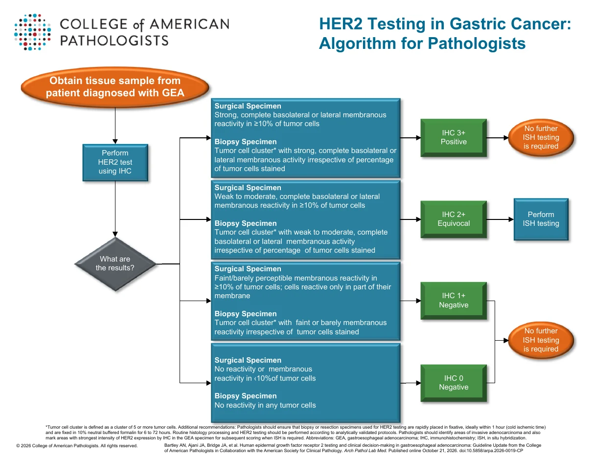
| IHC 结果 | 手术标本 | 活检标本 | 后续处理 |
|---|---|---|---|
| IHC 3+(阳性) | ≥10% 肿瘤细胞呈强、完整的基底侧或侧面膜着色 | 肿瘤细胞簇*呈强、完整的基底侧或侧面膜着色,不论着色肿瘤细胞所占比例 | 无需进一步 ISH 检测 |
| IHC 2+(不确定) | ≥10% 肿瘤细胞呈弱至中等、完整的基底侧或侧面膜着色 | 肿瘤细胞簇*呈弱至中等、完整的基底侧或侧面膜着色,不论着色肿瘤细胞所占比例 | 进行 ISH 检测 |
| IHC 1+(阴性) | ≥10% 肿瘤细胞呈微弱/勉强可见的膜着色;细胞仅部分胞膜着色 | 肿瘤细胞簇*呈微弱或勉强可见的膜着色,不论着色肿瘤细胞所占比例 | 无需进一步 ISH 检测 |
| IHC 0(阴性) | 无着色,或 <10% 肿瘤细胞膜着色 | 所有肿瘤细胞均无着色 | 无需进一步 ISH 检测 |
*肿瘤细胞簇定义为 5 个或以上肿瘤细胞组成的细胞簇。附加推荐:病理医生应确保用于 HER2 检测的活检或切除标本迅速置入固定液,理想情况下在 1 小时内(冷缺血时间),并以 10% 中性缓冲福尔马林固定 6~72 小时。常规组织学处理与 HER2 检测应按经分析验证的方案进行。病理医生应标出浸润性腺癌区域,并在 GEA 标本中标出 HER2 IHC 着色强度最强的区域,以便需要 ISH 时据此计数。缩写:GEA,胃食管腺癌;IHC,免疫组化;ISH,原位杂交。
常见问题
为何更新本指南?
CAP 会定期重新评估已发表的循证指南,以判断新证据或临床实践演变是否需要修订指导意见。自 2016 年以来,胃食管腺癌(GEA)的 HER2 检测受到治疗选择变化及替代标本类型使用增多的影响。分子/基因组检测手段的扩展也带来了新的实施问题,因而需要修订推荐。
2016 版指南是否仍然有效?
是。2016 版指南仍然有效,其中若干关键条目已被重申为良好实践声明。本次更新新增推荐并覆盖实践中不断演变的领域,但并不取代 2016 年确立的基础性指导。
何时适合做基因组检测?
基于组织和/或液体活检的基因组检测可同时或随后用于支持临床决策。但证据表明,IHC 加(必要时)反射性 ISH 仍是确定 HER2 状态的首选方法,因为与 IHC/ISH 相比,基因组方法的一致性不稳定、敏感性有限。
其他标本类型(包括转移灶组织和细胞学样本)的 HER2 结果与原发灶活检是否一致?
通常一致。活检与切除标本一致性高(一般 >90%),但肿瘤异质性偶可导致不一致。转移灶与原发瘤一般也一致(通常 >80%),但治疗后进展时可发生转换(HER2 阴性↔阳性)。其他组织不可得时可使用细胞学细胞块(如积液),但其一致性数据有限,判读也可能更困难。
疾病进展后是否应重复 HER2 检测?
是。在晚期 GEA 中,HER2 状态可在治疗后改变(包括丢失 HER2 阳性),这可能影响 HER2 靶向治疗是否仍然适用。这一点较 2016 版指南有所改变——2016 版主要建议对 HER2 状态未知或既往阴性的患者在进展时复检。本次更新中,既往 HER2 阳性病例在开始二线治疗前可重复 HER2 检测。首选基于组织的检测;无法再次取组织时可用液体活检。
有些推荐是强推荐,但证据级别有限或较低。专家组如何确定推荐强度?
CAP 专家组采用 GRADE(推荐分级的评估、制定与评价)证据到决策框架,这是一种结构化、标准化的临床推荐制定方法。证据的确定性(级别)是重要因素,但并非决定推荐强度的唯一因素。专家组还会权衡利弊,以及价值观与偏好、资源利用、健康公平、可及性和可行性等关键实施因素,并结合专家判断得出最终推荐强度。
病例示例
示例病例 1
72 岁男性,有 Barrett 食管病史,接受常规监测(上次内镜为 2018 年;每两年一次的监测因 COVID-19 疫情中断)。2025 年夏季因进行性干食吞咽困难及肩痛就诊。上消化道内镜联合超声内镜发现食管远端肿块,活检证实为腺癌。随后 PET/CT 显示符合转移性疾病的表现,包括多发高代谢肺结节、多发高代谢骨病灶及一个高代谢左背阔肌结节。
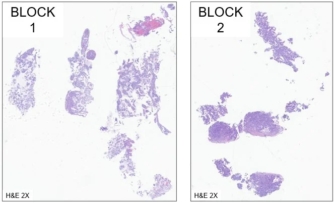
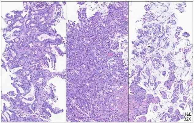
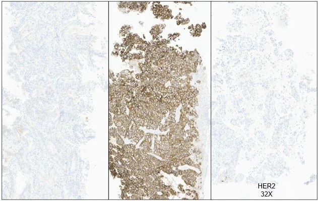
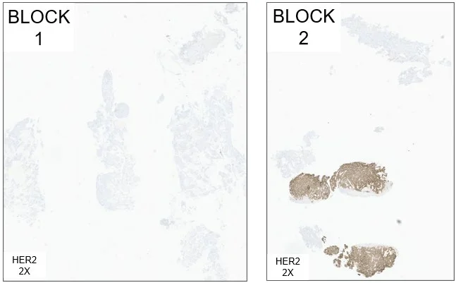
病例 1 讨论
本例强调两条重要原则。第一,所取肿瘤活检碎片的数量很重要。胃食管腺癌以异质性著称,多项研究表明每次取材至少获得 3~5 块活检碎片,可使活检与相应切除标本的 HER2 IHC 一致性超过 90%。部分研究提示 6~8 块活检碎片可能最理想。这与推荐 1 一致,强调充分的肿瘤取材以支持胃食管腺癌基于组织的准确 HER2 评估。
本例的第二个要点是:存在形态异质性时,须评估所有肿瘤形态。尽管 HER2 阳性更常见于低级别或高分化肿瘤成分以及肠型形态,本例表明确有例外。因此,与推荐 1 一致,对形态异质的 GEA 进行检查时,病理医生应考虑对一个以上蜡块做 HER2 染色,以尽量降低假阴性风险。
示例病例 2
73 岁女性,数月来间歇性腹痛伴恶心呕吐。腹部、盆腔及胸部 CT 显示胃后壁与胰体之间一 3 cm 边界不清肿块,以及累及骶骨和双侧髂骨的骨转移。其他所见包括胃肝韧带淋巴结肿大及纵隔淋巴结肿大。上消化道内镜见累及胃贲门及胃体的巨大浸润性、非环周性肿块,病灶中心位于胃食管交界远侧 2 cm 以上。
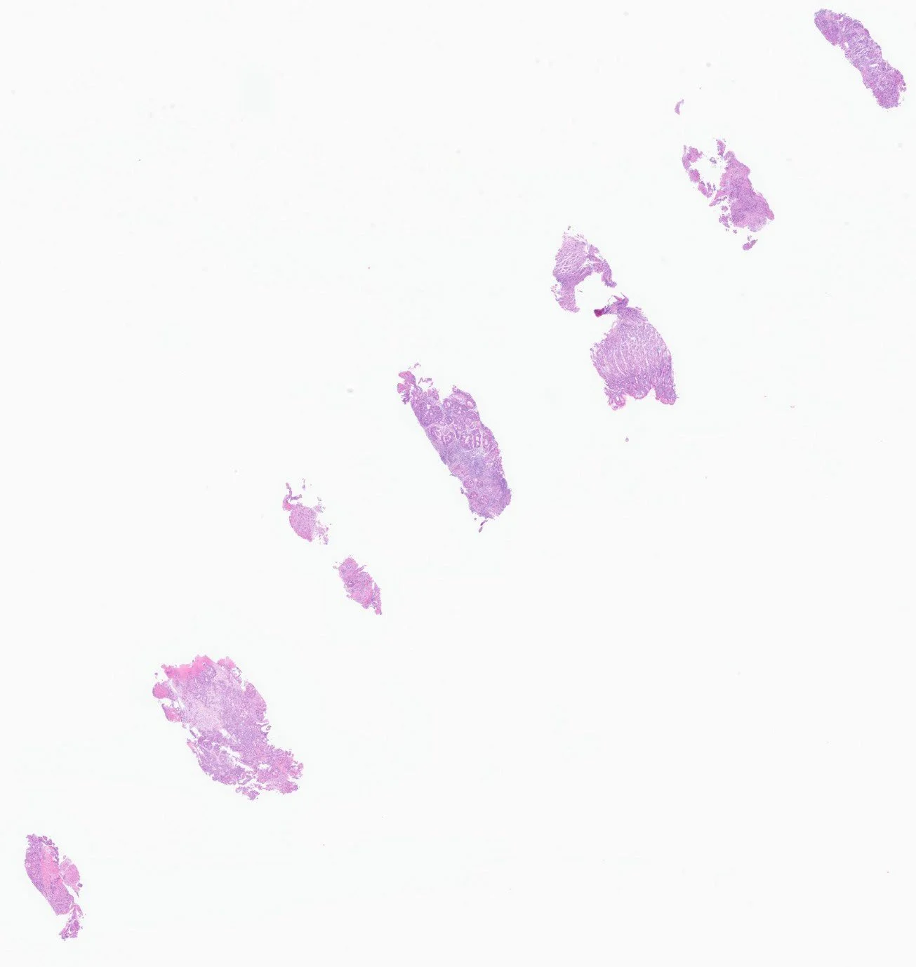
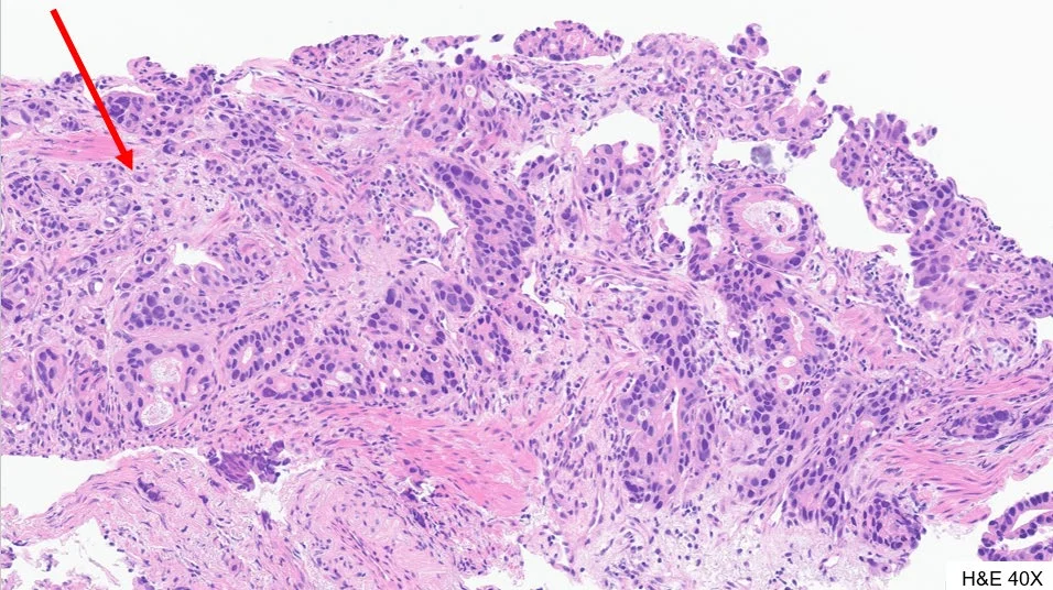
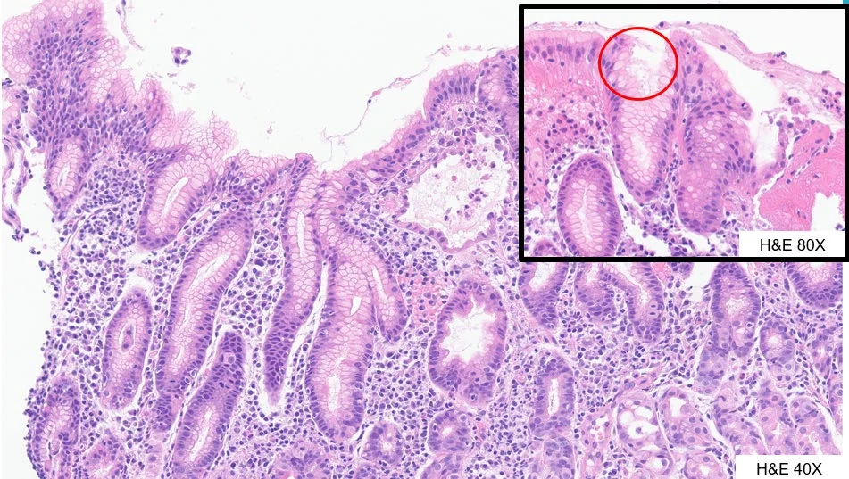
病例 2 讨论
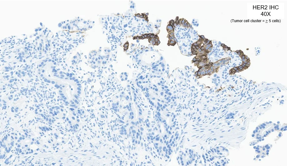
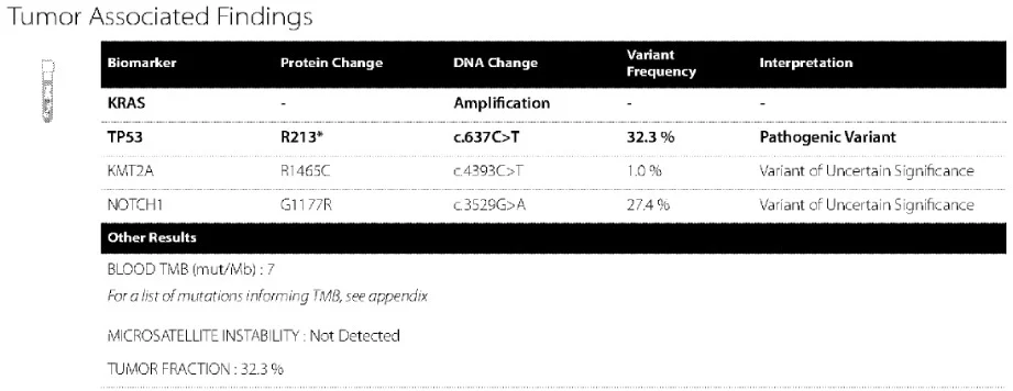
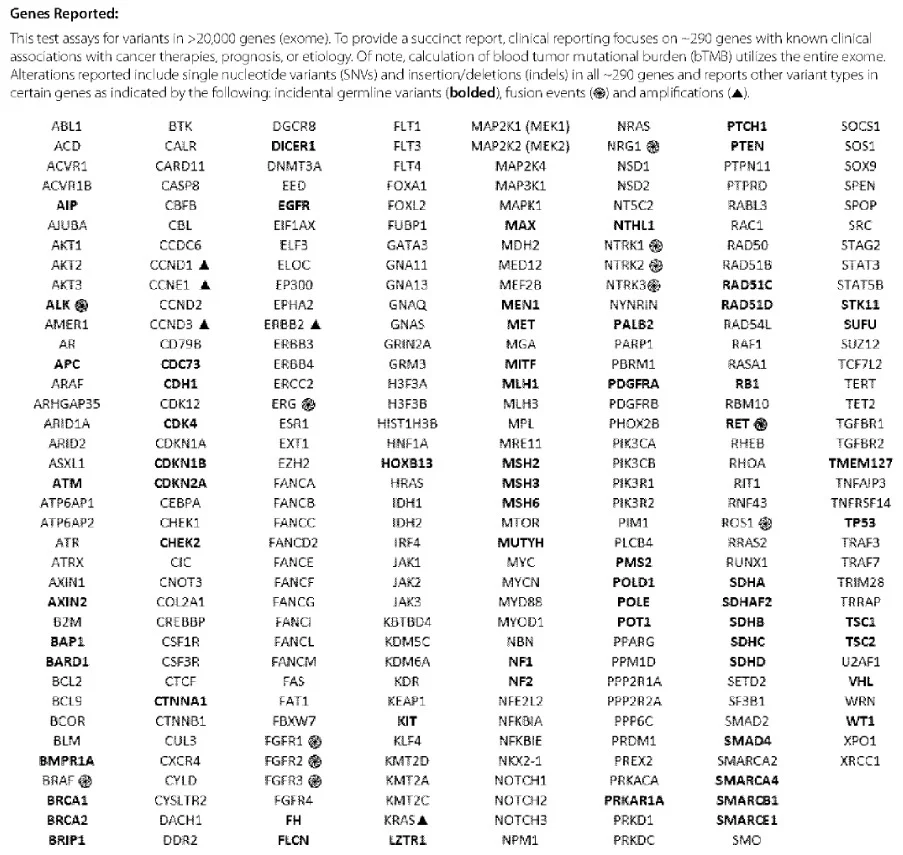
本例突出了晚期 GEA 中 HER2 评估检测方法选择的重要考量。尽管肿瘤组织 IHC 明确 HER2 阳性,组织活检后不久进行的液体基因组检测却未检出 ERBB2 异常,提示基因组方法在该情境下检出 HER2 阳性疾病的敏感性不稳定。正如本例所示,即使 IHC 显示 HER2 过表达,肿瘤异质性及检测靶点差异也可导致仅用基因组检测时出现 HER2 假阴性。这与更广泛的文献一致:与 IHC 加反射性 ISH 相比,组织及液体基因组检测 HER2 的敏感性和一致性差异很大。
因此,本例支持推荐 2,即以 IHC/ISH 作为晚期 GEA 患者 HER2 评估的主要方法。基因组检测可提供补充性分子信息,并可同时或随后用于指导临床决策,但不应取代 IHC/ISH 进行初始 HER2 评估。此外,IHC 与 ISH 在各类实践场所更易获得,这进一步巩固了其作为可靠、公平的 HER2 检测手段的地位,有助于确保患者一致地获得 HER2 靶向治疗。
缩写:COVID-19,2019 冠状病毒病;CT,计算机断层扫描;GEA,胃食管腺癌;HER2,人表皮生长因子受体 2;IHC,免疫组化;ISH,原位杂交;PET/CT,正电子发射断层扫描/计算机断层扫描。
- The CAP, with ASCP and ASCO, released online on October 21, 2026 in Arch Pathol Lab Med an update of the 2016 HER2 guideline for gastroesophageal adenocarcinoma (GEA): 3 strong recommendations plus 4 reaffirmed good practice statements; the 2016 guideline remains valid.
- Rec 1: at initial diagnosis of advanced GEA, test the highest quality specimen (primary or metastasis), choosing the block with the lowest grade morphology, and more than one block when patterns differ. Rec 2: IHC with ISH as needed is primary; genomic testing (liquid/solid) may be used concurrently or subsequently. Rec 3: in HER2-positive disease progressing toward subsequent therapy, HER2 may be reassessed on relapsed tumor, tissue preferred.
- Scoring: Rüschoff-Hofmann; ≥10% tumor cells for surgical specimens, clusters of ≥5 tumor cells irrespective of percentage for biopsies; only IHC 2+ needs ISH. Fixation: cold ischemia ≤1 hour, 10% NBF for 6–72 hours.
- Cases: in case 1 only the poorly differentiated component was HER2 3+ while glandular and mucinous components were negative; in case 2 a gastric biopsy was IHC-positive while liquid biopsy found no ERBB2 abnormality.
Scope: the CAP guideline web page (background, guideline information, FAQ) and the full text and images of three CAP tools — Summary of Recommendations, Algorithm for Pathologists and Case Studies. The Arch Pathol Lab Med article page returned a verification shell to our fetcher, so the guideline paper, methodology supplement and teaching presentation were not collected.
Background
The 2026 guideline, “HER2 Testing and Clinical Decision Making in Gastroesophageal Adenocarcinoma Update” addresses tumor heterogeneity, variable specimen quality, and evolving testing approaches, reflecting advances in testing since 2016.
Three strong recommendations and four statements reaffirmed as good practice statements are available for review as an early online release in the Archives of Pathology & Laboratory Medicine.
| Guideline status | Active |
|---|---|
| Originally published | December 2016 |
| Update published online ahead of print | October 21, 2026 |
| Collaborators | American Society for Clinical Pathology and American Society of Clinical Oncology |
Statements and Strengths of Recommendations
| Guideline Statement | Strength of Recommendation |
|---|---|
| 1. At initial diagnosis of advanced gastroesophageal adenocarcinoma (GEA), treating clinicians or pathologists should request human epidermal growth factor receptor 2 (HER2) testing on the highest quality tumor specimen (primary or metastasis). Pathologists should select the tissue block with the areas of lowest grade tumor morphology in biopsy, resection, or fine-needle aspiration (FNA) specimens. More than one tissue block may be selected if different morphologic patterns are present. Note: Highest quality tumor for HER2 testing in GEA: highest neoplastic cellularity, minimal necrosis, artifact or treatment effect. | Strong Recommendation |
| 2. In specimens from patients with advanced GEA, pathologists should use immunohistochemistry (IHC) with subsequent in situ hybridization (ISH) as needed for the primary assessment of HER2 status. Genomic testing (liquid and/or solid) may be used concurrently or subsequently for clinical decision making. | Strong Recommendation |
| 3. In patients with advanced HER2-positive GEA being considered for subsequent therapy after disease progression, HER2 assessment may be performed on relapsed/recurrent tumor sample. Tissue is preferred but if not available/feasible, liquid testing may be performed. | Strong Recommendation |
Good Practice Statements
- Laboratories/pathologists must specify the antibodies and probes used for the test and ensure that assays are appropriately validated for HER2 immunohistochemistry (IHC) and in-situ hybridization (ISH) on GEA specimens.
- When GEA HER2 status is being evaluated, laboratories/pathologists should perform/order IHC testing first, followed by ISH confirmation when IHC result is 2+ (equivocal). Positive (3+) or negative (0 or 1+) HER2 IHC results do not require further ISH testing.
- Pathologists should use the Rüschoff-Hoffmann method for HER2 IHC scoring in GEA and apply standard ISH interpretation criteria when indicated.
- Laboratories should incorporate GEA HER2 testing methods into their overall laboratory quality improvement program, following the requirements of applicable local regulatory bodies and accreditation organizations.
Algorithm for Pathologists
| IHC result | Surgical Specimen | Biopsy Specimen | Next step |
|---|---|---|---|
| IHC 3+ (Positive) | Strong, complete basolateral or lateral membranous reactivity in ≥10% of tumor cells | Tumor cell cluster* with strong, complete basolateral or lateral membranous activity irrespective of percentage of tumor cells stained | No further ISH testing is required |
| IHC 2+ (Equivocal) | Weak to moderate, complete basolateral or lateral membranous reactivity in ≥10% of tumor cells | Tumor cell cluster* with weak to moderate, complete basolateral or lateral membranous activity irrespective of percentage of tumor cells stained | Perform ISH testing |
| IHC 1+ (Negative) | Faint/barely perceptible membranous reactivity in ≥10% of tumor cells; cells reactive only in part of their membrane | Tumor cell cluster* with faint or barely membranous reactivity irrespective of tumor cells stained | No further ISH testing is required |
| IHC 0 (Negative) | No reactivity or membranous reactivity in <10% of tumor cells | No reactivity in any tumor cells | No further ISH testing is required |
*Tumor cell cluster is defined as a cluster of 5 or more tumor cells. Additional recommendations: Pathologists should ensure that biopsy or resection specimens used for HER2 testing are rapidly placed in fixative, ideally within 1 hour (cold ischemic time) and are fixed in 10% neutral buffered formalin for 6 to 72 hours. Routine histology processing and HER2 testing should be performed according to analytically validated protocols. Pathologists should identify areas of invasive adenocarcinoma and also mark areas with strongest intensity of HER2 expression by IHC in the GEA specimen for subsequent scoring when ISH is required. Abbreviations: GEA, gastroesophageal adenocarcinoma; IHC, immunohistochemistry; ISH, in situ hybridization.
Frequently Asked Questions
Why was this guideline updated?
The CAP periodically reevaluates published evidence-based guidelines to determine whether newer evidence or evolving clinical practice warrants revised guidance. Since 2016, HER2 testing in gastroesophageal adenocarcinoma (GEA) has been influenced by changes in treatment options and increased use of alternative specimen types. Expanded molecular/genomic testing approaches have also raised new implementation questions, supporting the need for revised recommendations.
Is the 2016 guideline still valid?
Yes. The 2016 guideline remains valid, and several of its key statements were reaffirmed as Good Practice Statements. This update adds new recommendations and addresses evolving areas of practice, but it does not replace the foundational guidance established in 2016.
When is genomic testing appropriate?
Genomic testing using tissue and/or liquid biopsy may be used concurrently or subsequently to support clinical decision-making. However, evidence indicates that IHC with reflex ISH (when indicated) remains the preferred approach for establishing HER2 status because genomic methods show variable concordance and limited sensitivity compared with IHC/ISH.
Are HER2 results from other specimen types (including metastatic tissue and cytology samples) concordant with the primary biopsy?
Yes, usually. Biopsy and resection specimens show high concordance (typically >90%), although tumor heterogeneity can lead to occasional discordance. Metastatic lesions are also generally concordant with the primary tumor (usually >80%), but conversion (HER2 negative↔positive) can occur after treatment progression. Cytology cell blocks (eg, effusions) may be used when other tissue is unavailable, though concordance data are limited and interpretation may be more challenging.
Should HER2 testing be repeated after disease progression?
Yes. In advanced GEA, HER2 status can change after treatment (including loss of HER2 positivity), which may affect whether HER2-targeted therapy remains appropriate. This is a change from the 2016 guideline, which recommended repeat testing at progression mainly for patients with unknown or previously negative HER2 status. In this update, repeat HER2 testing may be performed in previously HER2-positive cases before starting second-line therapy. Tissue-based testing is preferred; liquid biopsy may be used when tissue-based resampling is not feasible.
There are recommendations that are strong but have a limited or low level of evidence. How did the panel arrive at the strength of recommendations?
CAP expert panels use the GRADE (Grading of Recommendations Assessment, Development and Evaluation) Evidence to Decision Framework, a structured and standardized approach for developing clinical recommendations. While the certainty (level) of evidence is an important factor, it is not the only determinant of recommendation strength. The panel also considers the balance of benefits and harms, along with key implementation factors such as values and preferences, resource use, health equity, accessibility, and feasibility, drawing on expert judgment to reach the final strength of recommendation.
Case Studies
Example Case 1
A 72-year-old man with a history of Barrett’s esophagus undergoing routine surveillance (last endoscopy in 2018; biennial surveillance disrupted by the COVID-19 pandemic) presented in summer 2025 with progressive dysphagia to dry foods and shoulder pain. Upper endoscopy with endoscopic ultrasound revealed a distal esophageal mass, and biopsy confirmed adenocarcinoma. Subsequent PET/CT demonstrated findings consistent with metastatic disease, including numerous hypermetabolic pulmonary nodules, multiple hypermetabolic osseous lesions, and a hypermetabolic left latissimus dorsi muscle nodule.
Case Study 1: Discussion
This example case reinforces two important principles. First, the number of tumor biopsy fragments obtained matters. Gastroesophageal adenocarcinoma is a notoriously heterogeneous tumor, and multiple studies have demonstrated that obtaining at least 3–5 biopsy fragments per sampling results in greater than 90% concordance in HER2 IHC between biopsy and corresponding resection specimens. Some studies suggest that 6–8 biopsy fragments may be ideal. This aligns with Recommendation #1 by emphasizing the importance of adequate tumor sampling to support accurate tissue-based HER2 assessment in gastroesophageal adenocarcinoma.
The second key takeaway from this case is the importance of evaluating all tumor morphologies when morphologic heterogeneity is present. Although HER2 positivity is more frequently identified in low-grade or well-differentiated tumor components and in intestinal-type morphologies, this case highlights that exceptions do occur. Accordingly, and consistent with Recommendation #1, pathologists should consider staining more than one tissue block for HER2 when working up morphologically heterogeneous GEAs to minimize the risk of false-negative results.
Example Case 2
A 73-year-old woman presented with several months of intermittent abdominal pain accompanied by nausea and vomiting. Cross-sectional imaging with CT of the abdomen, pelvis, and chest demonstrated a 3 cm ill-defined mass located between the posterior aspect of the stomach and the pancreatic body, as well as bony metastases involving the sacrum and bilateral iliac bones. Additional findings included an enlarged gastrohepatic ligament lymph node and mediastinal lymphadenopathy. Upper endoscopy revealed a large, infiltrative, non-circumferential mass involving the gastric cardia and body, with the epicenter of the lesion located more than 2 cm distal to the gastroesophageal junction.
Case Study 2: Discussion
This case highlights important considerations regarding test selection for HER2 assessment in advanced GEA. Despite clear HER2 positivity by IHC in tumor tissue, liquid-based genomic testing performed shortly after tissue biopsy did not identify an ERBB2 abnormality, underscoring the variable sensitivity of genomic approaches for detecting HER2-positive disease in this setting. As demonstrated in this case, tumor heterogeneity and differences in analytic targets can result in false-negative HER2 results when genomic testing is used alone, even when HER2 overexpression is present by IHC. These findings are consistent with the broader literature, which shows wide variability in the sensitivity and concordance of both tissue-based and liquid-based genomic testing for HER2 compared with IHC with reflex ISH.
Accordingly, this case supports Recommendation #2, which recommends IHC/ISH as the primary method for HER2 assessment in patients with advanced GEA. While genomic testing may provide complementary molecular information and can be used concurrently or subsequently to inform clinical decision-making, it should not replace IHC/ISH for initial HER2 evaluation. In addition, the broader availability of IHC and ISH across practice settings reinforces their role as a reliable and equitable approach to HER2 testing, helping ensure consistent access to HER2-directed therapy.
Abbreviations: COVID-19, coronavirus disease 2019; CT, computed tomography; GEA, gastroesophageal adenocarcinoma; HER2, human epidermal growth factor receptor 2; IHC, immunohistochemistry; ISH, in situ hybridization; PET/CT, positron emission tomography/computed tomography.