- WHO/ISUP 分级正式用于透明细胞肾细胞癌(CCRCC)与乳头状肾细胞癌(PRCC)约十年后,Paner、Amin、Cheng、Moch 等系统复习文献,逐项梳理分级要素与应用中的争议,并在数据足够处给出实践建议。
- 分级要素:最高级别并不总是主要级别;1 级与 2 级预后有差异、不应合并;伴多形核的多核瘤细胞定为 4 级,合体型多核细胞与退变性异型多核细胞不宜定为 4 级;少见的「低级别样」梭形细胞行为未明;肉瘤样 4 级比非肉瘤样 4 级行为更差,肉瘤样比例有预后意义但最佳切点(10%/20%/30%)未定。
- 应用层面:观察者间一致性优于 Fuhrman 核分级;穿刺活检分级与肾切除标本分级显著不一致(多源于分级异质性);多灶肿瘤中最高级别可见于非主瘤,报告方式尚无推荐。PRCC 分级的效用有研究支持也有反对;≤1.5 cm 乳头状肿瘤的分级问题单独讨论。
- 新方向:将坏死纳入分级的改良系统,以及基于结构/模式的分级与风险评估。附表 1–6(含 CCRCC 与 PRCC 分级更新汇总表)与图 1–5 中英对照。
收录范围:PMC 开放获取全文(CC BY 4.0)——摘要、正文全部章节、表 1–6 与图 1–5 及图注中英对照;作者贡献、利益冲突、知情同意、数据可用性声明与参考文献未收录。
摘要
世界卫生组织(WHO)/国际泌尿病理学会(ISUP)分级系统正式用于透明细胞肾细胞癌(CCRCC)和乳头状肾细胞癌(PRCC)已历时十年,期间出现了新的分级概念、创新方法以及一些问题。本文对有关 CCRCC 和 PRCC 的 WHO/ISUP 分级系统及其他分级方法的文献进行了全面综述。介绍以下方面的进展与问题:(1)WHO/ISUP 分级的验证研究;(2)分级要素,包括异质性、1 级与 2 级的比较、多核肿瘤细胞的类型、梭形细胞的“分级谱”、肉瘤样改变对 4 级 RCC 的影响、高级别成分的百分比(%)、肉瘤样改变的百分比以及横纹肌样改变与肉瘤样改变的比较;(3)分级应用,包括观察者一致性、穿刺活检分级的准确性(与肾切除标本分级的相关性)及多灶性 RCC 的分级;(4)乳头状肿瘤的分级,包括异质性及小肿瘤(≤1.5 cm)的分级;(5)新的分级或风险分类,包括将坏死纳入分级及基于形态模式或结构的分级。在可行或数据充分的情况下,为部分问题提供实践指导。本综述重点介绍 WHO/ISUP 分级应用及 CCRCC 和 PRCC 新分级方法的近期进展与争议。本综述可作为解决部分分级问题的最佳实践指南,并有助于发现我们在理解和应用 CCRCC 和 PRCC 分级方面的不足,为未来研究提供依据。
引言
分级是肾细胞癌(RCC)治疗管理和预后判断中的关键病理因素。1 , 2在过去 >70 年中,RCC 分级的方式和适用性经历了显著演变,主要归因于分级组成要素的调整,以及后来为适应日益增多的 RCC 亚型所作的改变。3 , 4直到过去十年,世界卫生组织(WHO)/国际泌尿病理学会(ISUP)分级系统才正式被采用,用于透明细胞肾细胞癌(CCRCC)和乳头状肾细胞癌(PRCC)。5 , 6WHO/ISUP 分级现已纳入主要泌尿外科指南及 CCRCC 和 PRCC 的风险分层,1 , 7国际癌症报告协作组织(ICCR)和美国病理医师学会(CAP)也将其视为肾活检(RB)及肾切除标本中 RCC 报告的核心要素。8 , 9
CCRCC 和 PRCC 以外的其他 RCC 亚型的分级,包括 WHO/ISUP 分级系统的应用,仍在研究中。近期,三位作者(G.P.P.、M.B.A. 和 H.M.)介绍了较少见 RCC 亚型分级的进展,指出了分级方面的不足,并就这些肿瘤的分级考量为外科病理医师提供指导。4该实践指导也已纳入 2022 年 WHO 蓝皮书。10本综述将聚焦于 WHO/ISUP 分级应用已得到确立的 CCRCC 和 PRCC,涵盖分级应用及分级组成要素的进展与问题。此外,还将探讨在分级中纳入其他要素(如坏死、结构)等创新方法。我们也希望本综述能揭示我们对 CCRCC 和 PRCC 分级认识中的不足,为未来研究提供依据。
WHO/ISUP 分级系统的演变
历史上的综合分级
自 1949 年及其后的数十年间,从 Griffiths 和 Thackray 的分级开始,陆续提出了多种 RCC 的 3 级和 4 级分级方案11以及其他多种方案。3 , 12 , 13 , 14 , 15 , 16 , 17 , 18 , 19这些历史上的分级系统大多评估不同组合的组织学特征,包括肿瘤结构、细胞核特征、核分裂象、细胞类型或坏死等。这些分级方法显示了 RCC 生存结局的差异。然而,当时应用这些分级系统时,尚未认识到目前的 RCC 亚型;而且部分方案的标准模糊,难以获得可重复的应用结果。
Fuhrman 核分级
1968 年,Meyers 等 20提出了最早的以细胞核特征为重点的分级方法之一,此后又相继出现了许多此类方法。21 , 22 , 23 , 24 , 25 , 26 , 27 , 28 , 29在这些基于细胞核的分级中,获得广泛认可的是 4 级分级,由 Fuhrman 等 24于 1982 年提出。在 Fuhrman 最初的研究中,RCC 被分为 Fuhrman 核分级 1 级(14%)、2 级(50%)、3 级(26%)和 4 级(10%),以所见恶性程度最高或级别最高的成分为分级依据,即使该成分仅为局灶性。Fuhrman 核分级 1 级肿瘤与合并的 Fuhrman 核分级 2–4 级肿瘤之间,转移率存在显著差异;5 年生存曲线也存在统计学差异,但仅限于将 Fuhrman 核分级 2 级和 3 级肿瘤合并为一组时。有趣的是,在一项观察者间一致性研究中,Syrjänen 和 Hjelt 的核分级22优于 Fuhrman 核分级、Skinner 等 21的核分级以及 Arner 等 14基于肿瘤结构和细胞类型的分级。Fuhrman 核分级的难点在于需要同时评估三种不同的细胞核变量(大小、形状及核仁显著程度),而这些变量的级别并不总是一致。这也是由这三种细胞核特征构成的 Fuhrman 核分级无法严格应用于部分 CCRCC 的原因之一,30并且很可能导致应用该系统时观察者间一致性仅为一般至中等。
WHO/ISUP 分级
在 2000 年代前后,分级方法在实践中逐渐简化,1–3 级主要着眼于核仁的显著程度。尽管当时已有部分病理专家依据核仁进行 Fuhrman 核分级,但直到同一研究团队发表了两项研究,才确立了这种以核仁为基础、区别于 Fuhrman 核分级的分级系统。31 , 322006 年,Sika‐Paotonu 等 31对 PRCC 的研究显示,局灶性核仁分级与生存独立相关,而 Fuhrman 核分级中细胞核大小和形状这两项指标则不然。2011 年,Delahunt 等 32对 CCRCC 核仁分级 1–3 级的研究显示,最差核仁分级能够区分 2 级与 3 级肿瘤的生存情况。这种用于 CCRCC 和 PRCC 的基于核仁的分级随后在 2012 年 ISUP 共识会议上被采纳,作为 ISUP 分级,6经略作修改(核仁嗜酸性)后,以 WHO/ISUP 分级的名称纳入 2016 年 WHO 蓝皮书,5并在其后的 2022 年 WHO 版本中保留(表 1)。10建议在显示核改变程度最显著的单个高倍视野内确定 WHO/ISUP 分级。WHO/ISUP 分级系统也已被认为不适用于嫌色性肾细胞癌(ChRCC)。6目前,对 ChRCC 正在推荐不同的替代分级方法(2 级、3 级和 4 级分级)。4WHO 采纳该分级用于 CCRCC 和 PRCC 已历时十年,关于 WHO/ISUP 分级的研究仍在不断开展。
| 级别 | 描述 |
|---|---|
| 1 级 | 在 ×400 放大倍数下,核仁缺如,或不明显且呈嗜碱性 |
| 2 级 | 在 ×400 放大倍数下可见嗜酸性核仁,但在 ×100 放大倍数下不明显 |
| 3 级 | 在 ×100 放大倍数下核仁明显且呈嗜酸性 |
| 4 级 | 癌细胞表现为极度核多形性、瘤巨细胞和/或任意数量的肉瘤样或横纹肌样分化 |
WHO/ISUP 分级系统的应用
透明细胞肾细胞癌
WHO/ISUP 分级提出后,已有多项研究探讨其在 CCRCC 中的应用。Dagher 等 30显示,1–4 级之间的无癌生存期(CFS)存在差异;该研究纳入 376 例 CCRCC,平均随访 35 个月,这种差异在多变量分析中具有统计学意义。Delahunt 等 33对来自 Mayo Clinic 登记数据库的 3,017 例 CCRCC 队列进行研究,通过回归模型显示,在 10 年随访中各级别之间的肿瘤特异性生存期(CSS)均存在差异。Leibovich 等 7同样对来自 Mayo Clinic 登记数据库的大型 CCRCC 队列进行研究,多变量分析显示 WHO/ISUP 分级类别与疾病进展或 RCC 死亡显著相关,并更新了 CCRCC 的 Leibovich 预后模型,将 WHO/ISUP 分级纳入其中。其他研究在将 1/2 级和 3/4 级分别合并后,多变量分析显示分级与结局显著相关。34 , 35然而,Khor 等 36在 WHO 标准提出之前的一项研究中,采用 ISUP 分级(未考虑核仁嗜酸性)评估了 842 例连续 CCRCC,结果显示仅 4 级与总生存期(OS)显著相关,而 3 级与合并的 1/2 级相比则无显著相关性。总体而言,大多数研究证实了 WHO/ISUP 分级对 CCRCC 的预后价值。
乳头状肾细胞癌
WHO/ISUP 分级被采纳后,也开展了多项使用该系统对 PRCC 进行分级的研究。上述来自 Mayo Clinic 的 Leibovich 等 7的研究还对一个包含 607 例 PRCC 的队列进行了分级,多变量分析显示,3 层级 WHO/ISUP 分级(1/2、3 和 4)与疾病进展或 RCC 死亡显著相关。PRCC 的 Leibovich 预后模型也已更新,纳入了 WHO/ISUP 分级。Yang 等 37对 185 例 PRCC 的研究在多变量分析中显示,WHO/ISUP 高级别与无病生存期(DFS)和 OS 相关。Le 等 38对 52 例连续 PRCC 的研究在单变量分析中显示,WHO/ISUP 分级(1/2 级与 3/4 级相比)与无进展生存期(PFS)相关。Cornejo 等 39在 2016 年 WHO 标准提出之前的一项研究中,采用 ISUP 分级(未考虑嗜酸性)评估了 154 例 PRCC,多变量分析显示 ISUP 分级是 CSS 和 OS 的显著预测因素。然而,三项分别纳入 176、254 和 319 例 PRCC 患者的研究未能显示 WHO/ISUP 分级的预后价值。40 , 41 , 42有趣的是,Murugan 等 43对 199 例具有任何“1 型”特征的 PRCC 的研究显示,最高 WHO/ISUP 级别(推荐的方法)与结局无关,而占优势的 WHO/ISUP 级别在单变量分析中与 PFS 和 CSS 相关。使用 WHO/ISUP 分级研究 PRCC 得到的结果并不一致,但支持继续使用该分级系统的数据更多。
透明细胞肾细胞癌 WHO/ISUP 分级中的问题
WHO/ISUP 分级要素中的问题
分级异质性
CCRCC 中,分级、形态模式、遗传学和生物标志物表达的瘤内异质性并不少见。44 , 45 , 46 , 47 , 48 , 49 , 50 , 51 , 52 , 53已知嗜酸性细胞区域含有级别较高的细胞核(图 1)。44肺泡状、假乳头状、岛状或实性片状区域等肿瘤形态模式与较高的核分级相关。47当存在异质性分布时,BAP1, PTEN或SETD2的突变以及较高的肿瘤突变负荷更可能在高级别区域中检出。49 , 50 , 53同一肿瘤内分级的差异幅度可以很大;例如,肉瘤样改变(4 级)可见于 2 级背景中。54即使在小型(<4 cm)透明细胞肾细胞癌中,分级异质性也很常见,且在 37.5% 的样本中,最高级别并非占优势的级别。45有趣的是,Wu 等 46观察到,47% 的 CCRCC 原发肿瘤与静脉癌栓的 WHO/ISUP 分级不一致,并显示这种不一致是预测结局的独立危险因素。由于分级存在异质性,建议以所见最高级别为分级依据。6 , 10然而,最高级别所需的最佳最小面积仍待确定。在 2012 年 ISUP 会议上,未能就分级所需的最小面积达成共识。6目前建议,最高级别应至少占据一个高倍视野,其面积约为 0.23 mm2,适用于大多数现代显微镜。6 , 10为解决 CCRCC 的异质性问题,一些作者提出了改良的广泛肿瘤取材方法。55 , 56现阶段,确保肿瘤充分取材十分重要(传统上按肿瘤最大径每 cm 至少取 1 块组织,并确保所有肉眼表现不同的区域均已取材)。
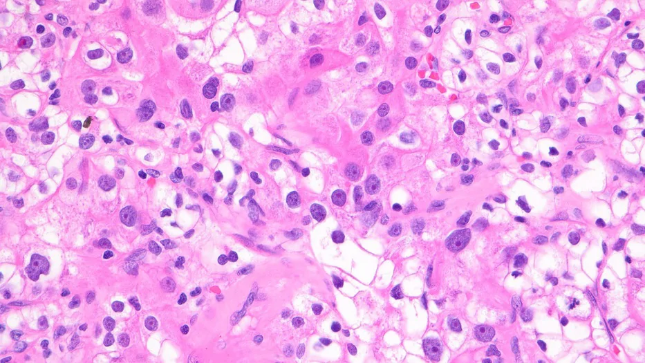
1–3 级的组成要素
一些研究将 WHO/ISUP 1 级和 2 级合并。鉴于 1 级肿瘤的数量可能较少,这引出了一个问题:是否应合并这两个较低级别(分为 1/2、3 和 4 级)。34 , 35目前 AUA 的 RCC 风险分层不区分 1 级与 2 级,2 , 57这与 CCRCC 的 Leibovich 评分不同。7然而,研究已显示 WHO/ISUP 1 级与 2 级之间的结局存在显著差异,应继续区分和使用这两个较低级别,包括用于 CCRCC 的风险预后判断。7 , 30 , 33分级异质性的高发生率也引出了另一个问题:3 级成分的范围或百分比(%)是否具有预后价值。一项研究显示,在 CCRCC 中,将 WHO/ISUP 3 级成分分为 <10%、10%–50% 和 >50%,或 ≤50 与 >50%,均与结局无关。58
4 级的组成要素
4 级成分的范围(%)
两项研究评估了 CCRCC 中高级别成分的百分比。Dagher 等 58将 WHO/ISUP 4 级成分分为 <10%、10%–50% 和 >50%,在 62 例 4 级 CCRCC 中进行研究;多变量分析显示,<10% 与 >50% 的 4 级成分组之间生存存在显著差异。而在同一研究中,3 级成分的百分比未显示生存差异。有趣的是,3 级 CCRCC 与含 <10% 的 4 级成分的 CCRCC 之间未显示结局差异。Serrano 等 59则将 Fuhrman 核分级 3 级和 4 级合并,并将 Fuhrman 核分级 3/4 级成分的百分比分为 1%–50% 和 51%–100%;Cox 比例风险多变量分析显示,Fuhrman 核分级 3/4 级成分的百分比与无转移生存期(MFS)、CSS 和 OS 显著相关。有限的数据提示,CCRCC 中 4 级成分的百分比具有预后价值;但仍需更多数据验证这一发现。
大型多核细胞
目前 WHO/ISUP 4 级的标准包括“瘤巨细胞”,即具有极度多形性或奇异细胞核的大型多核细胞。10然而,CCRCC 中还可见其他类型的大型多核细胞,即合体型多核细胞(STMC)及伴退变性异型性的多核细胞(MCDA)(图 2)。60 , 61 , 62 , 63 Williamson 等 60首次报道了少见的 STMC,在 1.2%–1.5% 的 CCRCC 中可见。62STMC 通常具有丰富的嗜酸性颗粒状胞质,其细胞核及免疫表型与背景中的单核透明细胞相似。多个细胞核可沿周边排列,白细胞伸入运动是常见特征。随后,Nova‐Camacho 和 Sangoi 的一项研究61报道,当存在 STMC 时,其可占肿瘤面积的高达 20%,且主要见于坏死区域周围。这两项研究均强调 STMC 与一部分侵袭性 CCRCC 的关联。在 Verine 等 64的结构分级研究中,STMC 在较高比例(15%)的 CCRCC 中被识别出;根据 DFS 的似然,STMC 被视为中等级别(第 2 级,共 3 级)。
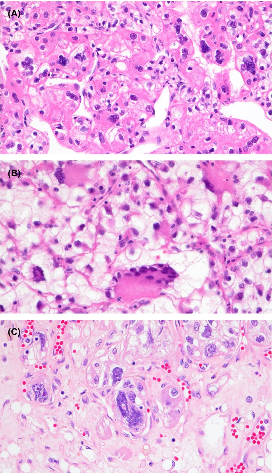
Pacheco 等 62描述了 28% 的 CCRCC 中散在出现 MCDA。这些大型细胞的特征是含有多个紧密聚集、相互重叠但未增大的细胞核,染色质致密成团。作者还描述了不伴间变的多核肿瘤细胞,但这些细胞很可能是双核或三核的 CCRCC 细胞,与其他多核细胞不同。与出现间变的多核细胞不同,含 MCDA 的肿瘤并未显示复发或转移风险增加,作者由此认为 MCDA 与分级无关。Cai 等 47显示,在 CCRCC 中,与存在 MCDA 相比,存在 STMC 的肿瘤具有更差的 DFS;而与这两种多核细胞均不存在的 RCC 相比,含 MCDA 的肿瘤 DFS 较差。
虽然典型的具有极度多形性的多核细胞应归为 WHO/ISUP 4 级,但 MCDA 不应被视为 4 级。STMC 的分级存在争议,这些细胞可能不属于 4 级。多核肿瘤细胞的鉴别诊断之一是巨核细胞,后者可罕见地见于伴髓外造血的 CCRCC。65
肉瘤样改变
梭形细胞的分级谱
长期以来已认识到,肉瘤样改变与 RCC 的不良结局相关,并具有特定的基因组和表观基因组改变,尤其涉及 Hippo 通路。54 , 66 , 67 , 68 , 69 , 70近期进展表明,免疫检查点抑制剂可使肉瘤样 RCC 患者在生存及治疗应答方面获益。71 , 72肉瘤样改变通常由类似未分化肉瘤的梭形细胞构成,罕见情况下可含有恶性异源性成分,如横纹肌肉瘤或骨肉瘤。54 , 66在肉瘤样 RCC 中,异源性成分的存在是否影响结局尚不明确。一项研究显示,在肉瘤样 RCC 中,恶性异源性成分与 RCC 死亡无关,但病例数有限。66
肉瘤样改变中的梭形细胞通常具有较高级别的细胞核,但少数情况下其细胞核可呈低级别外观(图 3)。Tanas Isikci 等 73描述了一系列 CCRCC 中的“低级别”梭形细胞增生,这些细胞的免疫组织化学及分子特征与常规 CCRCC 细胞相近,包括上皮–间质转化(EMT)标志物;作者认为这些细胞可能并不代表肉瘤样改变的初始阶段。Cai 等 47在 3.5% 的 CCRCC 中描述了区别于肉瘤样改变的“梭形细胞改变”,其 DFS 优于肉瘤样改变。然而,其他研究提示“低级别梭形细胞”具有更强的侵袭性行为。Cheville 等 66在 102 例以 CCRCC 为主的肉瘤样 RCC 中,将肉瘤样改变分为低级别(13.7%)和高级别(86.3%),分级与 RCC 死亡未显示相关性。Sirohi 等 74根据对 OS 的预测,将“低级别梭形细胞”和肉瘤样改变均归为不利的组织学生长模式。
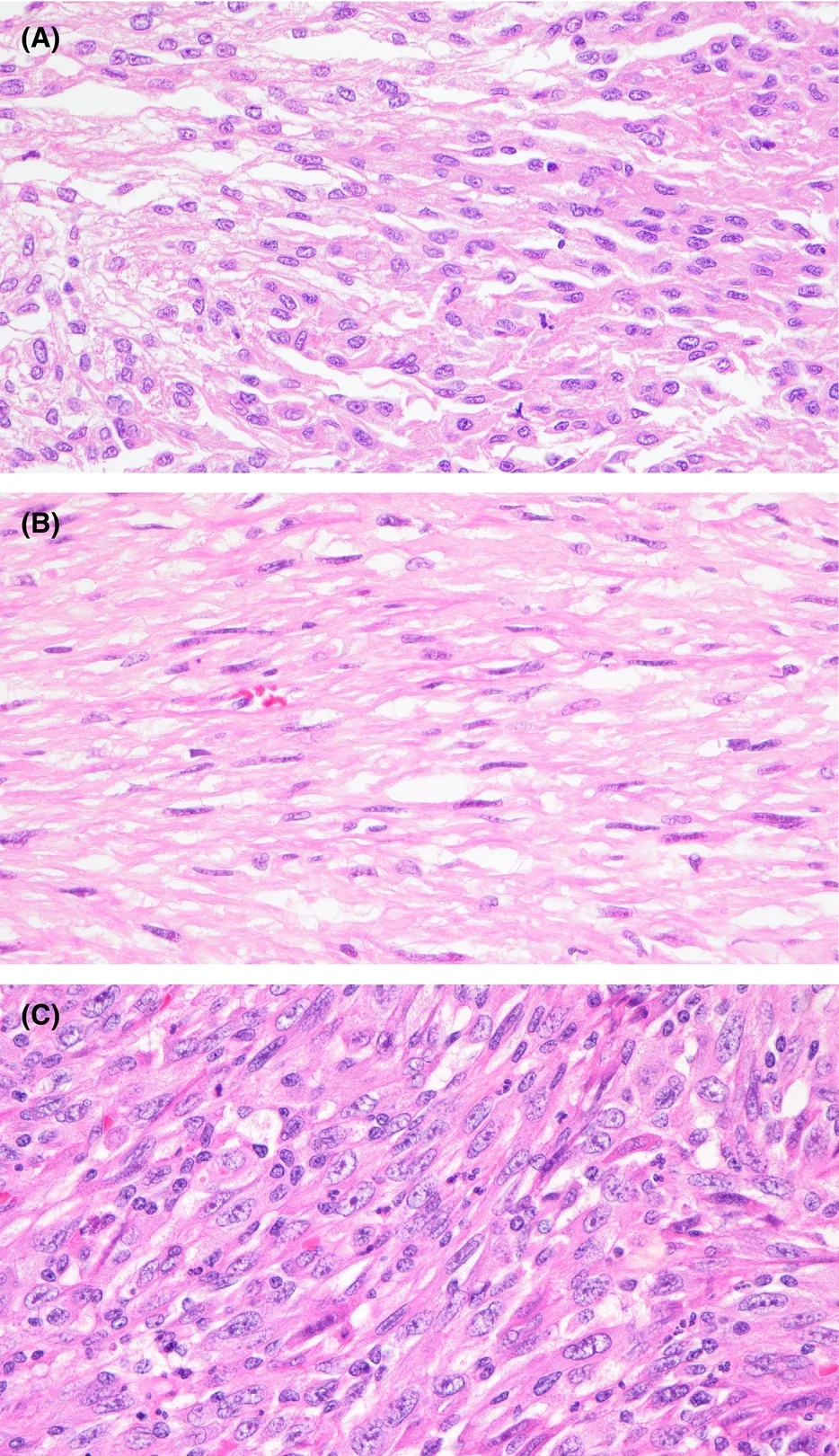
CCRCC 中可出现具有低级别细胞核的梭形细胞,需要进一步研究以阐明其预后意义。虽然分级应依据细胞核/核仁特征,但由于数据有限,将这一原则应用于这些呈低级别外观的梭形细胞仍存在不确定性。此外,必须解决观察者间可重复性的问题,以避免将确实存在、且具有不良预后影响的肉瘤样改变分级过低。
肉瘤样与非肉瘤样 4 级肿瘤的比较
约 35%–60% 的 4 级 CCRCC 可见肉瘤样改变。66 , 75 , 76 , 77研究显示,肉瘤样 4 级 RCC 的结局比非肉瘤样 4 级 RCC 更差。66 , 75 , 76 , 77 Cheville 等 66对 174 例 4 级 CCRCC 患者的研究报道,肉瘤样成分的存在(59.8%)与结局独立相关;肾切除术后 2 年 CSS 率,肉瘤样 RCC 为 32.9%,而 42.2% 为非肉瘤样 4 级 RCC 的相应值。Zhang 等 75随后在 411 例 ISUP 4 级 RCC 患者中(也来自同一机构)显示,肉瘤样改变的存在(占肿瘤的 49%)与 RCC 死亡风险增加 58% 相关。死亡风险增幅可进一步达到 82%,见于无转移的 4 级 RCC。Kara 等 77在 246 例 Fuhrman 核分级 4 级 RCC 患者中显示,无论有无转移,肉瘤样改变的存在均与更差的中位 CSS 相关。近期,Blum 等 76对 564 例无转移的 WHO/ISUP 4 级 RCC 的研究证实,在各个分期中,肉瘤样 RCC 的 CSS 均比非肉瘤样 4 级 RCC 更差。值得注意的是,局限性 pT1 肉瘤样 RCC 的 CSS 比 pT3 非肉瘤样 4 级 RCC 更差。
有充分证据表明,即使在 4 级 RCC 中,肉瘤样改变的存在也提示更差的结局。是否应将肉瘤样改变另设为高于 4 级的独立级别,仍需进一步考虑。重要的是,即使在 4 级 RCC 中,包括活检标本,也应报告肉瘤样改变的存在,因为它对结局具有不利影响,且关系到免疫检查点抑制剂治疗的选择。ICCR 和 CAP 均要求即使对于 4 级 RCC,也需报告是否存在肉瘤样改变。8 , 9
肉瘤样成分的范围(%)
RCC 中肉瘤样改变的范围可为 1% 至 100%。54多项研究显示,按 RCC 中肉瘤样成分的范围(%)进行划分具有预后价值(表 2)。66 , 75 , 78 , 79 , 80 , 81 , 82 , 83 , 84然而,肉瘤样成分百分比所用的界值各不相同,包括 10%、20% 和 30%。Zhang 等 75显示,肉瘤样成分每增加 10%,死亡风险相应增加 6%。Patel 等 81报道,肉瘤样成分每增加 10%,以及采用 ≥20% 的界值,均与更差的 OS 相关。Adibi 等 82显示,肉瘤样成分 >40% 时,1 年时仍存活的可能性最低。然而,一些较早的研究报道肉瘤样成分的百分比对结局无影响。66 Shuch 等 84将肉瘤样成分分为四分位组,而 Cheville 等 66将肉瘤样成分分为 5%–10%、15%–50% 和 >50%;这两项研究均未显示肉瘤样成分百分比对结局有影响。
| 研究 | 肉瘤样 RCC(透明细胞肾细胞癌占比 %) | 界值 | 终点 | 有统计学意义 | MVA | 说明 |
|---|---|---|---|---|---|---|
| Lucarelli 等 77 | 212 (100%) | 20% | CSS, RFS | 是 | 是 | |
| Soytas 等 78 | 201 (80%) | 10% | 转移、死亡 | 是 | 否 | 使用 5% 或 15% 界值时无相关性 |
| Salgia 等 79 | 104(占多数) | 10% | OS、MFS(局限于器官的肿瘤) | 是 | 是 | |
| Patel 等 80 | 128 (78%) | 20% | OS | 是 | 是 | 在 cM0 和 cM1 中均有统计学意义 |
| Adibi 等 81 | 186 (73%) | 10% | OS | 是 | 是 | >40% 时 1 年时仍存活的可能性最低 |
| Kim 等 82 | 59 (74%) | 25% | OS | 是 | 是 | 仅见于 cM0,而不见于 cM1。采用 5% 或 10% 界值时未观察到差异 |
| Zhang 等 74 | 215 (79%) | 30% | CSS | 是 | 是 | 肉瘤样成分增加 10% 与死亡风险增加 6% 相关 |
| Shuch 等 83 | 104 (65%) | <25%, 25%–<50%, 50%–<75%, ≥75% | 生存 | 否 | 是 | 将肉瘤样成分分为四分位组 |
| Cheville 等 65 | 120 (86%) | 5%–10%, 15–50%, >50% | RCC 死亡 | 否 | 否 |
缩写:CSS,肿瘤特异性生存期;MFS,无转移生存期;MVA,多变量分析;OS,总生存期;RFS,无复发生存期。
已有充分证据表明,肉瘤样成分的百分比影响 RCC 的预后,因此应报告这一要素。然而,肉瘤样成分百分比的最佳界值或分组(10%、20% 或 30%)尚未确立。值得注意的是,近期一项联合使用免疫检查点抑制剂阿替利珠单抗和抗 VEGF 药物贝伐珠单抗的 II 期临床试验显示,在肉瘤样成分 ≥20% 的 RCC 中出现了客观缓解。71ICCR 和 CAP 均建议将肾切除标本中肉瘤样成分的百分比作为可选要素报告,且不要求采用任何界值。8 , 9活检标本中出现肉瘤样成分时,不建议报告其百分比。
横纹肌样特征
RCC 中的横纹肌样改变被认为是不良预后因素,并与 SWI/SNF 染色质重塑复合物的改变相关。85 , 86横纹肌样改变的特征是大型上皮样细胞,具有空泡状核、明显核仁和大的核旁胞质内包涵体(图 4)。85据报道,在 4 级 RCC 中,仅有横纹肌样改变者占 20–28%,横纹肌样改变合并肉瘤样改变者占 7%–16%。77 , 87两者同时存在时,横纹肌样改变通常与肉瘤样改变相连续。
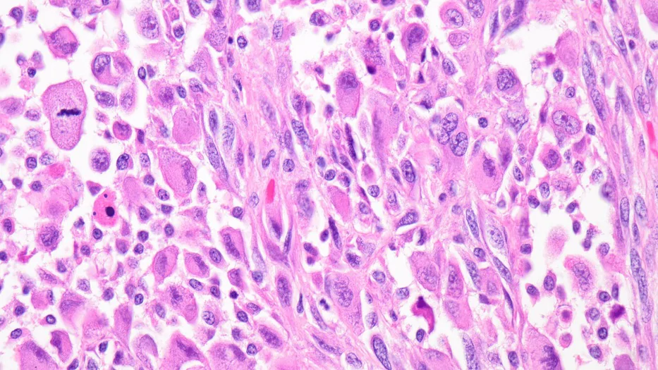
横纹肌样改变和肉瘤样改变均被归为 WHO/ISUP 4 级,这引出了一个问题:这两种形态模式对结局的影响是否相当。Zhang 等 87显示,与肉瘤样改变不同,ISUP 4 级 RCC 中的横纹肌样改变与 RCC 死亡无关。然而,与 3 级 RCC 患者相比,伴横纹肌样改变的 RCC 患者死于 RCC 的可能性显著更高。Kara 等 77也显示,与肉瘤样改变不同,横纹肌样改变并未增加 4 级 RCC 的死亡率。Verine 等 64显示,相比肉瘤样改变,横纹肌样改变的 DFS 似然较低,并在其结构分级中将这两种形态模式归入不同级别。
研究支持将横纹肌样改变归为 WHO/ISUP 4 级,且数据显示,横纹肌样改变对结局的不利影响与肉瘤样改变并不相同。因此,在报告时不应将这两种高级别形态模式合并。目前尚无数据支持报告 RCC 中横纹肌样改变的范围(%)。
WHO/ISUP 分级应用中的问题
分级的观察者一致性
由于 WHO/ISUP 分级提出时间相对较近,以往多数观察者间研究采用 Fuhrman 核分级;4 层级 Fuhrman 核分级的一致性为一般至中等(k= 0.22–0.44)。88 , 89 , 90 , 91一项由三位泌尿病理医师使用 WHO/ISUP 分级评估 CCRCC 的研究显示,在数字切片上达到较高一致性(κ= 0.62),在玻璃切片上达到中等一致性(κ= 0.50)。92在观察者自身一致性的研究中,病理医师 A、B 和 C 在两张玻璃切片之间的一致性 kappa 值分别为 0.69、0.69 和 0.81。在另两项各由三位病理医师参与的独立研究中,CCRCC 的 WHO/ISUP 分级分别显示较高及近乎完全的一致性(κ= 0.65 和 κ= 0.87–0.90)。64 , 93由于 WHO/ISUP 1–3 级的评估仅依赖单一变量,预期一致性会有所改善,现有少数研究也支持这一点。
穿刺活检分级的准确性
多数比较穿刺活检(NB)与肾切除标本分级的研究采用以往的 Fuhrman 核分级。一项荟萃分析显示,NB 与肾切除标本之间四层级分级的一致性一般(κ= 0.34),符合率为 62.5%,采用的是 NB 与肾切除标本之间的 4 层级分级比较。94一项系统综述报道,NB 与肾切除标本分级的符合率为 51.5%–75.9%。95 Proye 等 96报道,准确率为 52.6%,这是采用 4 层级 ISUP 分级评估 >1400 例 NB 的结果;准确率提高至 67.9%,则是在将 ISUP 分级二分为低级别(1/2)和高级别(3/4)之后。另一项来自大型中心的研究显示,将分级分为低级别和高级别两组时,NB 分级准确率为 94%。97RCC 的 NB 与肾切除标本分级存在明显不一致。大多数不一致是由于分级偏低(一项研究中占 92%),很可能归因于上述瘤内分级异质性。97 , 98ICCR 将活检报告中的分级视为暂定分级,因为异质性可能导致肾切除标本中的分级发生变化。8报告暂定分级具有价值,尤其是在级别较高时,因为其与肾切除标本分级的相关性更好,并有助于指导后续治疗管理。
多灶性肿瘤的分级
多灶性同时性 RCC 之间的分级可以不同。一项纳入 50 例多发散发性同时性肾肿块患者的研究显示,仅 62.5% 的病例中各肿瘤的分级一致。99另一项纳入 128 例散发性多灶性肿瘤患者的研究显示,77.3% 的病例中,最大(主要)肿瘤具有更具侵袭性的组织学表现。100还有一项纳入 97 例散发性单侧多灶性肾肿瘤患者的研究(肿瘤数量中位数为 3 个)显示,仅 51.5% 的病例中各肿瘤的分级(低级别或高级别)一致。101多灶性 RCC 的分级可以不同,最大肿瘤不一定总是具有最高级别。对于多灶性 RCC,尤其是分级不一致(最高级别位于较小 RCC)时,究竟仅报告最高级别还是逐一列出不同级别,目前尚无正式建议。仅报告最高级别(位于较小 RCC)的一个潜在问题是,可能无法准确反映最大或分期较高的 RCC(主要肿瘤);而在同时使用分级和分期的风险分层中,这会产生何种影响(若有)仍不明确。目前,ICCR 数据集和 CAP 规范均允许在一例肾切除标本中报告单一 WHO/ISUP 级别,而不论肿瘤是否为多灶性。8 , 9
乳头状肾细胞癌 WHO/ISUP 分级中的问题
与透明细胞肾细胞癌共有的分级问题
目前已有充分证据支持放弃将 PRCC 分为 1 型和 2 型;文献已充分讨论这一主题,本文不再赘述。37 , 102 , 103 , 104上文所述 CCRCC 的一些分级问题也可出现在 PRCC 中,如分级异质性、观察者间可重复性及活检分级偏低,尽管相关数据相对较少。Ball 等 45报道,小型(<4 cm)PRCC 中 Fuhrman 核分级异质性很常见,且在 25% 的样本中,较高级别并非占优势的(<50%)级别。六位泌尿病理医师对 30 例乳头状肾细胞癌进行核仁分级复核(2 级与 3 级),结果显示观察者间一致性仅为轻微(κ= 0.16)。105 Prendeville 等 106报道,四位病理医师评估 PRCC 时,NB 与肾切除标本分级的符合率为 64%,采用的是 4 层级 WHO/ISUP 分级。12% 的病例在肾切除标本中的分级较 NB 升高,无分级降低的病例,与 CCRCC 的 NB 情况相似。
小型(≤1.5 cm)乳头状肿瘤的分级
PRCC 和乳头状腺瘤在形态学、免疫组织化学及细胞遗传学上存在重叠。低级别细胞核(1 或 2 级)是区分乳头状腺瘤与 PRCC 的标准之一,其他标准为大小(≤1.5 cm)及无包膜。10这引出了一个问题:具有 WHO/ISUP 3 级细胞核的小型乳头状上皮性肿瘤,是否应诊断为 PRCC。尚无研究直接探讨这一问题;但两项分别基于瑞典国家肾癌登记数据库及美国和欧洲多中心数据的大型研究显示,转移率分别为 4% 和 <1%,研究对象为大小不超过 2 cm 的肾肿瘤。107 , 108有报道发现 Fuhrman 核分级 3 级(15.9%)和 4 级(0.5%),见于大小 <2 cm 的 RCC。108这些研究与来自 MSKCC 和 Mayo Clinic 的 >500 例患者的数据相矛盾,后者显示 <2 cm 的肾肿瘤未发生转移。109 , 110由于 <2 cm 肾肿瘤具有恶性潜能的发生率虽低但确实存在,诊断乳头状腺瘤应要求具有低级别细胞核;在获得更多数据之前,目前对于具有较高级别细胞核的 <2 cm 肿瘤,可考虑诊断为 PRCC。
纳入坏死的改良分级
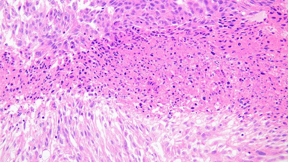
肿瘤坏死在 RCC 中常见,并在 CCRCC 中具有预后意义。6 , 70 , 111肿瘤相关坏死通常边界清楚,表现为结构消失,伴颗粒状细胞核及胞质碎屑(颗粒状坏死),不同于凝固性坏死(图 5)。112 Delahunt 等 33提出了一种用于透明细胞肾细胞癌的 4 级改良分级系统,将坏死纳入 ISUP 分级(表 3)。该研究显示,各改良级别之间的 CSS 存在显著差异。所提出的改良分级还具有更优的一致性指数,表现优于 ISUP 分级。
| 级别 | 描述 |
|---|---|
| 1 级 | ISUP 1 级 + 不伴坏死的 ISUP 2 级 |
| 2 级 | 伴坏死的 ISUP 2 级 + 不伴坏死的 ISUP 3 级 |
| 3 级 | 伴坏死的 ISUP 3 级 + 不伴坏死的 ISUP 4 级 |
| 4 级 | 伴坏死的 ISUP 4 级,或肉瘤样/横纹肌样肿瘤 |
随后,Dagher 等 111对 376 例 CCRCC 的研究显示,伴坏死的 WHO/ISUP 2 级与不伴坏死的 3 级(改良分级 2 级的组成部分)之间,以及伴坏死的 WHO/ISUP 3 级与不伴坏死的 4 级(改良分级 3 级的组成部分)之间,结局均无显著差异。Khor 等 36对 842 例 CCRCC 应用改良分级的研究显示,改良分级 1/2、3 和 4 级患者之间的生存差异具有统计学意义。作者进一步确定了三个具有统计学意义的预后组:(1)不伴坏死的 ISUP 1–3 级;(2)伴坏死的 ISUP 1–3 级及 ISUP 4 级且坏死 <10% 的肿瘤;(3)伴坏死或肉瘤样/横纹肌样改变的 ISUP 4 级。Sirohi 等 74显示,在 Kaplan–Meier 分析中,改良分级的区分效果更好;在 Cox 比例风险模型中,其统计学显著性也强于 WHO/ISUP 分级。Verine 等 64在 217 例局限性 CCRCC 中显示,就 DFS 和 CSS 而言,WHO/ISUP 分级与改良分级的 Harell 一致性指数相当。
纳入坏死增强了 WHO/ISUP 分级的预后预测能力。改良分级具有前景,但仍需对该方法开展更多研究,包括将其整合入风险模型。目前,ICCR 数据集和 CAP 规范均要求将坏死作为独立要素报告。8 , 9
基于结构或形态模式的分级与风险评估
基于形态模式的分级方法并非新概念,以往 RCC 的历史分级中已曾采用。11 , 12 , 13 , 14 , 15 , 16 , 17 , 18 , 19近期,基于形态模式的 CCRCC 分级及预后判断方案再次受到关注(表 4)。47 , 64 , 74 , 93 Verine 等 64提出了一种基于结构的 3 级评分,其表现优于 WHO/ISUP 分级及改良 WHO/ISUP 分级 + 坏死,并可独立预测 DFS 和 CSS。3 级结构评分的观察者间一致性优于 3 级 WHO/ISUP 分级(κ= 0.82,对比 κ= 0.71)。Cai 等 47在 CCRCC 的 9 种结构模式及 24 种细胞学和肿瘤微环境模式中发现,在考虑核仁分级后,管状结构、坏死、肾实质浸润及嫌色细胞样特征是 DFS 的独立特异性预测因素。基于这些变量,建立了用于 CCRCC 患者肾切除术后的预测列线图。通过组合 CCRCC 的 24 种高级别形态模式,Sirohi 等 74建立了 3 级和 2 级风险模型,在预测 OS 方面,其表现与改良 WHO/ISUP 分级 + 坏死同样良好,且优于 WHO/ISUP 分级。从九种结构模式出发,Ohe 等 93建立了一种基于血管的 3 类结构分类,对无复发生存期(RFS)的预测准确性最佳;将其纳入风险模型后,其预测准确性高于传统风险模型。这种基于血管分布的结构分类在 3 位病理医师之间显示出良好一致性(κ,基于血管分布的评分为 0.83–0.93;κ,WHO 评分为 0.87–0.90)。其他研究也显示,肾实质浸润或微结节性播散与透明细胞肾细胞癌的较差结局相关。113 , 114
| 研究 | 生长模式 |
|---|---|
| Verine 等 63 | 1 级*+:大囊性、微囊性、管囊性、致密型、腺泡状、透明细胞乳头状肾细胞癌样、退变型 |
| 2 级*+:大巢状、肺泡状或微乳头状、嫌色细胞/嗜酸细胞样、胞质内透明小球、瘤内炎症反应 | |
| 3 级*:横纹肌样、多核巨细胞、扩大的血管腔隙 | |
| 4 级*:肉瘤样、伴肾小球包埋的浸润性生长、淋巴血管侵犯(LVI) | |
| Cai 等 46 | 侵袭性**:肺泡状、乳头状、岛状、实性、瘤巨细胞、ChRCC 样、HLRCC 样、tRCC 样、透明小球、瘤内淋巴浆细胞浸润、中性粒细胞浸润、结节状生长、LVI、肿瘤浸润实质 |
| 有利**:微囊性、管状、出血性滤泡、致密巢状 | |
| 预测列线图:管状模式、坏死、肾实质浸润性边缘、ChRCC 样、分级 | |
| Sirohi 等 73 | 有利***:小巢状、膨胀性巢状、核质比高的巢状结构 |
| 中间型***:嗜酸细胞性乳头状、潘氏细胞样特征、实性假乳头状、嗜酸细胞性、肺泡状、退变型、管乳头状、钙化、乳头状、囊性、腺样、血管瘤样、内皮增生、管状、巨细胞 | |
| 不利***:浸润性、低级别梭形细胞、肉瘤样、融合巢状/片状、横纹肌样、坏死 | |
| 2 级风险模型:WHO/ISUP 4 级、存在坏死或任何不利形态模式者为高风险;其余均为低风险 | |
| 3 级风险模型:存在任何不利形态模式者为高风险;不符合高风险标准、存在任何有利形态模式,且模式总数不超过 5 种(其中不确定模式不超过 2 种)者为低风险;其余均为中等风险 | |
| Ohe 等 92 | 类别 1****:致密/小巢状、微囊/微囊、管状/腺泡状 |
| 类别 2****:肺泡状/大巢状、粗梁状 | |
| 类别 3****:实性片状、横纹肌样、肉瘤样 |
根据 DFS 的似然比;+ 将 1 级和 2 级合并,用于优选的 3 级分级系统。
依据 DFS 的单变量分析。
依据预测 OS 的风险比(HR)。
依据采用 CD31 免疫组织化学的血管网络评分。
近期报道了 PRCC 的几种不利结构。Yang 等 37发现 PRCC 中的实性、微乳头状或鞋钉样结构与较差的 DFS 和 OS 相关。Chan 等 105显示,PRCC 中的微囊、鞋钉样、实性及微乳头状结构与不良结局显著相关,但微囊更常见,且观察者间可重复性更佳。Murugan 等 43报道,PRCC 的片状结构与 CSS 显著相关。
初步研究提示,基于结构或形态模式的分级可提供有用的预后信息,其潜力可能优于 WHO/ISUP 分级及整合坏死的改良分级。然而,仍需其他作者开展验证研究,并可能需要对这些不同方案进行标准化。此外,分级时同时评估多种组织学和细胞学变量所带来的复杂性,可能使其难以用于常规实践。目前 WHO/ISUP 分级的一个优点是,1–3 级仅评估一个变量(核仁),因此在实践上优于 Fuhrman 核分级及其他同样采用结构和其他特征的历史综合分级。
结论
本综述介绍了 WHO/ISUP 分级应用的近期进展与问题,以及 CCRCC 和 PRCC 的组织学分级及风险评估新方法(表 5 和 6)。本综述未涵盖的其他创新方法包括在组织病理切片上应用人工智能或深度神经网络进行 RCC 分级,以及影像组学。115本综述可作为解决部分分级问题的最佳实践指南,并有助于发现潜在的研究空白,从而优化 CCRCC 和 PRCC 的分级。
| 问题 | 评述与建议 |
|---|---|
| I. WHO/ISUP 分级的验证 | |
| 1. CCRCC 的分级 | 多数研究证实其适用性 |
| 2. PRCC 的分级 | 部分研究证实其适用性,但其他研究结果与之矛盾 |
| 3. ChRCC 的分级 | WHO/ISUP 分级无效,正在推荐新的 2 级、3 级和 4 级分级方法 |
| II. CCRCC 的 WHO/ISUP 分级要素 | |
| 1. 分级异质性 | 最高级别*并非总是占优势的级别 |
| 2. 合并(最低的)1 级和 2 级 | 1 级与 2 级之间的结局存在差异,不应合并这两个级别 |
| 3. 3 级成分的百分比 | 有限的数据表明无预后价值 |
| 4. 4 级成分的百分比 | 有限的数据提示具有预后价值 |
| 5. 多核细胞 | 具有多形性细胞核的多核细胞归为 4 级 |
| 合体型多核细胞很可能不属于 4 级 | |
| 伴退变性异型性的多核细胞不应归为 4 级 | |
| 6. 梭形细胞 | 可出现少见的“低级别外观”梭形细胞;其行为是否相当于 4 级尚不明确 |
| 7. 肉瘤样与非肉瘤样 4 级的比较 | 肉瘤样肿瘤的生物学行为比非肉瘤样 4 级肿瘤更差 |
| 8. 肉瘤样成分的范围(%) | 肉瘤样成分的百分比具有预后价值,但最佳百分比界值(10%、20% 或 30%)尚未确定 |
| 建议免疫检查点抑制剂治疗采用 20% 界值 | |
| 9. 横纹肌样 | 归为 4 级,但生物学行为与肉瘤样不同 |
| III. WHO/ISUP 分级的应用 | |
| 1. 观察者间一致性 | 现有数据显示,其一致性优于 Fuhrman 核分级 |
| 2. 穿刺活检分级 | 与肾切除标本分级存在明显不一致,可能由分级异质性所致 |
| 3. 多灶性肿瘤 | 各肿瘤分级可不一致,最高级别可能出现在非主要肿瘤中 |
| 尚无报告建议(报告最高级别或所有级别) | |
至少占 1 个高倍视野;最佳面积尚未确定。
| 问题 | 评述与建议 |
|---|---|
| I. PRCC 的 WHO/ISUP 分级 | |
| 1. 分级异质性与活检 | 活检中可能出现与 CCRCC 相似的分级偏低,但数据有限 |
| 2. 小型(≤1.5 cm)乳头状肿瘤 | 诊断乳头状腺瘤要求具有低级别细胞核* |
| II. 新方法 | |
| 1. 纳入坏死的改良分级 | 预后预测能力优于 WHO/ISUP,但仍需更多研究 |
| 2. 基于形态模式的分级与风险评估 | 在预后判断方面显示出优于 WHO/ISUP 的潜力 |
| 需要验证,并可能需要标准化 | |
| 应用较 WHO/ISUP 分级更复杂 | |
具有较高级别细胞核时考虑 PRCC。
- About a decade after formal adoption of WHO/ISUP grading for clear cell RCC (CCRCC) and papillary RCC (PRCC), Paner, Amin, Cheng, Moch and colleagues review the literature, element by element, and offer practice guidance where data allow.
- Grade elements: the highest grade is not always the predominant grade; grades 1 and 2 differ in outcome and should not be combined; multinucleated cells with pleomorphic nuclei are grade 4, whereas syncytial-type and degenerative-atypia multinucleated cells likely are not; uncommon “low-grade-appearing” spindle cells have unclear behaviour; sarcomatoid grade 4 behaves worse than non-sarcomatoid grade 4, and % sarcomatoid is prognostic but the optimal cut-off (10%, 20% or 30%) is not settled.
- Applications: interobserver agreement is better than with Fuhrman grade; needle-biopsy grade is significantly discordant with nephrectomy grade (largely grade heterogeneity); in multifocal tumours the highest grade may sit in a non-index tumour, with no reporting recommendation yet. PRCC grading is supported by some studies and contradicted by others; small (≤1.5 cm) papillary tumours are discussed separately.
- New approaches: modified grading incorporating necrosis and architectural/pattern-based grading and risk assessment. Tables 1–6 (including summary tables for CCRCC and PRCC) and Figures 1–5 included.
Scope: PMC open-access full text (CC BY 4.0) — abstract, all body sections, Tables 1–6 and Figures 1–5 with captions. Author contributions, conflicts, consent, data availability statements and references are not included.
Abstract
A decade into the formal adoption of the World Health Organization (WHO)/International Society of Urological Pathology (ISUP) grading system for clear cell renal cell carcinoma (CCRCC) and papillary renal cell carcinoma (PRCC), newer grading concepts, innovative approaches and some issues have emerged. A comprehensive review of the literature on the WHO/ISUP grading system and other grading approaches for CCRCC and PRCC was conducted. Updates and issues are presented on the following: (1) validation studies on WHO/ISUP grading; (2) grade elements including heterogeneity, grades 1 versus 2, types of multinucleated tumour cells, ‘grade spectrum’ of spindle cells, impact of sarcomatoid change in grade 4 RCC, percent (%) high grade, % sarcomatoid change and rhabdoid change versus sarcomatoid change; (3) grade applications including observer agreement, needle biopsy grade accuracy (correlation with nephrectomy grade) and grading of multifocal RCCs; (4) grade in papillary tumours including heterogeneity and on small (≤1.5 cm) neoplasms; and (5) novel grading or risk categories including incorporation of necrosis into grading and pattern or architectural‐based grading. Practice guidance for some of the issues is provided when feasible or where data are sufficient. This review highlights the recent updates and controversies in the use of WHO/ISUP grading and on novel approaches to grading for CCRCC and PRCC. This review may serve as a best practice guide in addressing some of the grading issues and will help identify gaps in our understanding and use of grading for CCRCC and PRCC that can inform future research.
Introduction
Grade is a crucial pathological factor in the management and prognostication of renal cell carcinoma (RCC). 1 , 2 For >70 years, the manner and applicability of grading for RCC have considerably evolved largely due to adjustments in its components and to later adaptations to the growing number of RCC subtypes. 3 , 4 Only in the prior decade has the World Health Organization (WHO)/International Society of Urological Pathology (ISUP) grading system been formally adopted for clear cell RCC (CCRCC) and papillary RCC (PRCC). 5 , 6 The WHO/ISUP grade has now been integrated into major urological guidelines and risk stratifications for CCRCC and PRCC, 1 , 7 and is considered a core element when reporting RCC in renal biopsy (RB) and nephrectomy specimens by the International Collaboration on Cancer Reporting (ICCR) and the College of American Pathologists (CAP). 8 , 9
The grading of other RCC subtypes beyond CCRCC and PRCC, including the use of the WHO/ISUP grading system, is still under investigation. Recently, three authors (G.P.P., M.B.A. and H.M.) provided updates on grading the less common RCC subtypes that highlighted the gaps in grading and offered guidance for surgical pathologists on grading considerations for these tumours. 4 This practice guidance has also been incorporated into the 2022 WHO blue book. 10 This review will focus on CCRCC and PRCC, where the use of WHO/ISUP grading is established, and will cover updates and issues in the application of grading and the grade components. Further, innovative approaches including incorporation of other elements (e.g. necrosis, architecture) in grading will be explored. We also hope that this review will shed light on the gaps in our understanding of the grading of CCRCC and PRCC that can inform future research.
Evolution of WHO/ISUP grading system
Historical Composite Gradings
From 1949 and the ensuing decades, several 3‐ and 4‐tiered grading schemes for RCC were introduced beginning with the grading by Griffiths and Thackray 11 and several others. 3 , 12 , 13 , 14 , 15 , 16 , 17 , 18 , 19 Most of these historical grading systems assessed varying combinations of histological features that included tumour architecture, nuclear features, mitosis, cell type or necrosis, among others. These grading approaches showed differences in survival outcomes for RCC. However, these grading systems were applied then without the appreciation of the current RCC subtypes, and some of these schemes had vague criteria that could be challenging to use in a reproducible manner.
Fuhrman Nuclear Grade
In 1968, Meyers et al. 20 introduced one of the first of many several subsequent grading approaches that focused on nuclear features. 21 , 22 , 23 , 24 , 25 , 26 , 27 , 28 , 29 Among these nuclear‐based gradings, it was the 4‐tiered grading by Fuhrman et al. 24 in 1982 that gained wide recognition. In the original Fuhrman's study, RCCs were categorized into Fuhrman nuclear grades 1 (14%), 2 (50%), 3 (26%) and 4 (10%), with grading based on the most malignant or highest grade present, even if only focal. Metastatic rates were significantly different between Fuhrman nuclear grade 1 and combined Fuhrman nuclear grade 2–4 tumours, and 5‐year survival curves were statistically different but only when Fuhrman nuclear grade 2 and 3 tumours were combined as one group. Interestingly, in an interobserver agreement study, the nuclear grading by Syrjänen and Hjelt 22 outperformed Fuhrman nuclear grade, the nuclear grading by Skinner et al., 21 and the grading based on tumour architecture and cell type by Arner et al. 14 The Fuhrman nuclear grade suffered from the complexities of simultaneously assessing three different nuclear variables (size, shape and nucleolar prominence) that are not always concordant in their gradations. A reason that Fuhrman nuclear grade, as a composite of these three nuclear features, cannot be strictly applied in a subset of CCRCC, 30 and likely contributed to the fair to moderate interobserver agreements when applying this system.
WHO/ISUP Grade
Around the 2000s, the approach to grading was being simplified in practice to focus mainly on nucleolar prominence for grades 1–3. While nucleolar‐based grading was already being used by some expert pathologists then to derive Fuhrman nuclear grade, its use as a grading system distinct from Fuhrman nuclear grade was codified through two studies from the same group. 31 , 32 In 2006, the study by Sika‐Paotonu et al. 31 on PRCC showed that focal nucleolar grade was independently associated with survival, unlike the other nuclear measures of Fuhrman nuclear grade in size and shape. In 2011, the study by Delahunt et al. 32 for nucleolar grades 1–3 for CCRCC showed that the worst nucleolar grade discriminated in survival among grades 2 and 3 tumours. This nucleolar‐based grading for CCRCC and PRCC was subsequently adopted in the 2012 ISUP consensus conference as the ISUP grade, 6 and with slight modification (nucleolar eosinophilia), it was incorporated into the 2016 WHO blue book as the WHO/ISUP grade 5 and later retained in the 2022 WHO iteration (Table 1). 10 It is recommended that the WHO/ISUP grade should be assigned within a single high‐power field showing the greatest degree of nuclear changes. The WHO/ISUP grading system has also been recognized to be inapplicable for chromophobe RCC (ChRCC). 6 Currently, different alternative (2‐, 3‐ and 4‐tiered) grading approaches are being recommended for ChRCC. 4 A decade into its WHO adoption for CCRCC and PRCC, studies on WHO/ISUP grade are still unfolding.
| Grade | Description |
|---|---|
| Grade 1 | Nucleoli absent or inconspicuous and basophilic at ×400 magnification |
| Grade 2 | Eosinophilic nucleoli observed at ×400 but not prominent at ×100 magnification |
| Grade 3 | Nucleoli prominent and eosinophilic at ×100 magnification |
| Grade 4 | Carcinoma cells display extreme nuclear pleomorphism, tumour giant cells, and/or any amount of sarcomatoid or rhabdoid differentiation |
Applications of WHO/ISUP grading system
Clear Cell RCC
After its introduction, several studies were conducted on WHO/ISUP grading for CCRCC. Dagher et al. 30 showed a difference in cancer‐free survival (CFS) for grades 1–4 in 376 CCRCCs at mean follow‐up of 35 months that was significant on multivariate analysis. Delahunt et al., 33 on a cohort of 3,017 CCRCCs from the Mayo Clinic Registry, showed differences in cancer‐specific survival (CSS) across all grades on 10‐year follow‐up by regression models. Leibovich et al., 7 also on a large cohort of CCRCCs from the Mayo Clinic Registry, showed a significant association of WHO/ISUP grade categories with progression or death from RCC in multivariate analysis, and updated the Leibovich prognostic model for CCRCC to include WHO/ISUP grade. Other studies showed a significant association to outcome when grades 1/2 and 3/4 were combined in multivariate analysis. 34 , 35 However, the study by Khor et al. 36 that preceded the WHO criteria and used the ISUP grade (without accounting for nucleolar eosinophilia) on 842 consecutive CCRCCs showed a significant association only for grade 4, and not for grade 3 versus grades 1/2 combined, to overall survival (OS). Overall, most studies confirmed the prognostic value of WHO/ISUP grade for CCRCC.
Papillary RCC
Since the adoption of the WHO/ISUP grade, several grading studies using this system for PRCC were also conducted. The aforementioned study by Leibovich et al. 7 from Mayo Clinic also graded a cohort of 607 PRCCs and showed significant association with progression or death from RCC with 3‐tier WHO/ISUP grade (1/2, 3 and 4) in multivariate analysis. The Leibovich prognostic model for PRCC was also updated to incorporate the WHO/ISUP grade. Yang et al. 37 in a study of 185 PRCCs showed high WHO/ISUP grade to be associated with disease‐free survival (DFS) and OS in multivariate analysis. The study by Le et al. 38 on 52 consecutive PRCCs showed WHO/ISUP grade (grades 1/2 versus 3/4) to be associated with progression‐free survival (PFS) in univariate analysis. A study by Cornejo et al. 39 that preceded the 2016 WHO criteria and used the ISUP grade (not accounting for eosinophilia) on 154 PRCCs showed ISUP grade to be a significant predictor of CSS and OS in multivariate analysis. However, three studies with 176, 254 and 319 patients with PRCC failed to show the prognostic value of WHO/ISUP grading. 40 , 41 , 42 Interestingly, the study by Murugan et al. 43 on 199 PRCCs with any ‘type 1’ features showed that the highest WHO/ISUP grade (recommended approach) was not associated with outcome, whereas the predominant WHO/ISUP grade was associated with PFS and CSS in univariate analysis. Studies using WHO/ISUP grade on PRCC showed mixed results, but with more data supporting continued use of this grading system.
Issues in WHO/ISUP grading of clear cell RCC
Issues in WHO/ISUP Grade Elements
Grade heterogeneity
Intratumoral heterogeneity in grades, patterns, genetics and biomarker expressions is not uncommon in CCRCC. 44 , 45 , 46 , 47 , 48 , 49 , 50 , 51 , 52 , 53 Eosinophilic cell area is known to harbour higher grade nuclei (Figure 1). 44 Tumour patterns such as alveolar, pseudopapillary, insular or solid sheet‐like areas are associated with higher nuclear grade. 47 Mutations in BAP1, PTEN or SETD2 and higher tumour mutational burden when heterogeneously present are more likely to be detected in higher grade areas. 49 , 50 , 53 The range of variations in grade within a tumour can be marked; for example, sarcomatoid change (grade 4) can be seen with background of grade 2. 54 Grade heterogeneity is common even in small (<4 cm) clear cell RCC, and the highest grade is not the predominant grade in 37.5% of samples. 45 Interestingly, Wu et al. 46 observed discrepancies between WHO/ISUP grades at primary tumour and at venous tumour thrombus in 47% of CCRCCs and showed the discrepancy to be an independent risk factor in predicting outcome. Because of the heterogeneity in grades, it is recommended that grading should be based on the highest‐grade present. 6 , 10 However, the optimal minimum area for the highest grade remains to be established. In the 2012 ISUP conference, no consensus was reached regarding the minimum area for grading. 6 Currently, it is recommended that the highest grade should be present in at least one high power field, which is approximated to be about 0.23 mm2 in most modern microscopes. 6 , 10 To address the issue of heterogeneity in CCRCC, some authors proposed modified extensive tumour sampling techniques. 55 , 56 At this point, it is important to ensure adequate tumour sampling (traditionally at least 1 section per cm of tumour greatest dimension and to make sure that all macroscopically distinct areas are sampled).
Grades 1–3 components
Some studies combined WHO/ISUP grades 1 and 2. Given that the number of grade 1 tumours can be small, this begs the question of whether these two lower grades should be merged (grades 1/2, 3 and 4). 34 , 35 The current AUA risk stratification for RCC does not make a distinction between grades 1 and 2, 2 , 57 in contrast to the Leibovich score for CCRCC. 7 However, studies have shown a significant difference in outcome between WHO/ISUP grades 1 and 2, and continued separation and use of these two lower grades should be made including for risk prognostication for CCRCC. 7 , 30 , 33 The high prevalence of grade heterogeneity also raises the question if extent or percent (%) of grade 3 has any prognostic value. One study showed that dividing WHO/ISUP grade 3 into <10%, 10%–50% and >50% or ≤50 or >50% in CCRCC is not associated with outcome. 58
Grade 4 components
Extent (%) of grade 4
Two studies assessed % of high‐grade component in CCRCC. Dagher et al. 58 divided WHO/ISUP grade 4 component into <10%, 10%–50% and >50% in 62 grade 4 CCRCCs and showed a significant difference in survival between <10% and >50% grade 4 in multivariate analysis. Whereas in the same study, the difference in survival was not demonstrated with % grade 3. Interestingly, no difference in outcome was shown between grade 3 and <10% grade 4 CCRCCs. Serrano et al. 59 instead combined Fuhrman nuclear grades 3 and 4 and divided % Fuhrman nuclear grade 3/4 into 1%–50% and 51%–100% and by Cox proportional hazard multivariate analysis showed that % Fuhrman nuclear grade 3/4 is significantly associated with metastasis‐free survival (MFS), CSS and OS. Limited data suggest that % grade 4 in CCRCC has prognostic value; however, more data are needed to validate this finding.
Large multinucleated cells
The current criteria for WHO/ISUP grade 4 includes ‘tumour giant cells’ that encompass multinucleated large cells with extreme pleomorphism or bizarre nuclei. 10 However, there are other types of large multinucleated cells that can be seen in CCRCC, namely, syncytial‐type multinucleated cell (STMC) and multinucleated cell with degenerative atypia (MCDA) (Figure 2). 60 , 61 , 62 , 63 Williamson et al. 60 first reported the uncommon STMC, which can be seen in 1.2%–1.5% of CCRCCs 62 STMCs usually have voluminous eosinophilic granular cytoplasm and have nuclei and immunophenotype similar to the background mononuclear clear cells. The multiple nuclei can be arranged peripherally, and emperipolesis of leukocytes is a common feature. A subsequent study by Nova‐Camacho and Sangoi 61 reported that STMCs, when present, can be seen in up to 20% of tumour area and are mainly seen around areas of necrosis. Both studies highlighted the association of STMCs with a subset of aggressive CCRCCs. In the architectural grading study by Verine et al., 64 STMCs were identified in higher proportion (15%) of CCRCCs, and based on likelihood for DFS, STMC is considered as intermediate grade (grade 2 of 3).
Pacheco et al. 62 described the sporadic presence of MCDA in 28% of CCRCCs. These large cells are characterized by multiple tightly clustered and overlapping unenlarged nuclei with dense clumped chromatin. The authors also described multinucleated tumour cells with no anaplasia, although these cells are likely to be CCRCC cells that are bi‐ or trinucleated distinct from the other multinucleated cells. Tumours with MCDA did not show increased risk of recurrence or metastasis, unlike for multinucleated cells showing anaplasia, and the authors concluded that MCDA is not relevant for grading. Cai et al. 47 showed that the presence of STMCs had worse DFS compared to MCDA in CCRCCs and that tumours with MCDA had less favourable DFS compared to RCCs without both multinucleated cells.
While the classic multinucleated cells with extreme pleomorphism should be graded as WHO/ISUP grade 4, MCDA should not be considered as grade 4. Grading of STMC is controversial and these cells are possibly not grade 4. A differential diagnosis for multinucleated tumour cells is the megakaryocytes that can rarely occur in CCRCC with extramedullary haematopoiesis. 65
Sarcomatoid change
Grade spectrum of spindle cells
Sarcomatoid change is long recognized to be associated with poor outcome in RCC and shows specific genomic and epigenomic alterations, notably in the Hippo pathway. 54 , 66 , 67 , 68 , 69 , 70 Recent developments revealed that immune checkpoint inhibitors provide survival and response benefits in sarcomatoid RCC. 71 , 72 Sarcomatoid change is usually composed of spindle cells that resemble undifferentiated sarcoma, and rarely may contain malignant heterologous elements such as rhabdomyosarcoma or osteosarcoma. 54 , 66 It is unclear whether the presence of heterologous elements has an impact on outcome among sarcomatoid RCCs. One study showed no association of malignant heterologous elements with death from RCC among sarcomatoid RCCs, but with a limited number of cases. 66
The spindle cells in sarcomatoid change typically have higher grade nuclei, but uncommonly may have low‐grade‐appearing nuclei (Figure 3). Tanas Isikci et al. 73 described a series of ‘low‐grade’ spindle cell proliferation in CCRCC with comparable immunohistochemical and molecular profile to conventional CCRCC cells, including with epithelial–mesenchymal transition (EMT) markers, and concluded that these cells may not represent an initial phase of sarcomatoid change. Cai et al. 47 described ‘spindle cell change’ distinct from sarcomatoid change in 3.5% of CCRCCs that had better DFS than sarcomatoid change. However, other studies suggest a more aggressive behaviour for ‘low‐grade spindle cells’. Cheville et al. 66 divided sarcomatoid change in 102 sarcomatoid RCCs that were predominantly CCRCC into low grade (13.7%) and high grade (86.3%), and grade did not show correlation with death from RCC. Sirohi et al. 74 based on prediction of OS, categorized both ‘spindled low‐grade’ and sarcomatoid change to be unfavourable histological growth patterns.
Spindle cells with low‐grade nuclei can occur in CCRCC, and additional studies are needed to elucidate on its prognostic significance. While grading should be based on nuclear/nucleolar features, there is uncertainty on its application to these low‐grade appearing spindle cells because of limited data. Further, the issue of observer reproducibility must be addressed to avoid undegrading bona fide sarcomatoid change with poor prognostic effect.
Sarcomatoid versus non‐sarcomatoid grade 4
Sarcomatoid change is seen in about 35%–60% of grade 4 CCRCC. 66 , 75 , 76 , 77 Studies have shown a worse outcome of sarcomatoid grade 4 RCC compared to non‐sarcomatoid grade 4 RCC. 66 , 75 , 76 , 77 Cheville et al. 66 reported in 174 patients with grade 4 CCRCCs that the presence of a sarcomatoid component (59.8%) was independently associated with outcome; CSS at 2 years post‐nephrectomy was 32.9% for sarcomatoid RCC and 42.2% for non‐sarcomatoid grade 4 RCC. Zhang et al. 75 later showed in 411 patients with ISUP grade 4 RCC (also from the same institution) that the presence of sarcomatoid change (49% of tumours) was associated with a 58% increased risk of death from RCC. The increased risk of death was higher at 82% for non‐metastatic grade 4 RCCs. Kara et al. 77 showed that the presence of sarcomatoid change in 246 patients with Fuhrman nuclear grade 4 RCCs was associated with worse median CSS in both metastatic and non‐metastatic cases. Recently, the study by Blum et al. 76 on 564 non‐metastatic WHO/ISUP grade 4 RCCs demonstrated that at every stage, sarcomatoid RCC had worse CSS compared to non‐sarcomatoid grade 4 RCC. Notably, localized pT1 sarcomatoid RCC had worse CSS than pT3 non‐sarcomatoid grade 4 RCC.
There is convincing evidence that the presence of sarcomatoid change portends a worse outcome even for grade 4 RCCs. Whether sarcomatoid change warrants another higher‐grade distinction separate from grade 4 will need further consideration. Importantly, the presence of sarcomatoid change should be reported even in grade 4 RCCs, including in biopsy specimens, because of its negative effect on outcome and for immune checkpoint inhibitor treatment options. Both ICCR and CAP require reporting the presence of sarcomatoid change even for grade 4 RCC. 8 , 9
Extent (%) of sarcomatoid
The extent of sarcomatoid change in RCC can range from 1% to 100%. 54 Several studies showed the prognostic value of dividing extent (%) of sarcomatoid component in RCC (Table 2). 66 , 75 , 78 , 79 , 80 , 81 , 82 , 83 , 84 However, varying cut‐offs were used for % sarcomatoid including 10%, 20% and 30%. Zhang et al. 75 showed that for every 10% increase in sarcomatoid component there is an associated 6% increase in risk of death. Patel et al. 81 reported that sarcomatoid component was associated with worse OS per 10% increase and at ≥20% cut‐off. Adibi et al. 82 showed that presence of >40% sarcomatoid component had the least likelihood to be alive at 1 year. However, some older studies reported that % sarcomatoid component had no effect on outcome. 66 Shuch et al. 84 divided sarcomatoid component into quartiles while Cheville et al. 66 divided sarcomatoid component into 5%–10%, 15%–50% and >50% and both studies failed to show effect of % sarcomatoid component on outcome.
| Study | Sarcomatoid RCC (% clear cell RCC) | Cut‐off | End point | Significant | MVA | Comment |
|---|---|---|---|---|---|---|
| Lucarelli et al. 77 | 212 (100%) | 20% | CSS, RFS | Yes | Yes | |
| Soytas et al. 78 | 201 (80%) | 10% | Metastasis, death | Yes | No | No association using 5% or 15% cut‐offs |
| Salgia et al. 79 | 104 (predominant) | 10% | OS, MFS (organ‐confined tumours) | Yes | Yes | |
| Patel et al. 80 | 128 (78%) | 20% | OS | Yes | Yes | Significant for cM0 and cM1 |
| Adibi et al. 81 | 186 (73%) | 10% | OS | Yes | Yes | >40% least likely to be alive at 1 year |
| Kim et al. 82 | 59 (74%) | 25% | OS | Yes | Yes | In cM0 only and not with cM1. No difference observed using 5% or 10% cut‐offs |
| Zhang et al. 74 | 215 (79%) | 30% | CSS | Yes | Yes | 10% increase sarcomatoid associated with 6% increased risk of death |
| Shuch et al. 83 | 104 (65%) | <25%, 25%–<50%, 50%–<75%, ≥75% | Survival | No | Yes | Sarcomatoid divided into quartile |
| Cheville et al. 65 | 120 (86%) | 5%–10%, 15–50%, >50% | Death from RCC | No | No |
Abbreviations: CSS, cancer‐specific survival; MFS, metastasis‐free survival; MVA, multivariate analysis; OS, overall survival; RFS, recurrence‐free survival.
There is enough evidence to show that % sarcomatoid has an impact on prognosis of RCC, and thus, this element should be reported. However, the optimal cut‐off or divisions (10%, 20% or 30%) for % sarcomatoid has yet to be established. Of note, a recent phase II clinical trial combining immune checkpoint inhibitor atezolizumab and the anti‐VEGF bevacizumab showed objective response in RCC with ≥20% sarcomatoid component. 71 Both ICCR and CAP recommend reporting % sarcomatoid component in nephrectomy specimens as an optional element, and without requiring any cut‐offs. 8 , 9 Reporting % sarcomatoid component is not recommended when present in biopsy specimens.
Rhabdoid feature
Rhabdoid change in RCC is recognized as a poor prognostic factor and is associated with alterations in SWI/SNF chromatin remodelling complex. 85 , 86 Rhabdoid change is characterized by large epithelioid cells with vesicular nuclei, prominent nucleoli and large paranuclear intracytoplasmic inclusions (Figure 4). 85 In grade 4 RCCs, rhabdoid change (only) is reported in 20–28%, and rhabdoid in combination with sarcomatoid change in 7%–16%. 77 , 87 When present together, rhabdoid change is usually contiguous with sarcomatoid change.
Both rhabdoid and sarcomatoid change are graded as WHO/ISUP grade 4 and begs the question whether these two patterns are comparable in terms of effects on outcome. Zhang et al. 87 showed that rhabdoid change in ISUP grade 4 RCCs was not associated with death from RCC, in contrast to sarcomatoid change. However, patients with RCC with rhabdoid change are significantly more likely to die from RCC than patients with grade 3 RCC. Kara et al. 77 also showed that rhabdoid change was not associated with additional mortality in grade 4 RCCs, unlike sarcomatoid change. Verine et al. 64 showed a lower likelihood for DFS for rhabdoid change compared to sarcomatoid change and categorized these two patterns into different grade categories in their architectural grading.
Studies support the WHO/ISUP grade 4 designation of rhabdoid change, and data show that rhabdoid change does not convey an identical negative effect to outcome with sarcomatoid change. Therefore, these two high‐grade patterns should not be combined when reporting. There is no data to support reporting extent (%) of rhabdoid change in RCC.
Issues in WHO/ISUP Grade Applications
Observer agreement in grading
Since WHO/ISUP grade is relatively recent, most prior interobserver studies were performed with Fuhrman nuclear grade, and agreements on 4‐tier Fuhrman nuclear grade were fair to moderate (k = 0.22–0.44). 88 , 89 , 90 , 91 A study by three uropathologists using WHO/ISUP grade for CCRCC showed substantial agreement (κ = 0.62) on digital slides and moderate agreement (κ = 0.50) on glass slides. 92 For the intraobserver study, kappa agreement for pathologists A, B and C between two glass slides was 0.69, 0.69 and 0.81, respectively. In two separate studies by three pathologists, both studies showed substantial and almost perfect agreements (κ = 0.65 and κ = 0.87–0.90) on WHO/ISUP grade for CCRCC. 64 , 93 Since the assessment of WHO/ISUP grades 1–3 relies on a single variable, improvement in agreement is expected, and this is supported by the few studies available.
Accuracy of needle biopsy grade
Most studies comparing grades in NB and nephrectomy encompassed the prior Fuhrman nuclear grade. One meta‐analysis showed fair agreement (κ = 0.34) with concordance rate of 62.5% for 4‐tier grade between NB and nephrectomy. 94 One systematic review reported concordance of 51.5%–75.9% between NB and nephrectomy grades. 95 Proye et al. 96 reported accuracy of 52.6% for 4‐tier ISUP grade in >1400 NBs, and accuracy increased to 67.9% when ISUP grades were dichotomized to low (1/2) and high (3/4) grades. Another study from a large centre showed NB accuracy for grade was 94% for low and high grades grouping. 97 There is significant discordance between grades in NB and nephrectomy for RCC. Most discordant grades were due to under grading (92% in one study) and very likely attributed to intratumoral heterogeneity in grade discussed above. 97 , 98 The ICCR considers grade as provisional for reporting in biopsy because it can change in the nephrectomy specimen due to heterogeneity. 8 Reporting a provisional grade is of value, especially if it is of higher grade since correlation with nephrectomy grade is better, and can be helpful in guiding subsequent management.
Grading of multifocal tumours
The grades among multifocal synchronous RCCs can vary. In one study of 50 patients with multiple sporadic synchronous renal masses, grades among the tumours were concordant in only 62.5% of cases. 99 Another study on 128 patients with sporadic multifocal tumours showed that the largest (index) tumour had the more aggressive histology in 77.3% of cases. 100 In another study on 97 patients, this time with sporadic unilateral multifocal renal tumours (median 3 tumours), grade among the tumours was concordant (low or high) in only 51.5% of cases. 101 Multifocal RCCs can vary in grade and the largest tumour may not always have the highest grade. There is no official recommendation on how to report grades in cases of multifocal RCCs, especially with discordant grades (highest grade is in smaller RCC), whether to report the highest grade only or enumerate the different grades. A potential issue with reporting only the highest grade (in smaller RCC) is it may misrepresent the largest or higher stage RCC (index tumour), and its effect on risk stratifications if any, where both grade and stage are used, is still unclear. Currently, both ICCR dataset and CAP protocol allow reporting of a single WHO/ISUP grade in a nephrectomy case regardless of tumour multifocality. 8 , 9
Issues in WHO/ISUP grading of papillary RCC
Grading Issues in Common with Clear Cell RCC
There is now enough evidence to support the abandonment of subtyping PRCCs into types 1 and 2, a topic that is already well‐covered in the literature and will not be discussed here. 37 , 102 , 103 , 104 Some of the issues on grading described above in CCRCC can also occur in PRCC, such as issues on grade heterogeneity, observer reproducibility and biopsy undergrading, although comparatively with lesser data. Ball et al. 45 reported that heterogeneity in Fuhrman nuclear grade in small (<4 cm) PRCC is common, and in 25% of samples the higher grade is not the predominant (<50%) grade. A review by six urologic pathologists on 30 papillary RCCs for nucleolar grade (grades 2 vs 3) showed only slight interobserver agreement (κ = 0.16). 105 Prendeville et al. 106 reported grade concordance of 64% for four pathologists between NB and nephrectomy for 4‐tier WHO/ISUP grades in PRCC. Upgrading of NB grade in nephrectomy occurred in 12% with no cases of downgrading, a scenario similar in NB for CCRCC.
Grade in Small (≤1.5 cm) Papillary Tumours
PRCC and papillary adenoma overlap morphologically, immunohistochemically and cytogenetically. Low‐grade nucleus (1 or 2) is one criterion to distinguish papillary adenoma from PRCC in addition to size (≤1.5 cm) and absence of a capsule. 10 This begs the question of whether small papillary epithelial tumours with WHO/ISUP grade 3 nuclei should warrant a diagnosis of PRCC. No studies had directly addressed this issue; however, two large studies from the Swedish National Kidney Cancer Registry and multi‐institutional data of United States and European centres showed 4% and <1% metastasis in renal tumours of up to 2 cm in size. 107 , 108 Fuhrman nuclear grade 3 (15.9%) and 4 (0.5%) have been reported in RCCs <2 cm in size. 108 These studies were contradicted by data from MSKCC and Mayo clinic of >500 patients that showed no metastasis occurring in <2 cm renal tumours. 109 , 110 Because of the small incidence of malignant capability in <2 cm renal tumours, low‐grade nuclei should be required to diagnose papillary adenoma, and currently, PRCC may be considered for <2 cm tumours with higher grade nuclei until data become available.
Modified grading incorporating necrosis
Tumour necrosis is frequent in RCC and is of prognostic significance in CCRCC. 6 , 70 , 111 Tumour‐associated necrosis is typically well demarcated and shows loss of architecture with granular nuclear and cytoplasmic debris (granular necrosis) distinct from coagulative necrosis (Figure 5). 112 Delahunt et al. 33 proposed a 4‐tiered modified grading system for clear cell RCC wherein necrosis is incorporated into ISUP grade (Table 3). A significant difference in CSS between each of the modified grades was demonstrated in the study. The proposed modified grading also had a superior concordance index and performed better than ISUP grade.
| Grade | Description |
|---|---|
| Grade 1 | ISUP grade 1 + ISUP grade 2 without necrosis |
| Grade 2 | ISUP grade 2 with necrosis + ISUP grade 3 without necrosis |
| Grade 3 | ISUP grade 3 with necrosis + ISUP grade 4 without necrosis |
| Grade 4 | ISUP grade 4 with necrosis or sarcomatoid/rhabdoid tumours |
A subsequent study by Dagher et al. 111 on 376 CCRCCs showed no significant difference in outcome between WHO/ISUP grade 2 with necrosis and grade 3 without necrosis (modified grade 2 components), and between WHO/ISUP grade 3 with necrosis and grade 4 without necrosis (modified grade 3 components). The study by Khor et al. 36 on 842 CCRCCs using the modified grading showed statistically significant differences in survival between patients with modified grades 1/2, 3 and 4. The authors further identified three statistically significant prognostic groups: (1) non‐necrotic ISUP grade 1–3, (2) ISUP grade 1–3 with necrosis and ISUP grade 4 with <10% necrosis, and (3) ISUP grade 4 with necrosis or sarcomatoid/rhabdoid change. Sirohi et al. 74 showed better separation in Kaplan–Meier analysis for modified grade and stronger statistical significance than WHO/ISUP grade in Cox proportional hazard model. Verine et al. 64 showed comparable Harell concordance index on 217 localized CCRCCs for WHO/ISUP grade and modified grade for DFS and CSS.
By incorporating necrosis, the prognostic ability of WHO/ISUP grade is enhanced. Modified grading is promising; however, additional studies on this approach are needed including on its integration into risks models. Currently, both ICCR dataset and CAP protocol require reporting of necrosis as a separate element. 8 , 9
Architectural or pattern‐based grading and risk assessment
Pattern‐based approach in grading is not a new concept and has been used previously in historical gradings for RCCs. 11 , 12 , 13 , 14 , 15 , 16 , 17 , 18 , 19 Recently, there is resurgence in pattern‐based schemes for grading and prognostication of CCRCCs (Table 4). 47 , 64 , 74 , 93 Verine et al. 64 proposed a 3‐tiered architecture‐based scoring that outperformed WHO/ISUP grade and modified WHO/ISUP grade + necrosis and independently predicted DFS and CSS. Interobserver agreement for the 3‐tiered architectural score is better than the 3‐tiered WHO/ISUP grade (κ = 0.82 vs. κ = 0.71). Cai et al. 47 identified among 9 architectural and 24 cytological and tumour microenvironment patterns in CCRCC that tubular architecture, necrosis, renal parenchymal infiltration and chromophobe‐like feature are independent unique predictors of DFS when nucleolar grade was considered. From these variables, a predicted nomogram for CCRCC patients after nephrectomy was created. From combinations of 24 high‐grade patterns in CCRCC, Sirohi et al. 74 created 3‐tiered and 2‐tiered risk models that performed equally well as modified WHO/ISUP grade + necrosis and better than WHO/ISUP grade in predicting OS. From nine architectural patterns, Ohe et al. 93 created a 3‐category vascular‐based architectural classification that showed the best accuracy in predicting RFS and was integrated into a risk model with greater predictive accuracy than conventional risk models. This vascularity‐based architectural classification showed good agreement among 3 pathologists (κ 0.83–0.93 for vascularity‐based score and κ 0.87–0.90 for WHO score). Others also showed that renal parenchymal infiltration or micronodular spread are associated with poorer outcome in clear cell RCC. 113 , 114
| Study | Growth patterns |
|---|---|
| Verine et al. 63 | Grade 1 *+: macrocystic, microcystic, tubulocystic, compact, acinar, clear cell papillary RCC‐like, regressive |
| Grade 2 *+: large nest, alveolar or micropapillary, chromophobe/oncocytic cell‐like, intracytoplasmic hyaline globule, intratumoral inflammatory reaction | |
| Grade 3 *: rhabdoid, multinucleate giant cells, enlarge vascular space | |
| Grade 4 *: sarcomatoid, infiltrative growth with entrapped glomeruli, LVI | |
| Cai et al. 46 | Aggressive **: alveolar, papillary, insular, solid, tumour giant cells, ChRCC‐like, HLRCC‐like, tRCC‐like, hyaline globules, intratumoral lymphoplasmacytic, neutrophilic infiltrate, nodular growth, LVI, tumour infiltration into parenchyma |
| Favourable **: microcystic, tubular, bleeding follicles, compact nest | |
| Predictive nomogram: tubular pattern, necrosis, renal parenchymal infiltrative edge, ChRCC‐like, grade | |
| Sirohi et al. 73 | Favourable ***: small nests, expansile nests, nests with high nuclear to cytoplasmic ratio |
| Intermediate ***: oncocytic papillary, Paneth cell like features, solid pseudopapillary, oncocytic, alveolar, regressive, tubulopapillary, calcification, papillary, cystic, glandular, hemangiomatous, endothelial proliferation, tubular, giant cells | |
| Unfavourable ***: infiltrative, spindled low‐grade, sarcomatoid, fused nests/sheets, rhabdoid, necrosis | |
| 2‐tiered risk model: high risk if WHO/ISUP grade 4, necrosis or any unfavourable pattern; all others are low risk | |
| 3‐tiered risk model: high risk if with any unfavourable pattern, low risk if does not qualify as high risk and present with any favourable pattern and have up to 5 patterns including no more than 2 indeterminant patterns; all others at moderate risk | |
| Ohe et al. 92 | Category 1 ****: compact/small nested, microcyst/microcyst, tubular/acinar |
| Category 2 ****: alveolar/large nested, thick trabecular | |
| Category 3 ****: solid sheets, rhabdoid, sarcomatoid |
According to likelihood ratios for DFS; + Grade 1 and 2 combined for preferred 3‐tiered grading system.
By univariate analysis for DFS.
By HR predictive of OS.
By vascular network score with CD31 immunohistochemistry.
Several unfavourable architectures were recently reported for PRCC. Yang et al. 37 identified solid, micropapillary or hobnail architecture in PRCC to be associated with worse DFS and OS. Chan et al. 105 showed microcysts, hobnail, solid and micropapillary in PRCC as significantly associated with adverse outcomes, but microcysts are more common and have superior interobserver reproducibility. Murugan et al. 43 reported that sheet‐like architecture in PRCC was significantly associated with CSS.
Preliminary studies suggest that architectural or pattern‐based grading provides useful prognostic information that has the potential to be superior to the WHO/ISUP grading and modified grading that integrates necrosis. However, confirmation studies from other authors and possible standardization of these different schemes are needed. Further, the complexity in simultaneously assessing multiple histological and cellular variables for grading may become impractical for routine practice. An advantage of the current WHO/ISUP grade is that only a single variable (nucleoli) is assessed for grades 1–3, which provides practical advantage over Fuhrman nuclear grade and other historical composite gradings that also used architectural and other features.
Conclusion
This review presents recent updates and issues in use of WHO/ISUP grading and novel approaches to histological grading and risk assessments for CCRCC and PRCC (Tables 5 and 6). Other innovative approaches not covered in this review include artificial intelligence or deep neural networks in grading RCC on histopathological slides and radiomics. 115 This current review may serve as a best practice guide in addressing some of the grading issues and help identify potential gaps for research that will help optimize grading of CCRCC and PRCC.
| Issues | Comments and suggestions |
|---|---|
| I. WHO/ISUP grading validation | |
| 1. Grading of CCRCC | Most studies confirmed use |
| 2. Grading of PRCC | Some studies confirmed use but contradicted by others |
| 3. Grading of ChRCC | WHO/ISUP grade not useful and novel 2‐, 3‐ and 4‐tiered grading approach are being recommended |
| II. WHO/ISUP grade elements in CCRCC | |
| 1. Grade heterogeneity | Highest grade* is not always the predominant grade |
| 2. Combining (lowest) grades 1 and 2 | Differences in outcome shown between grades 1 and 2 and these grades should not be combined |
| 3. % Grade 3 | Limited data shows no prognostic value |
| 4. % Grade 4 | Limited data suggests prognostic value |
| 5. Multinucleated cells | Multinucleated cells with pleomorphic nuclei graded as 4 |
| Syncytial‐type multinucleated cells are likely not grade 4 | |
| Multinucleated cells with degenerative atypia should not be graded as 4 | |
| 6. Spindle cells | Uncommon ‘low‐grade‐appearing’ spindle cells occur; behaviour not clear as grade 4 |
| 7. Sarcomatoid vs non‐sarcomatoid grade 4 | Sarcomatoid has worse behaviour than non‐sarcomatoid grade 4 |
| 8. Extent (%) sarcomatoid | % sarcomatoid has prognostic value, but optimal % cut‐off (10%, 20% or 30%) is not yet identified |
| 20% cut‐off suggested for immune checkpoint inhibitor | |
| 9. Rhabdoid | Designate as grade 4 but behaviour is not similar with sarcomatoid |
| III. WHO/ISUP grade applications | |
| 1. Interobserver agreement | Data so far shows better agreement than Fuhrman nuclear grade |
| 2. Needle biopsy grade | Significant discordance with nephrectomy grade, likely due to grade heterogeneity |
| 3. Multifocal tumours | Grades can be discordant and highest grade may occur in non‐index tumour |
| No recommendation on reporting (report highest grade or all grades) | |
In at least 1 HPF; optimal area not established.
| Issues | Comments and suggestions |
|---|---|
| I. WHO/ISUP grading of PRCC | |
| 1. Grade heterogeneity and biopsy | Likely similar undegrading in biopsy as in CCRCC but limited data |
| 2. Small (≤1.5 cm) papillary tumours | Papillary adenoma requires low‐grade nuclei for diagnosis* |
| II. Novel approaches | |
| 1. Modified grading incorporating necrosis | Enhanced prognostic ability than WHO/ISUP but additional studies needed |
| 2. Pattern‐based grading and risk assessment | Shows potential to be better than WHO/ISUP for prognostication |
| Needs confirmation and possible standardization | |
| More complex to use than WHO/ISUP grade | |
Consider PRCC for higher grade nuclei.