- 原文:Case of the Month #567(讨论页;博客 2026-10-21)。贡献 Bin Xu(MSKCC),审阅 Marc Pusztaszeri(McGill)。
- 58 岁女性腮腺 3.0 cm 肿块:HE 呈鳞状分化、多灶角化珠,另见局灶腺腔与散在黏液细胞(成腺细胞 p40 阴性);NUT 斑点状核阳性,BRD4::NUTM1 融合。
- 要点:NUT 癌可出现腺样分化、乳头状鳞状形态、神经内分泌表型等非典型特征,少数 p40 阴性;头颈部低分化癌须常规考虑 NUT IHC 或 NUTM1 检测。附镜下图 4 幅、黏液卡红 / p40 / NUT 染色与两道测验题。
收录范围:Case of the Month #567 讨论页全文(临床病史、镜下图、诊断、测验题、染色、讨论及答案)中英对照;网站新闻与广告未收录。
感谢 Dr. Bin Xu(Memorial Sloan Kettering Cancer Center,美国纽约)贡献本病例与讨论,并感谢 Dr. Marc Pusztaszeri(McGill University,加拿大魁北克)审阅讨论。
临床病史
58 岁女性,腮腺内 3.0 cm 肿块。
镜下图像
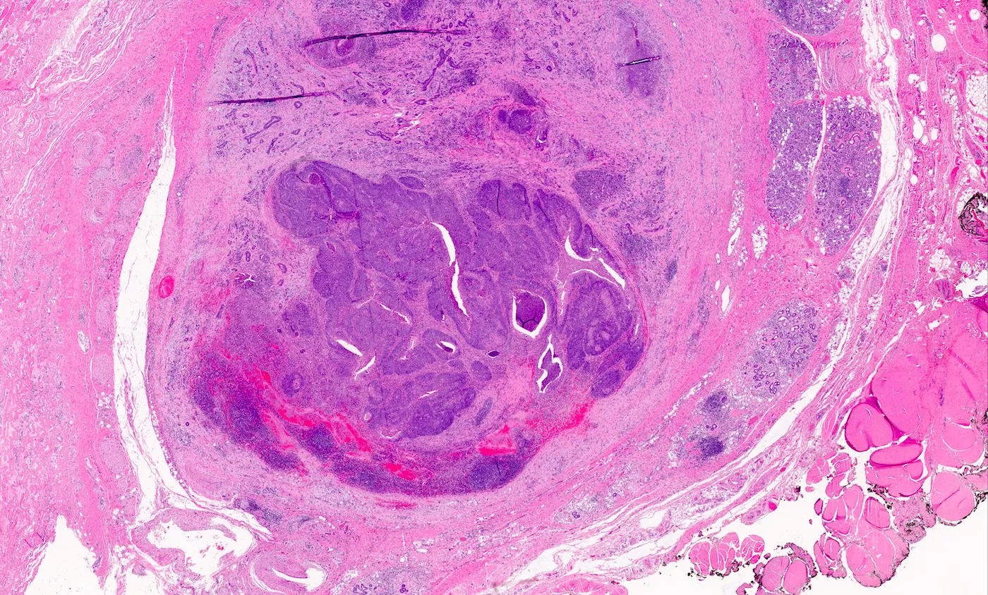
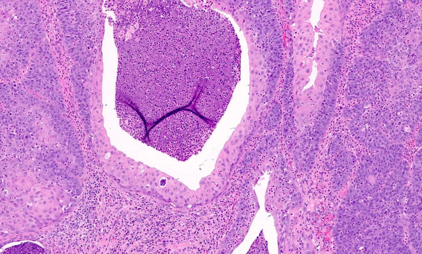
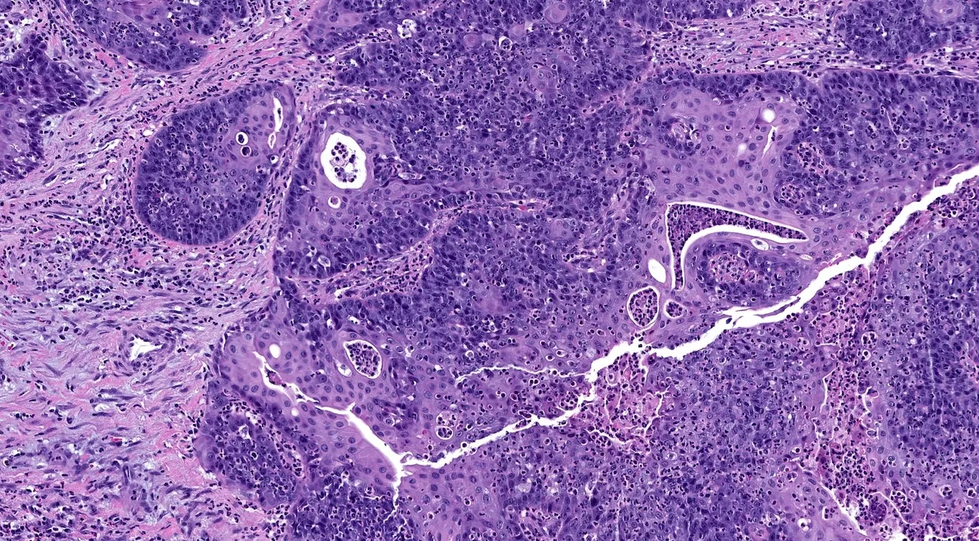
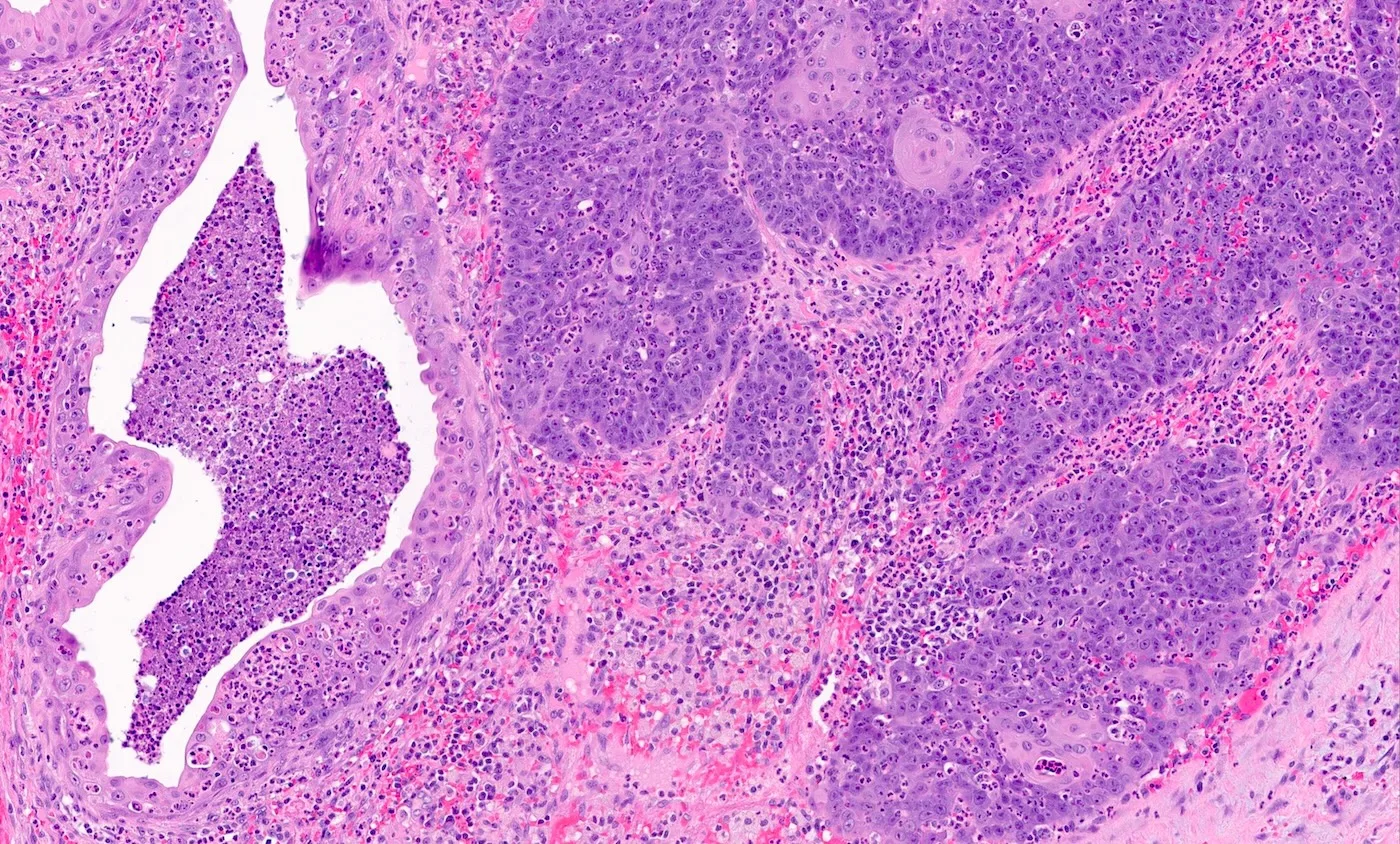
你的诊断是什么?
诊断
NUT 癌(NUT carcinoma)
测验题(答案见文末)
1. 头颈部 NUT 癌最常见的部位是?
- 口腔
- 腮腺
- 鼻腔鼻窦
- 下颌下腺
- 甲状腺
2. 下列哪项特征可确诊 NUT 癌?
- 癌表达神经内分泌标记(如 synaptophysin 与 chromogranin)
- 已除外 HPV 及 EBV 相关鳞状细胞癌
- 二代测序显示 NSD3::NUTM1 融合
- NUT 免疫组化仅见胞质着色
- p40 阳性、伴散在突然角化的癌
染色
讨论
NUT 癌是一种以 NUTM1 重排为特征的癌,可通过 NUT 免疫组化呈斑点状核着色,或通过分子检测(如荧光原位杂交、RT-PCR 或二代测序)显示 NUTM1 融合来证实。
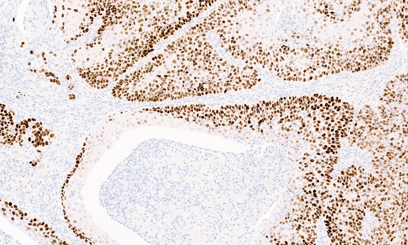
最常见的融合伙伴为 BRD4,其次为 BRD3 与 NSD3(Nat Rev Clin Oncol 2025;22:292)。其他融合伙伴包括 BRD2、ZNF592 与 ZNF532。本例肿瘤 NUT 呈斑点状核阳性,并检出 BRD4::NUTM1 融合,确诊为 NUT 癌。
NUT 癌最常见的部位是胸腔,其次为头颈部。头颈部以鼻腔鼻窦最常见(Histopathology 2024;85:317);但 NUT 癌亦可发生于其他部位,包括大涎腺、甲状腺、口腔、喉、鼻咽、泪腺、下咽、下颌骨及外耳道。
NUT 癌的典型组织学特征为:片状分布的单一形态、中等大小未分化细胞,核居中,核仁明显,胞质中等量、嗜酸性或嗜碱性。核分裂活跃、肿瘤性坏死及中性粒细胞浸润常见。约 55% 的病例可见突然角化与角化珠。
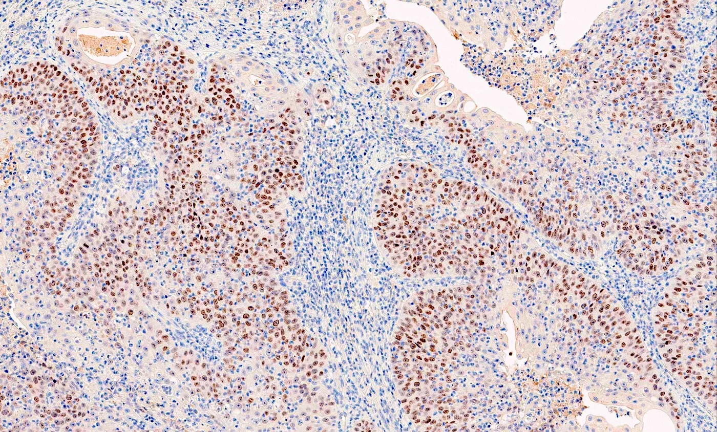
免疫组化上,NUT 癌通常表达鳞状标记,包括 p63、p40、角蛋白及高分子量角蛋白。不过,少数肿瘤可 p40 阴性(Arch Pathol Lab Med 2024;148:898)。
NUT 癌的非典型组织学与免疫表型包括:伴乳头状结构的分化型鳞状形态(Head Neck Pathol 2024;18:9,Histopathology 2024;85:317)、腺样分化(如本例所示)(Histopathology 2024;85:317),以及表达神经内分泌标记(如 synaptophysin、chromogranin 与 INSM1)而非鳞状标记(Arch Pathol Lab Med 2024;148:898,Histopathology 2024;85:317)。文献描述的其他少见特征还有基底样、横纹肌样及透明细胞形态。
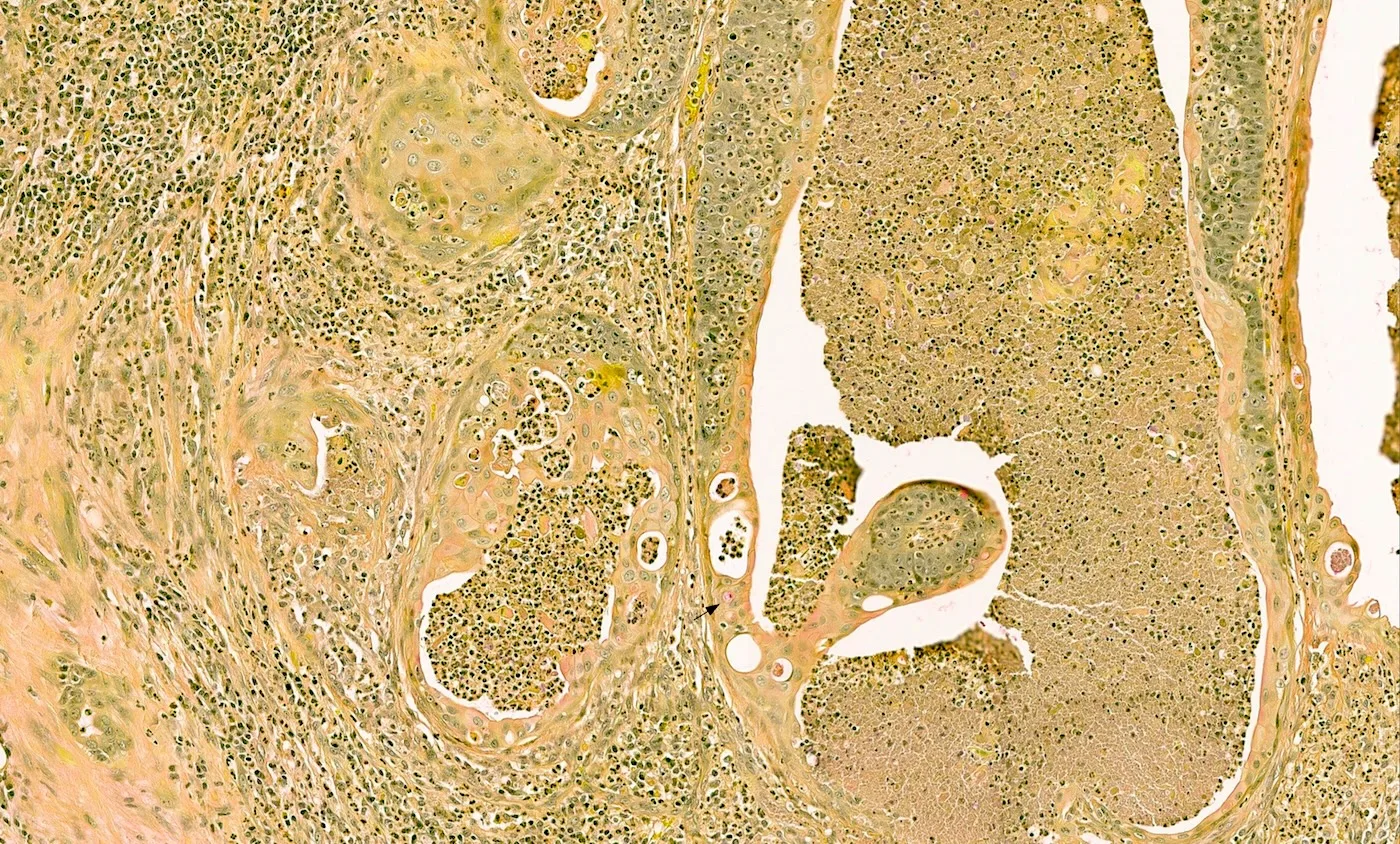
本例 HE 上呈鳞状分化,多灶见角化珠,鳞状标记弥漫阳性;同时存在局灶腺样分化,表现为腺腔形成及散在黏液细胞。形成腺体的肿瘤细胞 p40 阴性。主要鉴别诊断为转移至腮腺的腺鳞癌及高级别黏液表皮样癌。NUT 癌易被漏诊和误诊,因此对头颈部及胸部的低分化癌,NUT 免疫组化或 NUTM1 重排分子检测对诊断至关重要(Nat Rev Clin Oncol 2025;22:292)。
临床上,NUT 癌典型见于 20~39 岁的年轻成人;但报道病例的年龄跨度很大(18 天至 86 岁)(JNCI Cancer Spectr 2019;4:pkz094,Histopathology 2024;85:317)。肿瘤临床侵袭性强,中位生存期约 10 个月(JNCI Cancer Spectr 2019;4:pkz094)。
测验题答案
1. C. 头颈部 NUT 癌最常见的部位是鼻腔鼻窦,其次为大涎腺。
2. C. 二代测序检出 NSD3::NUTM1 融合。NUT 癌可通过 NUT 核阳性或分子检测证实 NUTM1 融合而确诊。D 不正确:NUT 阳性为核着色而非胞质着色。A、B、E 不正确:这些特征可见于 NUT 癌,但不能据以确诊 NUT 癌。
- Source: Case of the Month #567 (discussion page; blog 21 October 2026). Contributed by Bin Xu (MSKCC), reviewed by Marc Pusztaszeri (McGill).
- A 58 year old woman with a 3.0 cm parotid mass: squamous differentiation with keratin pearls plus focal glandular lumina and mucocytes (gland-forming cells p40 negative); speckled nuclear NUT positivity and BRD4::NUTM1 fusion.
- Takeaway: NUT carcinoma can show glandular, papillary squamous or neuroendocrine features and may be p40 negative; NUT IHC or NUTM1 testing is essential for poorly differentiated head and neck carcinomas. Four microscopic images, mucicarmine / p40 / NUT stains and two test questions.
Scope: the full Case of the Month #567 discussion page (clinical history, images, diagnosis, test questions, stains, discussion and answers); website news and advertising are not included.
Thanks to Dr. Bin Xu, Memorial Sloan Kettering Cancer Center, New York, New York, USA for contributing this case and discussion and to Dr. Marc Pusztaszeri, McGill University, Quebec, Canada for reviewing the discussion.
Clinical history
A 58 year old woman presents with a 3.0 cm mass in the parotid gland.
Microscopic images
What is your diagnosis?
Diagnosis
NUT carcinoma
Test question (answer at the end)
1. What is the most common site of NUT carcinoma in the head and neck region?
- Oral cavity
- Parotid gland
- Sinonasal tract
- Submandibular gland
- Thyroid gland
2. Which of the following features is diagnostic for NUT carcinoma?
- Carcinoma shows neuroendocrine marker expression (e.g., synaptophysin and chromogranin)
- Exclusion of HPV and EBV related squamous cell carcinoma
- Next generation sequencing shows NSD3::NUTM1 fusion
- NUT immunohistochemistry shows cytoplasmic staining only
- p40 positive carcinoma with scattered abrupt keratinization
Stains
Discussion
NUT carcinoma is a carcinoma characterized by NUTM1 rearrangement, demonstrated by nuclear NUT immunohistochemical staining with a speckled staining pattern or by molecular testing (e.g., fluorescence in situ hybridization, RT-PCR or next generation sequencing) showing NUTM1 fusion.
The most common fusion partners are BRD4, followed by BRD3 and NSD3 (Nat Rev Clin Oncol 2025;22:292). Other fusion partners include BRD2, ZNF592 and ZNF532. This specific tumor shows speckled NUT nuclear positivity and BRD4::NUTM1 fusion, confirming the diagnosis of NUT carcinoma.
The most common location of NUT carcinoma is the thoracic cavity, followed by the head and neck region. Within the head and neck region, the sinonasal tract is the most common site of origin (Histopathology 2024;85:317); however NUT carcinoma can also arise at other sites, including major salivary glands, the thyroid gland, oral cavity, larynx, nasopharynx, lacrimal gland, hypopharynx, mandibular bone and ear canal.
The typical histologic features of NUT carcinoma include sheets of monotonous intermediate sized undifferentiated cells with centrally located nuclei, prominent nucleoli and moderate amounts of eosinophilic or basophilic cytoplasm. Brisk mitotic activity, tumor necrosis and neutrophilic infiltrate are common. Abrupt keratinization and keratin pearls are seen in ~55% of cases.
By immunohistochemistry, NUT carcinoma typically expresses squamous markers, including p63, p40, keratins and high molecular weight keratins. However, a small subset of tumors may be negative for p40 (Arch Pathol Lab Med 2024;148:898).
Aberrant histologic and immunohistochemical features of NUT carcinoma include a differentiated squamous morphology with papillary architecture (Head Neck Pathol 2024;18:9, Histopathology 2024;85:317), glandular differentiation (as shown in this case) (Histopathology 2024;85:317) and expression of neuroendocrine rather than squamous markers (e.g., synaptophysin, chromogranin and INSM1) (Arch Pathol Lab Med 2024;148:898, Histopathology 2024;85:317). Other rare features that have been described include basaloid, rhabdoid and clear cell morphology.
This case shows squamous differentiation on H&E, with multifocal keratin pearls and diffuse positivity for squamous markers, as well as focal glandular differentiation characterized by glandular lumina and scattered mucocytes. Tumor cells forming glands are p40 negative. The key differential diagnoses include metastatic adenosquamous carcinoma to the parotid gland and high grade mucoepidermoid carcinoma. NUT carcinoma is prone to underdiagnosis and misdiagnosis. Therefore, NUT immunohistochemistry or molecular testing for NUTM1 rearrangements is essential for diagnosis of poorly differentiated carcinoma of head and neck and thoracic regions (Nat Rev Clin Oncol 2025;22:292).
Clinically, NUT carcinoma typically occurs in young adults in their 20s and 30s; however, cases have been reported across a wide age range (from 18 days to 86 years) (JNCI Cancer Spectr 2019;4:pkz094, Histopathology 2024;85:317). The tumor is clinically aggressive with a median survival of ~10 months (JNCI Cancer Spectr 2019;4:pkz094).
Test question answers
1. C. The most common site of NUT carcinoma in the head and neck region is the sinonasal tract, followed by the major salivary glands.
2. C. Next generation sequencing identifies an NSD3::NUTM1 fusion. NUT carcinoma can be diagnosed by demonstrating nuclear NUT immunopositivity or an NUTM1 fusion by molecular testing. Answer D is incorrect because NUT positivity is nuclear rather than cytoplasmic. Answers A, B and E are incorrect because these are features that may occur in NUT carcinoma but are not diagnostic for NUT carcinoma.