- 多中心研究(Bode、Suwala、Northcott、Kool、Schüller 等)报道 27 例松果体区肿瘤:DNA 甲基化谱自成一簇,与所有已知 CNS 肿瘤类别及现有松果体肿瘤类型均不匹配(分类器 v12.8 评分 <0.9),暂命名为「伴神经内分泌和上皮分化的松果体肿瘤」(PINNED)。
- 临床:男女比例均衡,平均 35 岁(7–75 岁);不足半数获全切除,随访中 4 例死于肿瘤、7 例进展,5 年无进展生存不足 25%。
- 形态与免疫组化:细胞丰富,可见坏死、核分裂、乳头、菊形团、上皮样细胞、印戒样细胞及「假腺样」结构;CK18、synaptophysin、OTX2 全部阳性,chromogranin A 多数阳性(常呈点状),CRX 恒定阴性,SPDEF 仅 1 例局灶阳性;ATRX 缺失/突变 4/16(25%)。
- 诊断陷阱:52% 原诊为 PTPR 或松果体区高级别神经上皮肿瘤,另有误作松果体母细胞瘤(PNET)、脉络丛癌或转移性神经内分泌肿瘤者。snRNA-seq 最接近 PTPR 但自成一组,投射到小鼠松果体发育图谱提示起源于松果体细胞祖细胞。附图 1–4 中英对照。
收录范围:Springer 开放获取全文(CC BY 4.0)——摘要、引言、材料与方法、结果、讨论及图 1–4 与图注中英对照;数据可用性、致谢、资助、作者信息、伦理声明、补充材料与参考文献未收录。
摘要
松果体区肿瘤是一类罕见肿瘤,可发生于各年龄段,在成人中占所有中枢神经系统(CNS)肿瘤的比例不足 1%,但在儿童中更为常见。在此,我们描述一种此前尚未被认识的松果体起源肿瘤类型,其特征为独特且一致的 DNA 甲基化谱,与任何已知的 CNS 肿瘤类别均不匹配,并有别于目前所有已被认识的松果体肿瘤实体。本研究队列(n = 27)的性别分布均衡,患者平均年龄为 35 岁(范围为 7–75 岁)。组织病理学分析显示,这是一种细胞密度高的肿瘤,可见不同程度的坏死、核分裂活性、乳头状结构、菊形团、上皮样细胞形态或印戒样细胞。这些肿瘤均表达细胞角蛋白 18、synaptophysin、chromogranin A 和 OTX2,而不表达 CRX。16 例肿瘤中有四例(25%)存在 ATRX 突变和/或免疫组织化学所显示的蛋白表达缺失。单细胞核 RNA 测序证实其在分子层面有别于其他松果体肿瘤,但显示其与松果体区乳头状肿瘤(PTPR)最为相似。将转录组数据投射至小鼠松果体发育的细胞轨迹,提示其细胞起源为松果体细胞祖细胞。综上,我们的研究结果界定了一种新的松果体区肿瘤类型,我们暂将其命名为“伴神经内分泌和上皮分化的松果体肿瘤”(PINNED)。认识这一肿瘤类型似乎对准确诊断松果体区肿瘤具有重要意义,并可能对未来的肿瘤分类和治疗策略产生影响。
引言
松果体是位于上丘脑的神经内分泌器官,主要负责产生和分泌褪黑素,后者调节昼夜节律 [1, 4]。在发育过程中,包括 OTX2 在内的齿同源盒基因对松果体发育起着至关重要的作用,并调控参与松果体细胞分化的转录因子 CRX 的表达 [12, 17, 30, 32]。
松果体区肿瘤是一组罕见肿瘤,约占儿童脑肿瘤的 3–11%,而在成人中占颅内中枢神经系统(CNS)肿瘤的比例不足 1% [10, 25]。2021 年世界卫生组织(WHO)分类认可了五种不同的松果体区肿瘤实体:松果体细胞瘤(PC)、中间分化型松果体实质肿瘤(PPTID)、松果体母细胞瘤(PB)、松果体区乳头状肿瘤(PTPR),以及松果体区促纤维增生性黏液样肿瘤(SMARCB1 突变型)[26]。
PC 是一种 WHO 1 级肿瘤,其特点是生长缓慢、预后良好。PC 可发生于各年龄段,但最常见于 30 至 60 岁的患者 [9, 10]。这一谱系的另一端是 PB,一种具有原始神经外胚层性质、高度侵袭性的 CNS WHO 4 级肿瘤 [24]。PB 主要发生于幼儿,尤其是 2 岁以下的儿童。DNA 甲基化谱分析已识别出五个不同的分子亚组(PB-GRP1A、PB-GRP1B、PB-GRP2、PIN-RB 和 PB-MYC/FOXR2),各自具有不同的分子谱、遗传驱动因素、人口学特征和临床结局 [5, 10, 12, 35]。
PPTID 因其中间型的组织学表现而带来诊断挑战。其定义为具有弥漫性或不明显分叶状结构的肿瘤 [26]。KBTBD4 基因中的小片段框内插入是 PPTID 的特征,可见于多达 80% 的病例 [21, 23, 39, 42]。PPTID 被归类为 WHO 2 至 3 级肿瘤。PPTID 包含两个不同的甲基化组,但其与组织学或临床参数的相关性仍不明确 [10, 25, 31]。
PTPR 是一种罕见的神经上皮肿瘤 [26]。其被归类为 WHO 2 级或 3 级肿瘤,发病年龄范围广,从 15 个月至 67 岁不等 [10]。组织学上,PTPR 的特征为上皮样的乳头状生长模式,以及菊形团样结构的存在 [10]。免疫组织化学上,其强表达 S100、细胞角蛋白、SPDEF、vimentin 和神经元特异性烯醇化酶(NSE)[15]。与松果体实质肿瘤不同,其始终不表达 CRX [34]。目前已识别出三个不同的 DNA 甲基化亚组,它们在基因组 CNV 负荷及无进展生存(PFS)方面似乎存在差异 [15, 40]。
最后,松果体中还报道了两种非常罕见的肿瘤类型。其中一种是松果体区促纤维增生性黏液样肿瘤(SMARCB1 突变型)(DMT SMARCB1 突变型)[26, 28],另一种则呈现出已知见于 WNT 型髓母细胞瘤的分子特征 [24]。
总体而言,由于组织病理学特征相互重叠及分子多样性,松果体区肿瘤的诊断仍具有挑战性。尽管如此,全基因组 DNA 甲基化谱分析已成为这一领域的重大进展,不仅用于肿瘤实体的亚分类,也提高了诊断准确性。
在此,我们描述一种此前尚未被认识、发生于松果体区的肿瘤类型(n= 27),其特征是独特且高度一致的全基因组 DNA 甲基化谱。这种表观遗传特征与任何已确立的 CNS 肿瘤类别均不相关,并有别于目前所有已被认识的松果体肿瘤实体。
材料与方法
患者队列
我们的队列由来自 27 例患者的 27 例松果体肿瘤组成。纳入标准为具有可用的 DNA 甲基化数据以及基本临床和神经病理学注释;分子数据质量不足的病例被排除于后续分析之外。本研究纳入的病例均在上传至 www.molecularneuropathology.org 后,通过分类器数据库获取。相应的临床和组织病理学数据由提交机构提供,并通过与送检神经病理中心直接沟通获取额外材料或信息。
参考样本包括 166 例既往已发表的松果体肿瘤病例 [3, 22, 24, 36, 37, 40],以及 76 例此前未发表的参考病例。所有参考病例均匹配中枢神经系统肿瘤甲基化分类器(v12.8)所定义的某一肿瘤组,校准评分为 0.9 或以上。
组织学与免疫组织化学(IHC)
我们按照标准流程,对所有可用肿瘤的石蜡切片进行苏木精-伊红(H&E)染色。评估依据 2021 年 WHO 中枢神经系统肿瘤分类进行 [26]。
采用 Ki-67 标记指数对肿瘤细胞增殖率进行半定量评估。按照制造商说明,使用 Ventana 系统(Roche)进行 IHC。IHC 分析使用针对 OTX2(Thermo Fisher,1H12C4B5,1:2000)、细胞角蛋白 18(Dako,GA618,即用型)、synaptophysin(Dako,M7315,1:500)、CRX(Santa Cruz,sc377138,1:200)、desmin(Dako,M0760,1:200)、chromogranin A(Dako,M0869,1:800)、SPDEF(OriGene,TA324209,1:200)和 ATRX(Atlas antibodies,HPA001906,1:300)的抗体。还对组织学切片进行目视检查,以观察坏死和钙化。
DNA 甲基化谱分析
按照制造商说明,使用 ReliaPrepTM FFPE gDNA Miniprep System(Promega)从福尔马林固定、石蜡包埋(FFPE)组织中分离 DNA。将约 100–500 ng DNA 进行亚硫酸氢盐转化(EZ DNA Methylation Kit;Zymo Research)。随后使用 DNA Clean & Concentrator-5(Zymo Research)和 Infinium HD FFPE DNA Restore Kit(Illumina)对转化后的 DNA 进行纯化和修复。最后,在 iScan 设备(Illumina)上使用以下 Illumina 芯片之一分析样本:HumanMethylation450 BeadChip array、Infinium MethylationEPIC BeadChip Kit 或 Infinium MethylationEPIC v2.0 Kit(均为 Illumina 产品)。我们使用中枢神经系统肿瘤甲基化分类器 [3],获得样本归属于已确立的 CNS 肿瘤类型的预测结果,该分类器可通过 https://app.epignostix.com 获取。该工具采用随机森林分类器评估全局 DNA 甲基化模式。根据该监督分类器评分,系统返回一个经实体校正的所谓校准评分,旨在指示样本与每一种已训练类型的基础参考数据集之间的相似性。我们的分析使用分类器 v12.8 版本,并按照该算法作者的建议,将 ≥0.9 的截断值视为显著匹配。我们来自 Capper 等人 [3] 数据集的参考样本包括用于德国癌症研究中心(DKFZ)中枢神经系统肿瘤甲基化分类器 v11b2 版本的病例,随后使用 v12.5 版本对其重新分类。
生物信息学
数据处理在 R 中完成(Microsoft Windows 下的 v4.3.2)。我们的肿瘤样本和参考队列的甲基化数据使用 1.48.0 版本的“minfi”软件包处理 [19]。该软件包利用越界探针估计并去除背景噪声,而染料偏倚归一化则使用部分对照探针 [38]。检测 p 值低于 0.01 截断点,点和重复病例通过基因型匹配被识别并去除。使用“getBeta”函数计算 beta 值,随后使用“DMRcate”软件包的“rmSNPandCH”函数过滤探针 [29]。该函数剔除在 CpG 位点两个核苷酸范围内含有 SNP 的任何探针,与性染色体相关的探针及不在 BeadChip 上的位点也被弃除。为进行可视化,根据标准差选取变异最大的 10,000 个甲基化探针。使用“Rtsne”软件包(v0.17)[20],设定 dims = 2、perplexity = 15、iterations = 10,000,绘制 t-SNE 图和热图。使用“conumee”(v1.36.0)和“conumee2.0”(v2.0)软件包分析拷贝数谱中的缺失和扩增 [6]。选择 ±0.4 的阈值识别相关的杂合性缺失或增益,使用 ±0.8 的截断值判定纯合性缺失或扩增。对我们所有样本的拷贝数变异(CNV)谱均进行了目视检查以作验证。
二代测序
从 FFPE 肿瘤组织中提取 RNA 和 DNA,并按照制造商说明,使用 TruSight Oncology 500 Assay(TSO500,Illumina)进行分析。DNA 分析的序列数据比对至人类参考基因组 hg19,RNA 分析的序列数据比对至 hg38。另对 RNA 测序数据进行基因融合评估。使用 1000 Genomes Project、dbSNP、ClinVar 和 COSMIC 的信息进行变异注释。仅将等位基因频率 ≥ 5% 且总靶标覆盖深度 ≥40× 的变异纳入进一步分析。被归类为良性、可能良性或已知多态性的变异不再进行进一步评估。
单细胞核 RNA 测序(snRNA-seq)
使用 Next-GEM Chromium Single Cell Flex Gene Expression Assay(10× Genomics)对福尔马林固定石蜡包埋(FFPE)组织进行 snRNA-seq。按照“从 FFPE 组织切片中分离细胞以进行 Chromium 固定 RNA 谱分析”方案(文档编号 CG000632 Rev B,10× Genomics),采用 gentleMACS Octo Dissociator(#130–134-029,Miltenyi Biotec)工作流程分离细胞。使用含 Liberase TH(#5401151001,MilliporeSigma)的酶混合物解离 FFPE 组织卷。用 4′,6-二脒基-2-苯基吲哚(DAPI,1:5000)对解离后的细胞核悬液染色。使用 BD FACSAria-Fusion™ 流式细胞仪对 DAPI 阳性细胞核进行荧光激活细胞分选,以尽量减少红细胞和碎屑污染。探针杂交在德国汉堡-埃彭多夫大学医学中心内部完成,使用 Chromium Fixed RNA Human Transcriptome kit PN-1000476(4 rxns × 16 BC),按照“用于多重化样本的 Chromium 固定 RNA 谱分析试剂盒”方案(文档编号 CG000527 Rev D,10× Genomics)操作。共处理了四例 PINNED 肿瘤和一例 PB 样本。文库制备在 UKE 单细胞核心平台(汉堡)完成。所得多重化文库由 CeGaT(德国图宾根)测序。我们的目标是每个文库回收 80,000 个细胞。使用 Cell Ranger v8.0.1,以默认参数运行 cellranger“multi”命令,生成特征-条形码矩阵。使用 GRCh38 参考基因组(refdata-gex-GRCh38-2020-A)和 Chromium Human Transcriptome Probe Set v1.0.1 进行比对和定量。审查 Cell Ranger 测序指标报告,以评估样本和测序质量。
后续分析在 R(v4.3.2)中进行。除非另有说明,snRNA 测序数据使用“Seurat”软件包(v4.4.0)联合“SeuratObject”(v4.1.4)进行分析 [14]。质量控制后,检测到的特征数在 200 至 5000 之间的细胞予以保留。排除每个样本中在少于 10 个细胞内检测到的特征,以及线粒体转录本含量超过 5% 的细胞。使用“AggregateExpression”函数生成每个样本的伪总体表达谱。按照此前所述方法 [12],将 PINNED 肿瘤细胞通过 UMAP 投射至参考的发育中松果体。
结果
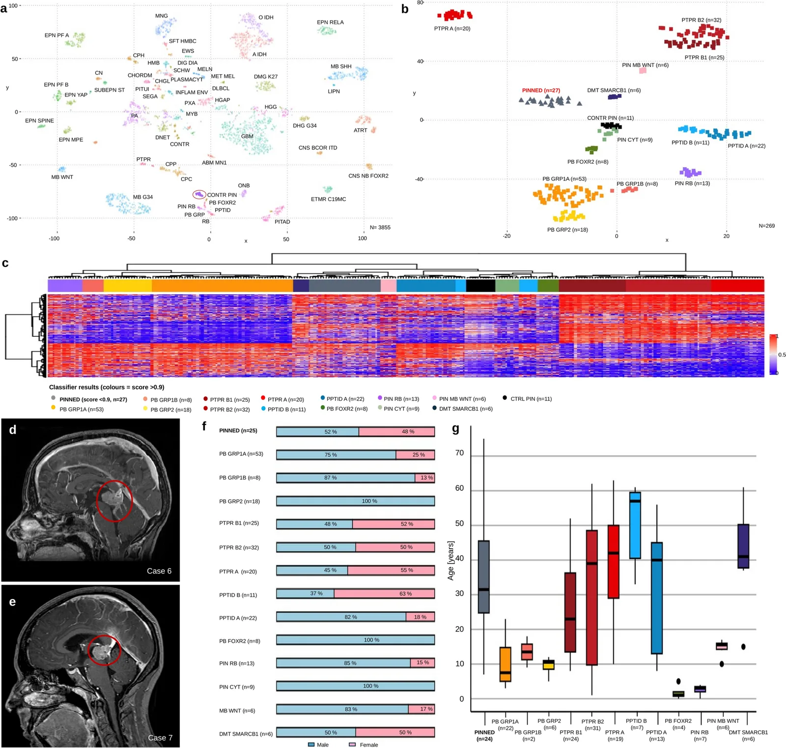
我们研究了一个由 27 例肿瘤组成的队列,基于全局 DNA 甲基化谱分析,这些肿瘤与所有参考松果体肿瘤样本分别聚类,t-SNE 和层次聚类的可视化结果显示了这一点。此外,使用中枢神经系统肿瘤甲基化分类器时,这些肿瘤不匹配任何此前描述的肿瘤实体(评分 < 0.9,版本 v12.8,图 1a–c,补充表 1)。这些肿瘤被暂命名为“伴神经内分泌和上皮分化的松果体肿瘤”(PINNED,详见下文)。为进一步表征这些肿瘤,我们评估了其解剖部位以及患者的年龄分布和性别均衡性。如代表性病例所示,肿瘤位于松果体区内(图 1d, e)。患者平均年龄为 35 岁(范围:7–75 岁),52% 为男性(图 1f, g)。仅有不足一半的患者实现了全切除。现有随访数据显示,四例患者死于该疾病,另有七例疾病进展,5 年无进展生存低于 25%(补充表 1)。总之,这些结果描述了一种局限于松果体、具有独特 DNA 甲基化聚类模式的肿瘤,其患者年龄分布广、性别分布均衡,临床病程可能包括疾病进展甚至死亡。
接下来,我们更仔细地考察了这些肿瘤的组织病理学和免疫组织化学特征。图 2a–j 展示了五个代表性病例,显示整个队列的形态具有相当大的异质性(图 2a–e:低倍;图 2f–j:高倍)。除明显的核分裂象外,病例中反复出现实性结构,与邻近神经组织分界清楚,并可见钙化(2/11 例肿瘤(18%),图 2a)、坏死(5/16 例肿瘤(31%),图 2f)或巢状生长模式(图 2b)。部分病例出现的印戒样形态(图 2g)弥漫可见于肿瘤组织中,与特定结构无明显关系。其他特征包括具有真性腔隙、紧密排列的管状结构,形成“假腺样”结构(图 2c, h)、上皮样细胞形态(图 2d, i),或排列致密的未分化肿瘤组织(图 2e, j)。在部分标本中,整个肿瘤内可见多核、空泡化细胞或横纹肌样细胞,横纹肌样细胞与假乳头状结构存在一定关联。如果存在真性菊形团,则数量较多且遍布肿瘤各部分。
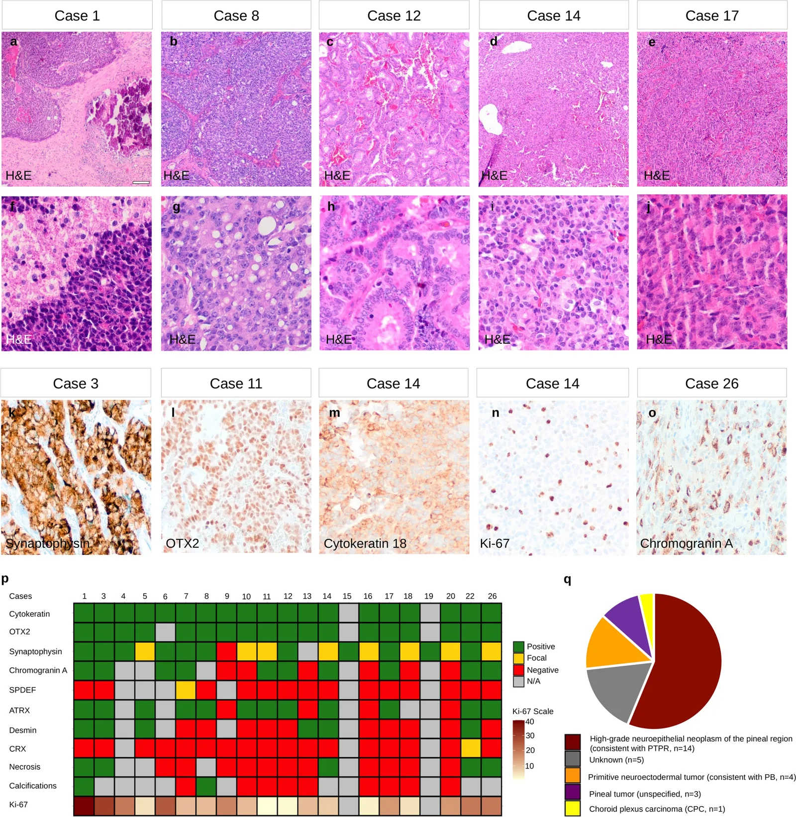
免疫组织化学上,这些肿瘤显示出特征性表达谱,均表达 synaptophysin(图 2k)、OTX2(图 2l)和细胞角蛋白 18(图 2m),即使在分化较差的肿瘤细胞中,这些标志物也全部表达。平均 Ki-67 增殖指数为 10%(图 2n)。synaptophysin 在包括印戒样细胞在内的部分肿瘤细胞胞质中呈强而弥漫的表达,可见于单个细胞或呈阳性细胞灶,有时表现为围腔性分布。其他肿瘤则表现为广泛表达。OTX2 和细胞角蛋白 18 在所有病例中均为阳性。相比之下,chromogranin A 在大多数病例中表达(图 2o),且常呈点状或颗粒状模式。在肿瘤的某些区域,chromogranin A 表达更强、范围更广。其他肿瘤仅有散在单个细胞显示强胞质染色而无顶端增强,或可见含空泡的肿瘤细胞呈偏心性 chromogranin A 表达(印戒样细胞)。部分肿瘤的组织中还可见散在单个 desmin 阳性肿瘤细胞,而 CRX 表达均缺失。仅一例肿瘤显示局灶性 SPDEF 表达,该标志物此前被描述为 PTPR 的标志物 [15];其余所有肿瘤均为阴性。免疫组织化学分析显示,4/16 例肿瘤(25%)存在 ATRX 蛋白表达缺失。综上,组织病理学特征表明其具有上皮性和神经内分泌组织学特征,均表达细胞角蛋白 18、synaptophysin 和 OTX2,并常为 chromogranin A 阳性。所有组织学结果的汇总见图 2p。基于这些特征,我们暂将这一肿瘤组命名为:“伴神经内分泌和上皮分化的松果体肿瘤”(PINNED)。
送检机构最初给出的组织病理学诊断见图 2q。27 例肿瘤中有十四例(52%)最初被诊断为 PTPR、松果体区高级别神经上皮肿瘤,或以其他与 PTPR 一致的描述命名。四例肿瘤被描述为原始神经外胚层肿瘤,与 PB 一致。三例肿瘤仅被归类为松果体肿瘤,一例肿瘤被描述为最接近脉络丛癌。至少四例病例曾讨论过松果体区转移性神经内分泌肿瘤的诊断。
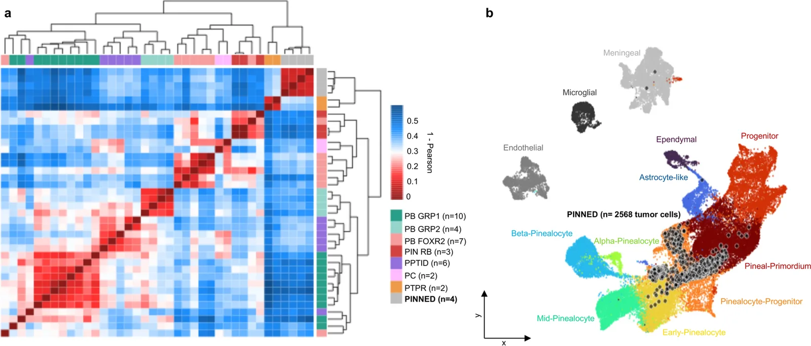
为了解 PINNED 的转录组,将这些肿瘤的转录组与其他松果体区肿瘤进行比较,并更好地理解肿瘤的细胞起源,我们生成并分析了四例 PINNED 肿瘤的 snRNA-seq 数据。基于相似性热图所呈现的基因共表达模式,将肿瘤细胞核的伪总体数据与另外 34 例松果体和松果体区肿瘤样本 [12] 进行比较时,PINNED 与 PTPR 的转录组相似性最高,但形成一个独立的肿瘤组(图 3a)。将转录组数据投射至小鼠松果体发育图谱 [12] 后发现,PINNED 肿瘤细胞与松果体细胞祖细胞最为接近(图 3b)。
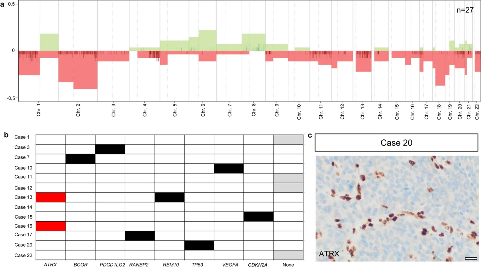
最后,我们尝试识别这些肿瘤潜在的遗传驱动因素。基因组谱分析显示多种拷贝数改变,但无反复出现的局灶扩增或缺失(图 4a)。对 13 例肿瘤使用 TSO500 面板进行了靶向测序(图 4b)。在这些病例中,两例肿瘤检出 ATRX 突变,两者的免疫组织化学也均显示 ATRX 蛋白表达缺失(图 4c)。此外,在通过总体 RNA 测序分析的 17 例肿瘤中,未检出具有临床相关性的基因融合(数据未展示)。总之,ATRX 改变是我们队列中识别出的最常见分子异常,见于 25% 的肿瘤。
讨论
我们的研究结果支持在松果体区肿瘤谱系中存在一种此前尚未被认识的肿瘤实体。使用中枢神经系统肿瘤分类器时,这些肿瘤均被分类为“不匹配”,其最高校准评分常被赋予髓肌母细胞瘤(MB_MYO)或非 WNT/非 SHH 型髓母细胞瘤(MB_G34)[3]。值得注意的是,没有任何一例肿瘤匹配中枢神经系统肿瘤甲基化分类器(v12.8)中任何已确立的甲基化类别,提示现有参考队列未充分涵盖这些肿瘤。无监督分析表明,它们确实在松果体区肿瘤谱系中形成了一个明显独立的组,但从表观遗传学角度不应被纳入髓母细胞瘤谱系。这些聚类结果在多次 t-SNE 和热图可视化中保持一致,并在不同规模的参考队列中均表现稳健。综上,这些发现表明,本文描述的肿瘤代表了一个在生物学上独立的组。
从临床角度看,该队列也不同于几种已确立的松果体肿瘤实体。我们的队列性别分布均衡、年龄跨度广,中位年龄为 35 岁。在松果体区肿瘤范围内,这种人口学特征更接近 PTPR [40, 41],而非主要影响幼儿且常以男性为主的 PB [10, 40]。尽管我们的队列规模有限,其 5 年无进展生存似乎低于 PTPR 和 PB。
此外,我们队列中的许多肿瘤最初被神经病理医师分类为 PTPR,很可能是由于其局灶性乳头状生长模式,或存在偶见的菊形团和上皮样细胞形态。这种相似性还体现在缺乏 CRX 表达以及细胞角蛋白 18 的强表达:CRX 是视网膜和松果体的特异性标志物 [11],通常不在 PTPR 中表达 [7]。然而,若干发现不支持将其分类为 PTPR 的一个亚组。首先,我们的队列均显示明确的 synaptophysin 和 chromogranin A 表达,并且大多不表达 SPDEF,而 Heim 等人报道 79% 的 PTPR 表达 SPDEF [15]。此外,经集中复核,这些肿瘤表现出独特的组织病理学特征。与 PTPR 相比,其肿瘤结构大多为实性。乳头状结构缺如或仅局灶存在,且轮廓较不清晰。重要的是,尽管形态存在部分重叠,免疫组织化学与组织病理学的综合结果支持将其与目前已被认识的 PTPR 肿瘤实体区分开来。
尽管这些样本在转录层面与两例可用的 PTPR 参考肿瘤最为相似,但四例 PINNED 的伪总体 snRNA-seq 分析表明,从无监督层次聚类来看,它们在分支进入任何已确立的 PTPR 亚组之前即已分离。
此外,拷贝数谱分析未发现反复出现的染色体增益或缺失,这与 PTPR 不同:所有接受分析的 PTPR 肿瘤均出现染色体 10 缺失,同时还出现反复出现的染色体 3 和 22q 缺失及染色体 8p 增益 [15]。类似地,PTPR、PB 或 PPTID 中观察到的反复出现的突变,例如 PTEN, RB1, DROSHA 或 KBTBD4, 的变异,在 PINNED 中均不存在。值得注意的是,16 例接受分析的肿瘤中有四例检出 ATRX 表达缺失,这是我们队列中最常见的分子改变。此前曾在独立于我们队列、被诊断为 PPTID 的两例松果体肿瘤中报道 ATRX 改变 [27]。然而,这类改变尚未成为已确立的松果体肿瘤实体中反复出现的定义性特征,因此,其在我们队列中相对较高的发生频率尤其耐人寻味。
分泌性和神经内分泌结构特征的存在,包括巢状生长模式、菊形团形成和印戒样细胞,提示这些肿瘤具有神经内分泌分化。原发性颅内神经内分泌肿瘤极为罕见,文献中仅报道了少量病例 [2, 5, 8, 13, 16, 18, 33]。值得注意的是,此前报道的两例松果体神经内分泌肿瘤均表达 synaptophysin、chromogranin A 和细胞角蛋白 [5, 13],这些发现与我们队列中观察到的表型明显重叠。识别 PINNED 至关重要,因为其组织学表现和细胞角蛋白表达也可能类似转移性癌,使后者成为成人患者中的一项重要鉴别诊断。
尽管具有这些神经内分泌和上皮特征,使用小鼠松果体参考数据集进行的起源细胞分析表明,其与松果体发育存在明确关联,与松果体细胞祖细胞的相似性最高 [12]。不同亚型的 PB 也有类似观察,而 PTPR 在转录层面与松果体原基相似,这进一步支持 PINNED 很可能并非 PTPR 的一个子集的观点。松果体原基和 PTPR 中均未观察到 CRX 表达。另一方面,松果体细胞祖细胞虽较成熟松果体细胞表达程度低,但仍表达 CRX;尽管 PB 和 PINNED 均与松果体细胞祖细胞的相似性最高,为何 PB 中 CRX 表达相当强,而 PINNED 中却不表达,仍不清楚。尽管如此,我们的发现提供了证据,表明 PINNED 可能起源于松果体发育谱系内部,并提示我们的队列是一种松果体肿瘤实体,而非位于松果体区的异位肿瘤。
本研究存在若干局限性。尽管就一种罕见且此前尚未被认识的甲基化类别而言,我们报道的队列规模相对较大,但病例总数仍然有限,仍需独立的外部验证队列。此外,肿瘤发生的遗传驱动因素在很大程度上仍不明确,由于随访数据有限,目前尚无法界定受累患者的临床病程和长期预后。虽然整合的表观遗传、组织病理学和分子数据均支持识别出一种独立的肿瘤实体,但仍需在规模更大且独立的队列中开展进一步研究,以证实这些发现的可重复性,并更好地界定该队列完整的生物学、遗传学和临床谱系。
总之,我们的研究结果支持存在一种具有独特临床、组织病理学和分子特征的新松果体区肿瘤类型,我们暂将其命名为 PINNED。
- A multicentre study (Bode, Suwala, Northcott, Kool, Schüller and colleagues) of 27 pineal region tumours that form a distinct DNA methylation cluster, matching no known CNS tumour class or recognized pineal entity (classifier v12.8 score <0.9), provisionally named Pineal Neoplasm with Neuroendocrine and Epithelial Differentiation (PINNED).
- Clinical: balanced sex distribution, mean age 35 years (7–75); gross total resection in fewer than half; on follow-up 4 died of disease and 7 progressed, with 5-year progression-free survival below 25%.
- Morphology and IHC: highly cellular tumours with necrosis, mitoses, papillae, rosettes, epithelioid and signet-ring cells and “pseudoglandular” tubes; cytokeratin 18, synaptophysin and OTX2 positive in all, chromogranin A in most (often dot-like), CRX uniformly absent, SPDEF focal in one; ATRX loss/mutation in 4/16 (25%).
- Pitfall: 52% were originally diagnosed as PTPR or high-grade neuroepithelial pineal tumour; others as PNET/pineoblastoma, choroid plexus carcinoma or metastatic neuroendocrine tumour. snRNA-seq: closest to PTPR yet distinct; mapping to murine pineal development points to pinealocyte progenitors. Figures 1–4 included.
Scope: Springer open-access full text (CC BY 4.0) — abstract, introduction, materials and methods, results, discussion and Figures 1–4 with captions. Data availability, acknowledgements, funding, author information, ethics declarations, supplementary material and references are not included.
Abstract
Pineal region tumors are rare neoplasms that occur in all ages and account for less than 1% of all central nervous system (CNS) tumors in adults but are more frequent in children. Here, we describe a previously unrecognized tumor type of pineal origin characterized by a unique and consistent DNA methylation profile that does not match any known CNS tumor class and is distinct from all currently recognized pineal tumor entities. The presented cohort (n = 27) displayed a balanced sex distribution with a mean patient age of 35 years (range 7–75 years). The histopathological analyses demonstrated a highly cellular tumor showing various degrees of necrosis, mitotic activity, papillary structures, rosettes, epithelioid cytology, or signet-ring cells. The tumors consistently expressed cytokeratin 18, synaptophysin, chromogranin A, and OTX2, whereas CRX expression was absent. Four out of 16 tumors (25%) showed ATRX mutations and/or loss of protein expression as revealed by immunohistochemistry. Single-nucleus RNA sequencing confirmed molecular distinctiveness from other pineal neoplasms but revealed the closest similarities to papillary tumors of the pineal region (PTPR). Projection of the transcriptome data onto the cellular trajectory of the developing murine pineal gland suggests that pinealocyte progenitors are the cellular origins. Taken together, our findings define a novel type of pineal region tumor that we provisionally named ‘Pineal Neoplasm with Neuroendocrine and Epithelial Differentiation’ (PINNED). Recognition of this tumor type appears to be important for the accurate diagnosis of pineal region tumors and may have implications for future tumor classification and treatment strategies.
Introduction
The pineal gland is a neuroendocrine organ located in the epithalamus and primarily responsible for the production and secretion of melatonin, which regulates the circadian rhythm [1, 4]. During development, orthodenticle homeobox genes, including OTX2, play an essential role in pineal development and regulate the expression of CRX, a transcription factor involved in pinealocyte differentiation [12, 17, 30, 32].
Pineal region tumors represent a rare group of neoplasms, accounting for approximately 3–11% of pediatric brain neoplasms, while constituting less than 1% of intracranial central nervous system (CNS) tumors in adults [10, 25]. The 2021 World Health Organization (WHO) classification recognizes five distinct tumor entities of the pineal region: pineocytoma (PC), pineal parenchymal tumor of intermediate differentiation (PPTID), pineoblastoma (PB), papillary tumor of the pineal region (PTPR), and desmoplastic myxoid tumor of the pineal region, SMARCB1-mutant [26].
PC is a WHO grade 1 tumor characterized by slow growth and a favorable prognosis. PC can occur at all ages but is most frequently found in patients between 30 and 60 years [9, 10]. At the opposite end of the spectrum is PB, a highly aggressive CNS WHO grade 4 tumor of primitive neuroectodermal nature [24]. PBs predominantly affect young children, particularly those under 2 years of age. DNA methylation profiling has identified five distinct molecular subgroups (PB-GRP1A, PB-GRP1B, PB-GRP2, PIN-RB, and PB-MYC/FOXR2), which are characterized by distinct molecular profiles, genetic drivers, demographic features, and clinical outcomes [5, 10, 12, 35].
PPTID presents a diagnostic challenge due to its intermediate histological appearance. It is defined as a neoplasm with a diffuse or vaguely lobulated architecture [26]. Small in-frame insertions in the KBTBD4 gene are characteristic for PPTID and are found in up to 80% of cases [21, 23, 39, 42]. PPTIDs are classified as WHO grade 2 to 3 tumors. PPTID comprises two distinct methylation groups, although correlation with histological or clinical parameters remains unclear [10, 25, 31].
PTPR is a rare neuroepithelial tumor [26]. It is classified as a WHO grade 2 or 3 neoplasm and can occur in a wide age range, from 15 months to 67 years [10]. Histologically, PTPRs are characterized by an epithelial-like, papillary growth-pattern, and the presence of rosette-like structures [10]. Immunohistochemically, they show a strong expression of S100, cytokeratin, SPDEF, vimentin, and neuron-specific enolase (NSE) [15]. In contrast to pineal parenchymal tumors, CRX expression is consistently absent [34]. Three distinct DNA methylation subgroups have been identified, which appear to vary in genomic CNV load as well as progression-free survival (PFS) [15, 40].
Finally, two very rare tumor types have been described in the pineal gland. One of them was a desmoplastic myxoid tumor of the pineal region, SMARCB1-mutant (DMT SMARCB1-mutant) [26, 28], and the other presented molecular features known from WNT medulloblastoma [24].
Together, the diagnosis of pineal region tumors remains challenging due to their overlapping histopathological features and molecular diversity. Nevertheless, genome-wide DNA methylation profiling has represented a major advance in this field, not only in the subclassification of tumor entities but also in improving diagnostic accuracy.
Here, we describe a previously unrecognized tumor type arising in the pineal region (n= 27) characterized by a unique and highly consistent genome-wide DNA methylation profile. This epigenetic signature does not correlate with any established CNS tumor class and is distinct from all currently recognized pineal tumor entities.
Materials and methods
Patient cohort
Our cohort consisted of 27 pineal tumors from 27 patients. Inclusion criteria were the availability of DNA methylation data and basic clinical and neuropathological annotations; cases with insufficient molecular data quality were excluded from further analyses. The cases included in this study were accessed through the classifier database following upload to www.molecularneuropathology.org. Corresponding clinical and histopathological data were obtained from the submitting institutions, with additional material or information retrieved through direct communication with the referring neuropathology centers.
The reference samples included 166 previously published cases of pineal tumors [3, 22, 24, 36, 37, 40], along with 76 previously unpublished reference cases. All reference cases matched a tumor group as defined by the CNS Tumor Methylation Classifier (v12.8) with a calibrated score of 0.9 or higher.
Histology and immunohistochemistry (IHC)
We performed hematoxylin and eosin (H&E) stains on paraffin sections of all available tumors, following standard procedures. The evaluation was conducted per the 2021 WHO classification of tumors of the CNS [26].
The tumor cell proliferation rate was semi-quantitatively assessed using the Ki-67 labelling index. IHC was performed using a Ventana System (Roche) according to the manufacturer’s specifications. IHC analyses were conducted using antibodies against OTX2 (Thermo Fisher, 1H12C4B5, 1:2000), cytokeratin 18 (Dako, GA618, ready-to-use), synaptophysin (Dako, M7315, 1:500), CRX (Santa Cruz, sc377138, 1:200), desmin (Dako, M0760, 1:200), chromogranin A (Dako, M0869, 1:800), SPDEF (OriGene, TA324209, 1:200), and ATRX (Atlas antibodies, HPA001906, 1:300). Histological sections were also visually inspected for necrosis and calcifications.
DNA methylation profiling
DNA was isolated from formalin-fixed, paraffin-embedded (FFPE) tissue using the ReliaPrepTM FFPE gDNA Miniprep System (Promega) according to the manufacturer’s instructions. About 100–500 ng of DNA were bisulfite converted (EZ DNA Methylation Kit; Zymo Research). Afterwards, the DNA Clean & Concentrator-5 (Zymo Research) and the Infinium HD FFPE DNA Restore Kit (Illumina) were employed to clean and restore the converted DNA. Finally, samples were analyzed using one of the following Illumina arrays on an iScan device (Illumina): HumanMethylation450 BeadChip array, Infinium MethylationEPIC BeadChip Kit, or Infinium MethylationEPIC v2.0 Kit (all Illumina). We obtained the affiliation predictions for established tumor types of the CNS by using the CNS Tumor Methylation Classifier [3], available at https://app.epignostix.com. This tool evaluates the global DNA methylation pattern using a random forest classifier. From this supervised classifier score, an entity-adjusted, so-called calibrated score is returned that intends to indicate the similarity between the sample and the underlying reference dataset for each trained type. We used the classifier version v12.8 for our analyses and considered the cut-off value of ≥0.9, as suggested by the authors of the algorithm, to indicate a significant match. Our reference samples from the Capper et al. [3] dataset encompass cases used for the v11b2 version of the German Cancer Research Center (DKFZ) CNS Tumor Methylation Classifier, which were then reclassified using the v12.5 version.
Bioinformatics
Data processing was performed in R (v4.3.2 in Microsoft Windows). Our tumor samples and the reference cohort methylation data were processed using the “minfi” package in version 1.48.0 [19]. This package estimates and removes the background noise from out-of-bound probes, while the dye-bias normalization uses a subset of the control probes [38]. Detection p values were below the 0.01 cut-off, point and duplicate cases were recognized and removed using genotype matching. The beta values were calculated using the ‘getBeta’ function, followed by filtering probes using the ‘rmSNPandCH’ function of the “DMRcate” package [29]. This function eliminated any probes containing SNPs within two nucleotides of the CpG sites, probes associated with the sex chromosomes and sites not on the BeadChips were discarded. For visualization, the 10,000 most variable methylation probes were chosen based on standard deviation. The package “Rtsne” (v0.17) [20], dims = 2, perplexity = 15, and iterations = 10,000 was used to create t-SNE and Heatmap plots. Losses and amplifications in the copy number profiles were analyzed using the packages “conumee” (v1.36.0) and “conumee2.0” (v2.0) [6]. To identify relevant heterozygous losses or gains, a threshold of ±0.4 was chosen and a cut-off of ±0.8 was used to determine homozygous losses or amplifications. All of our samples’ copy-number variation (CNV) profiles were visually examined for validation.
Next-generation sequencing
RNA and DNA were extracted from FFPE tumor tissue and analyzed using the TruSight Oncology 500 Assay (TSO500, Illumina) according to the manufacturer’s instructions. The sequence data were aligned to the human reference genome hg19 for DNA analyses and hg38 for RNA analyses. RNA sequencing data were additionally evaluated for gene fusions. The variants were annotated using information from the 1000 Genomes Project, dbSNP, ClinVar, and COSMIC. Only variants with an allele frequency ≥ 5% and a total target coverage ≥40× were considered for further analysis. Variants classified as benign, likely benign, or representing known polymorphisms were excluded from further evaluation.
Single-nucleus RNA sequencing (snRNA-seq)
SnRNA-seq was performed on formalin-fixed paraffin-embedded (FFPE) tissue using the Next-GEM Chromium Single Cell Flex Gene Expression Assay (10× Genomics). Cells were isolated according to the protocol “Isolation of Cells from FFPE Tissue Sections for Chromium Fixed RNA Profiling” (Document Number CG000632 Rev B, 10× Genomics) using a gentleMACS Octo Dissociator (#130–134-029, Miltenyi Biotec) workflow. FFPE tissue scrolls were dissociated with an enzyme mix containing Liberase TH (#5401151001, MilliporeSigma). The dissociated nuclei suspension was stained with 4′,6-diamidino-2-phenylindole (DAPI, 1:5000). A BD FACSAria-Fusion™ Flow Cytometer was used to perform fluorescence-activated cell sorting of DAPI-positive nuclei to minimize erythrocytes and debris contamination. Probe hybridization was performed in-house in the University Medical Center Hamburg-Eppendorf (Germany) using the Chromium Fixed RNA Human Transcriptome kit PN-1000476 (4 rxns × 16 BC) according to the protocol “Chromium Fixed RNA Profiling Reagent Kits for Multiplexed Samples” (Document Number CG000527 Rev D, 10× Genomics). In total, four PINNED tumors and one PB sample were processed. Library preparation was performed at the UKE Single Cell Core Facility (Hamburg). The resulting multiplexed libraries were sequenced by CeGaT (Tübingen, Germany). We aimed for a target recovery of 80,000 cells per library. Cell Ranger v8.0.1 was used to generate feature-barcode matrices using the cellranger ‘multi’ command with default parameters. The GRCh38 reference genome (refdata-gex-GRCh38-2020-A) and Chromium Human Transcriptome Probe Set v1.0.1 were used for alignment and quantification. Cell Ranger sequencing metrics reports were reviewed to assess sample and sequencing quality.
Subsequent analyses were performed in R (v4.3.2). Unless otherwise specified, snRNA-sequencing data were analyzed using the ‘Seurat’ package (v4.4.0) together with ‘SeuratObject’ (v4.1.4) [14]. The cells were retained following quality control if they contained between 200 and 5000 detected features. Features detected in fewer than 10 cells per sample and cells with more than 5% mitochondrial transcript content were excluded. Pseudobulk expression profiles for each sample were generated using the ‘AggregateExpression’ function. UMAP projections of PINNED tumor cells into the reference developmental pineal gland have been performed as previously described [12].
Results
We investigated a cohort of 27 tumors that clustered separately from all reference pineal tumor samples based on global DNA methylation profiling, as visualized by t-SNE and hierarchical clustering. Furthermore, these tumors did not match any previously described tumor entity using the CNS Tumor Methylation Classifier (score < 0.9, version v12.8, Fig. 1a–c, Suppl. Table 1). The tumors were provisionally named ‘Pineal Neoplasm with Neuroendocrine and Epithelial Differentiation’ (PINNED, see further below). To further characterize these tumors, we assessed their anatomical location, as well as the patients’ age distribution and sex balance. As shown in representative examples, tumors were located within the pineal region (Fig. 1d, e). The patients had a mean age of 35 years (range: 7–75 years), and 52% were male (Fig. 1f, g). Only for less than half of the patients, gross total resections were achieved. Available follow-up data showed death of disease in four cases and progressive disease in an additional seven cases with a 5-year progression-free survival below 25% (Suppl. Table 1). Together, these results describe a pineal gland–localized tumor with a distinct DNA methylation clustering pattern occurring in patients with a broad age distribution, a balanced sex distribution, and a clinical course that may include progressive disease or even death.
In the next step, we took a closer look at the histopathological and immunohistochemical profile of the tumors. Figure 2a–j shows five representative examples that reveal a relatively heterogeneous morphology throughout the cohort (Fig. 2a–e: low power; Fig. 2f–j: high power). Apart from overt mitotic figures, cases recurrently demonstrated solid architecture with sharp demarcation from the adjacent nervous tissue as well as calcifications (2/11 tumors (18%), Fig. 2a), necrosis (5/16 tumors (31%), Fig. 2f), or nested-growth pattern (Fig. 2b). Signet-ring morphology (Fig. 2g) that occurred in some of the cases was diffusely visible in the tumor tissue without overt relation to distinct architectures. Further features included packed tubes with true lumina resulting in a “pseudoglandular” architecture (Fig. 2c, h), epithelioid cytology (Fig. 2d, i), or densely-packed undifferentiated tumor tissue (Fig. 2e, j). Multinucleated, vacuolated cells or rhabdoid cells were visible in some specimens throughout the tumor with rhabdoid cells showing some association with pseudopapillary structures. If present, true rosettes were frequent and in all parts of the tumor.
Immunohistochemically, the tumors displayed a characteristic profile with consistent expression of synaptophysin (Fig. 2k), OTX2 (Fig. 2l), cytokeratin 18 (Fig. 2m), with all of these markers being expressed even in less differentiated tumor cells. Mean Ki-67 proliferation index was 10% (Fig. 2n). Synaptophysin showed strong and diffuse expression in the cytoplasm of a fraction of tumor cells including signet-ring cells, either in single cells or as foci of positive cells, sometimes with a periluminal appearance. Other tumors showed widespread expression. OTX2 and cytokeratin 18 were positive throughout all cases. In contrast, chromogranin A was expressed in the majority of cases (Fig. 2o) and often presented as a dot-like or granular pattern. In some areas of the tumors, chromogranin A expression was stronger and broader. Other tumors presented with only single cells that showed a strong cytoplasmatic staining without apical enhancement or with tumor cells that harbored vacuoles displaying eccentric chromogranin A expression (signet-ring cells). A subset of tumors also showed single desmin-positive tumor cells in the tissue, whereas CRX expression was uniformly absent. Only one tumor demonstrated focal SPDEF expression, which had been described as a marker for PTPRs [15]; all remaining tumors were negative. Immunohistochemical analysis revealed loss of ATRX protein expression in 4/16 tumors (25%). Taken together, the histopathological characteristics indicate an epithelial and neuroendocrine histology with consistent expression of cytokeratin 18, synaptophysin, and OTX2, and frequent positivity for chromogranin A. A summary of all histological findings is provided in Fig. 2p. Because of these features, we provisionally named this tumor group: ‘Pineal Neoplasm with Neuroendocrine and Epithelial Differentiation’ (PINNED).
The original histopathological diagnoses assigned by the referring institutions are shown in Fig. 2q. Fourteen of 27 tumors (52%) had originally been diagnosed as PTPR or as a high-grade neuroepithelial neoplasm of the pineal region or other descriptions consistent with PTPR. Four tumors were described as a primitive neuroectodermal tumor, consistent with PB. Three tumors were only classified as pineal tumors, and one tumor was described to most closely resemble a choroid plexus carcinoma. In at least four cases, the diagnosis of a metastatic neuroendocrine tumor in the pineal region has been discussed.
To understand the transcriptome of PINNED, compare the transcriptome of these tumors with other tumors of the pineal region, and to better understand the cellular origin of the tumors, we generated and analyzed snRNA-seq data from four PINNED tumors. When comparing pseudobulk neoplastic nuclei data with 34 other pineal and pineal region tumor samples [12] based on gene co-expression patterns visualized in a similarity heatmap, PINNED demonstrated the greatest transcriptomic similarity to PTPR but formed a distinct tumor group (Fig. 3a). Projection of the transcriptomic data onto an atlas of the developing murine pineal gland [12] revealed the closest proximity of PINNED tumor cells with pinealocyte-progenitors (Fig. 3b).
We finally sought to identify potential genetic drivers of these tumors. Genomic profiling revealed multiple copy number alterations but no recurrent focal amplifications or deletions (Fig. 4a). Targeted sequencing using the TSO500 panel was performed in 13 tumors (Fig. 4b). Among these cases, ATRX mutations were identified in two tumors, both of which also exhibited loss of ATRX protein expression by immunohistochemistry (Fig. 4c). Furthermore, no clinically relevant gene fusions were detected among the 17 tumors analyzed by bulk RNA sequencing (data not shown). In conclusion, ATRX alterations were the most common molecular abnormality identified in our cohort, being present in 25% of tumors.
Discussion
Our findings support the existence of a previously unrecognized tumor entity within the spectrum of pineal region tumors. Using the CNS tumor classifier, the tumors were consistently classified as “no match”, with the highest calibrated scores often assigned to medullomyoblastoma (MB_MYO) or Non WNT/Non SHH medulloblastoma (MB_G34) [3]. Notably, none of the tumors matched any established methylation class in the CNS Tumor Methylation Classifier (v12.8), suggesting they are not adequately represented in existing reference cohorts. Unsupervised analyses demonstrated that they indeed formed a clearly separate group within the spectrum of pineal region tumors but should epigenetically not be included in the spectrum of medulloblastoma. These clustering results remained consistent across multiple t-SNE and Heatmap visualizations and were robust with reference cohorts of different sizes. Together, these findings indicate that the tumors described here represent a biologically distinct group.
From a clinical perspective, the cohort also differs from several established pineal tumor entities. Our cohort showed a balanced sex distribution and a broad age range, with a median age of 35 years. This demographic profile is—within the range of pineal region tumors—more similar to PTPR [40, 41] than to PB, which predominantly affects young children and often shows a male predominance [10, 40]. Although our cohort is limited in sized, it appears that 5-year progression-free survival is lower than both for PTPRs and PBs.
Additionally, many tumors in our cohort were originally classified by neuropathologists as PTPR, most likely due to their focal papillary growth pattern or the presence of occasional rosettes and epithelioid cytology. This similarity is underpinned by the absence of CRX expression, a specific marker of the retina and pineal gland [11], which is typically not expressed in PTPR [7], as well as strong expression of cytokeratin 18. However, several findings argue against a classification as a PTPR subgroup. First, our cohort consistently showed convincing synaptophysin and chromogranin A expression, and mostly lacked SPDEF expression, which was reported in 79% of PTPRs by Heim et al. [15]. Furthermore, upon central review, the tumors displayed distinctive histopathological features. Compared with PTPRs, the tumoral architecture was mostly solid. Papillary structures were absent or only focal, and less well-delineated. Importantly, despite partial morphological overlap, the combined immunohistochemical and histopathological findings support distinction from the currently recognized PTPR tumor entity.
Although these samples showed their greatest transcriptional similarity to the two available PTPR reference tumors, Pseudobulk snRNA-seq analysis of four PINNEDs demonstrated that they diverged before branching into any established PTPR subgroup when looking at an unsupervised hierarchical clustering.
Furthermore, the copy-number profiling revealed no recurrent chromosomal gains or losses, in contrast to PTPR, which exhibited chromosome 10 loss on all analyzed tumors, as well as recurrent losses of chromosomes 3 and 22q and gains of chromosome 8p [15]. Similarly, recurrent mutations observed in PTPR, PB, or PPTID, such as variants in PTEN, RB1, DROSHA, or KBTBD4, were absent in PINNED. Notably, the loss of ATRX expression was identified in four of 16 analyzed tumors, representing the most frequent molecular alteration in our cohort. ATRX alterations have previously been reported in two pineal tumors diagnosed as PPTID that were independent from our cohort [27]. However, such alterations have not emerged as a recurrent defining feature of established pineal tumor entities, making their relatively high frequency in our cohort particularly intriguing.
The presence of secretory and neuroendocrine architectural features, including nested growth patterns, rosette formation, and signet-ring cells, suggests neuroendocrine differentiation in these tumors. The primary intracranial neuroendocrine neoplasms are exceedingly rare, with only a limited number of cases reported in the literature [2, 5, 8, 13, 16, 18, 33]. Notably, both previously reported neuroendocrine tumors of the pineal gland demonstrated expression of synaptophysin, chromogranin A, and cytokeratin [5, 13], findings that overlap noticeably with the phenotype observed in our cohort. Distinguishing PINNEDs is crucial, as their histological appearance and cytokeratin expression may also resemble metastatic carcinoma, making this an important differential diagnosis for adult patients.
Despite these neuroendocrine and epithelial features, cell-of-origin analyses using a murine pineal gland reference dataset indicated a clear relationship to pineal gland development, with the strongest similarities observed to pinealocyte progenitor cells [12]. A similar observation has been made for PBs of different subtypes, whereas PTPRs show transcriptional similarities to the pineal primordium, further strengthening the notion that PINNED are most likely not a subset of PTPRs. The expression of CRX is neither observed in the pineal primordium nor in PTPRs. On the other hand, pinealocyte progenitor cells, although to a lesser extent than mature pinealocytes, demonstrate CRX expression, and it remains unclear why CRX is rather strong in PB but absent in PINNED although both entities show the highest similarities to pinealocyte progenitor cells. Nevertheless, our findings provide evidence that PINNED may arise from within the pineal developmental lineage and suggest that our cohort is a pineal tumor entity rather than an ectopic neoplasm located in the pineal region.
This study has several limitations. Although we report a relatively large cohort for a rare and previously unrecognized methylation class, the total number of cases remains limited and independent external validation cohorts are warranted. Moreover, the genetic drivers underlying tumor development remain largely unclear and the clinical course and long-term prognosis of affected patients cannot yet be defined due to limited follow-up data. While the integrated epigenetic, histopathological, and molecular data consistently support the identification of a distinct tumor entity, further studies in larger and independent cohorts will be necessary to confirm the reproducibility of these findings and to better define the full biological, genetic, and clinical spectrum of this cohort.
In conclusion, our findings support the existence of a novel type of pineal region tumor with distinct clinical, histopathological, and molecular features that we provisionally named PINNED.