- Brain Pathology 读片病例(罗马 Bambino Gesù 儿童医院):9 岁男孩,第四脑室/小脑蚓部不均质强化肿块,全切除后按高危胚胎性肿瘤方案治疗,2 个月进展、确诊后 9 个月死亡。
- 形态:ETMR 样原始细胞血管周聚集与神经毡样区交替、局灶髓上皮瘤样假复层神经上皮小管,另见横纹肌母细胞(desmin/myogenin/MYOD1 阳性)与软骨样(S100 阳性)异源成分;LIN28、SALL4 局灶阳性,H3K27me3 全片缺失,DICER1 热点无变异。
- 分子:甲基化归为 ETMR,非 C19MC 改变型(评分 0.99,v12.8),无 C19MC 扩增;13q31.3 MIR17HG/GPC5 与 13q33.3–q34 MYO16/IRS2 扩增,RNA-seq 检出 IRS2::GPC5、GPC5::MIR17HG 融合,另有 1q、2、7 获得与 4p 部分缺失。
- 要点:年龄 ≥5 岁和间叶性异源成分都不典型(大宗系列中 ≥5 岁仅 4.4%),须与伴恶性成分的畸胎瘤、髓母细胞瘤、ATRT 鉴别(INI1/BRG1 保留可排除 ATRT)。附 ISN 虚拟玻片链接与图 1–2 中英对照。
收录范围:PMC 开放获取全文(CC BY-NC-ND 4.0)——虚拟玻片说明、病例资料、检查所见、最终诊断、讨论及图 1–2 与图注中英对照;作者贡献、利益冲突与伦理声明及参考文献未收录(正文中 [1]–[3] 为原文文献序号)。
框 1 虚拟玻片。访问地址:https://isn‐slidearchive.org/?&col=ISN&fol=Archive&file=ID‐6893255.svs;https://neuro2.pathology.pitt.edu/isn/brainpathology/underyourmicroscope?&col=ISN&fol=Archive&file=ID‐6893255‐2.svs。
病例资料
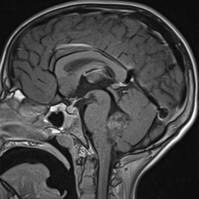
9 岁男孩,头晕、恶心进行性加重 3 周,左侧颈部疼痛与僵硬 1 周。磁共振成像显示后颅窝一不均质实性肿块(26 × 20 × 33 mm),累及第四脑室、左侧 Luschka 孔及小脑蚓部,呈轻度不均匀强化(图 1)。肿瘤获全切除,随后按 AIEOP 高危中枢神经系统胚胎性肿瘤方案行多模式治疗。2 个月后疾病进展;患者于确诊后 9 个月死亡。
检查所见
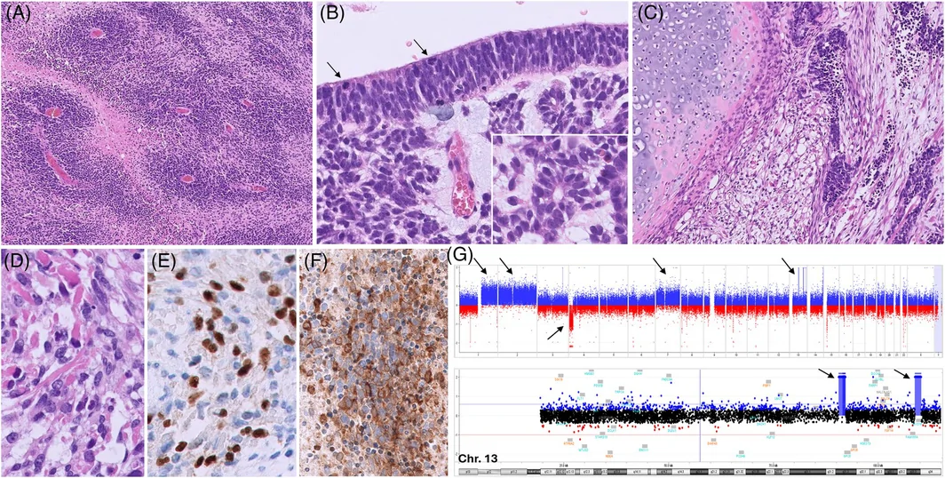
组织学检查见肿瘤成分不均一:(i)原始未分化细胞形成的致密血管周聚集灶,与纤维性神经毡样区交替分布,这是多层菊形团胚胎性肿瘤(ETMR)常见的模式(图 2A);(ii)局限区域含囊性结构及由假复层肿瘤性神经上皮被覆的小管,全层可见核分裂,类似原始神经管(髓上皮瘤模式)(图 2B);(iii)异源性横纹肌母细胞区和软骨样区(图 2C、D)。肿瘤在 ETANTR 区和髓上皮瘤区局灶表达 OLIG2、synaptophysin、LIN28 和 SALL4;GFAP 阴性;横纹肌母细胞区表达 desmin、myogenin 和 MYOD1,软骨样成分表达 S100(图 2E、F);H3K27me3 表达全片缺失。据此,初步诊断为具有多层菊形团胚胎性肿瘤(ETMR)特征并伴间叶性异源分化的胚胎性肿瘤。PCR 分析在 DICER1 RNase IIIb 热点区未检出变异。甲基化谱分析将肿瘤归为 ETMR,非 C19MC 改变型(novel)(评分 0.99,v12.8),无 19q13.42 染色体 microRNA 簇(C19MC)扩增。染色体微阵列显示 1.7 Mb(13q31.3;MIR17HG/miR-17-92/部分 GPC5 基因)与 1.5 Mb(13q33.3q34;MYO16、IRS2、部分 TNFSF13B 基因)扩增(图 2G)。RNA 测序在同一区域发现新的 IRS2::GPC5 与 GPC5::MIR17HG 融合;综合提示存在复杂的 13q 重排。此外,嵌合性改变包括 1q、2 号和 7 号染色体获得及 4p 部分缺失(图 2G)。未发现致病/可能致病变异(TruSight™ Oncology 500)(框 1)。
最终诊断
具有 ETMR 样特征并伴 MIR17HG 扩增的胚胎性肿瘤(CNS WHO 4 级)。
讨论
本例呈现 ETMR 的多项标志性特征,包括原始未分化细胞的血管周聚集灶与纤维性神经毡样区交替分布、局灶髓上皮瘤样形态、局灶 LIN28 表达,以及与 ETMR 甲基化谱相符。此外,拷贝数改变(CNA)谱也很典型:2 号、1q 和 7 号染色体获得,属 ETMR 中最常见的 CNA [1]。对多模式治疗反应差的侵袭性临床经过也与 ETMR 一致。然而,其他特征对该实体而言并不寻常,偏离了经典 ETMR 谱系。首先,患者年龄超出通常所见范围(0–4 岁)。在分子证实的最大宗 ETMR 系列中,仅 7/159 例(4.4%)发生于 ≥5 岁患者 [1]。其次,表型中出现间叶性异源区,这在典型 ETMR 中罕见。这一所见使鉴别诊断须考虑其他儿童实体,如伴恶性间叶/神经外胚层成分的畸胎瘤、髓母细胞瘤或非典型畸胎样/横纹肌样肿瘤(ATRT)。LIN28 免疫阳性支持 ETMR 诊断,但并不特异,少数情况下可见于其他肿瘤,包括 ATRT。此时 INI1 与 BRG1 表达保留可排除 ATRT。有意思的是,曾有一例婴儿 DICER 驱动的 ETMR 报道出现横纹肌母细胞区和软骨样区 [2]。结合 SALL4 表达与 H3K27me3 缺失,这提示可能为 DICER1 相关的发病机制。然而,分子分析并未证实这一假设。
第三个特殊之处是 13q31.3 区扩增,该区包含 miR-17-92 簇(MIR17HG)和部分 GPC5 基因。据我们所知,MIR17HG 扩增曾报道于两例缺乏 C19MC 扩增和 DICER1 突变的 ETMR,以及一例 C19MC 阳性 ETMR,均发生于婴儿 [1]。本例中 MIR17HG 还与 Glypican-5(GPC5)融合,可能使致癌性 miR-17–92 簇处于 GPC5 调控之下。这与 C19MC 阳性 ETMR 中所见相似——后者 C19MC 扩增之前被认为先发生与 Tweety Family Member 1(TTYH1)或较少见伙伴基因的易位 [1]。有意思的是,涵盖 MIR17HG 与 GPC5 的 13q31-32 扩增已见于不同组织学类型,包括横纹肌肉瘤,主要是 PAX3 重排的腺泡状横纹肌肉瘤 [3]。横纹肌肉瘤和子宫癌肉瘤的体外与体内研究提示 GPC5 在促进肉瘤样分化中起作用。可以推测,13q31.3 改变连同定位于相邻 13q33.3–q34 区的 MYO16 扩增,通过激活异常的发育程序而导致间叶性异源区。既往报道的 MIR17HG 扩增病例未提供组织学特征的详细描述,因此无法与本例比较。
本例为伴 MIR17HG 扩增的胚胎性肿瘤这一有限文献增添了资料。尽管部分特征对 ETMR 而言不典型(发病年龄、异源成分),但总体所见支持其与该实体在生物学上关系密切,拓宽了其临床病理谱系。
- Brain Pathology quiz case (Bambino Gesù Children’s Hospital, Rome): a 9-year-old boy with a heterogeneously enhancing fourth ventricle/vermis mass; gross total resection and high-risk embryonal tumour therapy, progression at 2 months and death 9 months after diagnosis.
- Morphology: ETMR-like perivascular aggregates of primitive cells alternating with neuropil-like areas, focal medulloepithelioma-like pseudostratified tubules, plus heterologous rhabdomyoblastic (desmin/myogenin/MYOD1) and chondroid (S100) areas; focal LIN28 and SALL4, global H3K27me3 loss, no DICER1 hotspot variant.
- Molecular: methylation class ETMR, non-C19MC-altered (score 0.99, v12.8) without C19MC amplification; 13q31.3 MIR17HG/GPC5 and 13q33.3–q34 MYO16/IRS2 amplifications with IRS2::GPC5 and GPC5::MIR17HG fusions; gains of 1q, 2 and 7 and partial 4p loss.
- Takeaway: age ≥5 years and mesenchymal heterologous elements are atypical (only 4.4% of the largest series were ≥5 years); differentials include teratoma with malignant components, medulloblastoma and ATRT (retained INI1/BRG1 excludes ATRT). ISN virtual slide links and Figures 1–2 included.
Scope: PMC open-access full text (CC BY-NC-ND 4.0) — virtual slide box, case presentation, findings, final diagnosis, discussion and Figures 1–2 with captions. Author contributions, conflict of interest and ethics statements and references are not included (bracketed numbers refer to the original reference list).
BOX 1 Virtual glass slide. Access at https://isn‐slidearchive.org/?&col=ISN&fol=Archive&file=ID‐6893255.svs; https://neuro2.pathology.pitt.edu/isn/brainpathology/underyourmicroscope?&col=ISN&fol=Archive&file=ID‐6893255‐2.svs.
CASE PRESENTATION
A 9‐year‐old boy presented with a 3‐week history of worsening dizziness and nausea and a 1‐week history of left‐sided neck pain and stiffness. Magnetic resonance imaging revealed a heterogeneous solid mass in the posterior fossa (26 × 20 × 33 mm), involving the fourth ventricle, left foramen of Luschka, and cerebellar vermis with mild heterogenous contrast enhancement (Figure 1). Gross total resection was obtained, followed by multimodal therapy, according to AIEOP protocols for high‐risk CNS embryonal tumors. Disease progression occurred 2 months later; the patient died at 9 months post‐diagnosis.
FINDINGS
At histological examination, the neoplasm was heterogeneous with: (i) densely cellular perivascular aggregates of primitive undifferentiated cells alternating with fibrillary neuropil‐like areas, a pattern commonly seen in embryonal tumor with multilayered rosettes (ETMR) (Figure 2A); (ii) limited areas containing a cystic structure and tubules lined by pseudostratified neoplastic neuroepithelium, showing mitoses through the entire thickness, resembling the primordial neural tube (medulloepithelioma pattern) (Figure 2B); and (iii) heterologous rhabdomyoblastic and chondroid areas (Figure 2C,D). The tumor showed focal expression of OLIG2, synaptophysin, LIN28, and SALL4 in the ETANTR and medulloepithelioma areas; GFAP was negative; desmin, myogenin, and MYOD1 were expressed in the rhabdomyoblastic areas and S100 in chondroid components (Figure 2E,F); H3K27me3 expression was lost throughout. Based on these findings, the preliminary diagnosis was embryonal tumor with features of embryonal tumor with multilayered rosettes (ETMR) and mesenchymal heterologous differentiation. PCR analysis detected no variants in DICER1 RNase IIIb hotspot regions. Methylation profiling classified the tumor as ETMR, non‐C19MC‐altered (novel) (score 0.99, v12.8), without amplification of the microRNA cluster on chromosome 19q13.42 (C19MC). Chromosomal microarray revealed 1.7‐Mb (13q31.3; MIR17HG/miR‐17‐92/part of GPC5 gene) and 1.5‐Mb (13q33.3q34; MYO16, IRS2, part of TNFSF13B gene) amplifications (Figure 2G). RNA‐seq identified novel IRS2::GPC5 and GPC5::MIR17HG fusions in the same region; together these findings suggest a complex 13q rearrangement. Additionally, mosaic alterations included gain of 1q, 2, and 7, and partial loss of 4p (Figure 2G). No pathogenic/likely pathogenic variants were identified (TruSight™ Oncology 500) (Box 1).
FINAL DIAGNOSIS
Embryonal tumor with ETMR‐like features and MIR17HG amplification (CNS WHO Grade 4).
DISCUSSION
The case presented exhibits several hallmark features of ETMR, including perivascular aggregates of primitive undifferentiated cells alternating with fibrillary neuropil‐like areas, focal medulloepithelioma‐like morphology, focal LIN28 expression, and alignment with the ETMR methylation profile. Additionally, the CNA profile was typical, with gains of chromosome 2, 1q, and 7, among the most common CNAs identified in ETMR [1]. The aggressive clinical course, characterized by poor response to multimodal therapy, was also consistent with ETMR. However, other features were unusual for this entity, deviating from the classical ETMR spectrum. First, the patient's age exceeded the range typically observed (0–4 years). In the largest series of molecularly proven ETMR, only 7/159 (4.4%) cases occurred in patients ≥5 years [1]. Second, the phenotype showed mesenchymal heterologous areas, rarely seen in typical ETMR. This finding raises the differential diagnosis with other pediatric entities such as teratomas with malignant mesenchymal/neuroectodermal components, medulloblastomas, or atypical teratoid/rhabdoid tumors (ATRT). LIN28 immunopositivity supports the diagnosis of ETMR but is not specific and may rarely occur in other tumors, including ATRT. In such cases, retained INI1 and BRG1 expression excludes ATRT. Interestingly, rhabdomyoblastic and chondroid areas have been reported in one infantile DICER‐driven ETMR [2]. Together with SALL4 expression and H3K27me3 loss, this suggested a possible DICER1‐related pathogenesis. However, molecular analysis did not confirm this hypothesis.
A third peculiar feature was amplification of the 13q31.3 region containing the miR‐17‐92 cluster (MIR17HG) and part of the GPC5 gene. To our knowledge, MIR17HG amplification has been reported in two ETMR lacking C19MC amplification and DICER1 mutation, and in one C19MC‐positive ETMR, all occurring in infants [1]. In our case, MIR17HG was also fused with Glypican‐5 (GPC5), likely placing the oncogenic miR‐17–92 cluster under GPC5 regulation. This resembles what is observed in C19MC‐positive ETMR, where C19MC amplification is supposed to be preceded by translocation with Tweety Family Member 1 (TTYH1) or rarer partner genes [1]. Interestingly, amplification of the 13q31‐32, encompassing MIR17HG and GPC5, has been reported in different histotypes, including rhabdomyosarcoma, mainly PAX3‐rearranged alveolar rhabdomyosarcoma [3]. In vitro and in vivo studies on rhabdomyosarcomas and uterine carcinosarcomas suggest a role for GPC5 in promoting sarcomatous differentiation. It may be speculated that the alterations of the 13q31.3, together with amplification of MYO16, mapping to the adjacent 13q33.3–q34 region, account for the mesenchymal heterologous areas by activating aberrant developmental programs. A detailed description of the histological features of previously reported MIR17HG‐amplified cases was not provided, preventing comparison with our case.
This case adds to the limited literature on embryonal tumors with MIR17HG amplification. Although some features are atypical for ETMR (age at onset, heterologous components), the overall findings support a close biological relationship to this entity, broadening its clinicopathological spectrum.