- PathologyOutlines 2026-10-26 教科书更新专题(Sydney Rashid、Bushra Zafar Sayeed、Reima El Naili;审阅 Rola M. Saleeb):脐尿管或脐尿管残余发生的囊性黏液性上皮肿瘤,低级别细胞异型、无浸润,形态类似 LAMN 与卵巢黏液性交界性肿瘤。
- 诊断要点:外渗的无细胞间质黏液很常见,不应误判为浸润(破裂可致腹膜假黏液瘤);结构复杂性及有无浸润比单纯细胞异型更有生物学意义,须广泛取材排除小灶癌或微浸润。
- 免疫组化:CK20、CDX2 弥漫阳性,SATB2 常阳性,CK7 约 30% 阳性,β-catenin 膜/胞质着色而核阴性,GATA3、ER/PR、PSA、AMACR、p63 阴性;须结合影像与临床排除结直肠或阑尾黏液性肿瘤继发累及。
- 预后与治疗:完整切除(膀胱部分切除并整块切除脐尿管残余)后通常无复发转移,浸润性脐尿管腺癌 5 年生存约 45%。附 10 张镜下/免疫组化图、病理报告示例与 1 道练习题。
收录范围:PathologyOutlines 专题「Urachal mucinous cystic tumor of low malignant potential」全文与全部 10 张图(图注中英对照)。同期 2026-10-26 Textbook Updates 另含 Factor VIII deficiency(血友病 A)、Appendiceal neoplasms(卵巢转移)、涎腺多形性腺癌与睾丸表皮样囊肿等专题,未收录。
定义 / 概述
- 起源于脐尿管或脐尿管残余的腺上皮肿瘤,以囊性结构和衬覆具有低级别细胞异型性的黏液性上皮为特征 (Am J Surg Pathol 2014;38:1033)
- 形态上类似于阑尾低级别黏液性肿瘤和卵巢黏液性交界性肿瘤 (Case Rep Gastrointest Med 2019;2019:1434838)
要点
- 脐尿管腺性肿瘤,具有低级别肠型黏液性上皮,无浸润性侵犯
- 可见乳头状 / 绒毛状上皮增生
- 间质中外渗的无细胞黏液非常常见,不应误判为浸润
- 破裂可导致腹膜假黏液瘤 (Curr Oncol 2014;21:e155, Exp Ther Med 2023;26:555)
- 结构复杂程度及有无浸润似乎比单纯的细胞异型性具有更重要的生物学意义 (Adv Anat Pathol 2016;23:71)
- 广泛取材对于排除小灶癌或局灶性微浸润至关重要 (Am J Surg Pathol 2014;38:1033)
术语
- 低度恶性潜能黏液性囊性肿瘤(MCTLMP)(首选)
- 首选“低度恶性潜能”这一名称,因为这些肿瘤的上皮异型性超过良性囊腺瘤,但缺乏诊断腺癌所必需的破坏性间质浸润 (Am J Surg Pathol 2014;38:1033)
- 脐尿管交界性黏液性囊性肿瘤
- 脐尿管低级别黏液性囊性肿瘤
- 脐尿管黏液性交界性肿瘤
- 在现代亚分类建立之前,历史上曾将其归入脐尿管黏液性囊腺瘤或黏液腺癌 (Adv Anat Pathol 2016;23:71, Arch Pathol Lab Med 2019;143:258)
ICD 编码
- 与脐尿管黏液性腺性肿瘤存在重叠;编码可能取决于各机构的分类
- ICD-O: 8472/1 - 交界性黏液性囊性肿瘤
- ICD-10: Q64.4 - 脐尿管先天性异常;临床上可能与之相关
- ICD-11: 2F77 & XH2FF0 - 男性生殖器官动态未定的肿瘤 & 交界性黏液性囊性肿瘤
- 目前尚无专门针对 MCTLMP 的统一标准化专用编码
流行病学
- 见于成年人,年龄范围广:26 - 80 岁 (Am J Surg Pathol 2014;38:1033, Int Cancer Conf J 2022;11:104)
- 脐尿管肿瘤总体占膀胱恶性肿瘤的 < 1% (J Urol 2012;188:1102)
- 肿瘤大小差异较大,报道的大小范围为约 2 至 8 cm (Am J Surg Pathol 2014;38:1033, Int Cancer Conf J 2022;11:104)
- 尚未发现性别与 MCTLMP 风险之间存在明确关联 (Int Cancer Conf J 2022;11:104)
部位
- 脐尿管 / 脐尿管残余
- 最常累及膀胱顶或膀胱与脐之间的膀胱上方中线区域
- 可向脐或腹前壁延伸 (Adv Anat Pathol 2016;23:71)
病理生理
- 脐尿管持续存在,遗留有上皮衬覆的残余组织,易发生肿瘤性转化
- 认为起源于脐尿管残余内的腺性 / 肠上皮化生 (Adv Anat Pathol 2016;23:71, Am J Clin Pathol 2022;158:263)
- 表现出类似卵巢黏液性上皮肿瘤的形态学进展
- 黏液性囊腺瘤 → MCTLMP → 伴上皮内癌的 MCTLMP → 黏液性囊腺癌(伴微浸润或明确浸润) (Adv Anat Pathol 2016;23:71, Am J Surg Pathol 2014;38:1033)
病因
- 与脐尿管持续存在(尿囊的胚胎性残余)相关
- 可继发于尿液对脐尿管残余上皮的慢性刺激 (Radiographics 2025;45:e240122, Adv Anat Pathol 2016;23:71)
- 有人提出,持续存在的脐尿管残余内的慢性尿液淤滞、炎症和感染是促进肿瘤性转化的因素
示意图 / 表格
托管于其他服务器的图像:
临床特征
- 最常在影像学检查中偶然发现
- 可表现为血尿、黏液尿、腹痛及可触及的肿块 (Am J Surg Pathol 2014;38:1033, Cureus 2025;17:e90905, Arch Pathol Lab Med 2019;143:258)
- 少数病例在破裂或黏液溢出后表现为腹膜假黏液瘤 (Int Cancer Conf J 2022;11:104, Exp Ther Med 2023;26:555, Curr Oncol 2014;21:e155)
- 现行分类方案参照卵巢黏液性上皮肿瘤,将脐尿管黏液性囊性肿瘤分为以下类别 (Adv Anat Pathol 2016;23:71, Am J Surg Pathol 2014;38:1033)
- 黏液性囊腺瘤
- 低度恶性潜能黏液性囊性肿瘤(MCTLMP)
- 伴上皮内癌的 MCTLMP
- 伴微浸润的黏液性囊腺癌
- 伴明确浸润的黏液性囊腺癌
诊断
- 可通过活检作出诊断
- 腹部 / 盆腔 CT 显示从脐延伸至膀胱顶的囊性病变 (Int Cancer Conf J 2022;11:104, Medicine (Baltimore) 2017;96:e8740)
- 必须进行全面的组织学检查和广泛取材,以排除浸润性侵犯或微浸润 (Am J Surg Pathol 2014;38:1033, Arch Pathol Lab Med 2019;143:258)
- 与良性脐尿管囊肿及浸润性腺癌的鉴别至关重要
实验室检查
- 部分病例的血清 CEA 和 CA 19-9 水平升高 (Exp Ther Med 2023;26:555)
影像学描述
- 增强 CT 常显示膀胱上方无强化的单房性或多房性囊性病变 (Urol Case Rep 2025;61:103104, Exp Ther Med 2023;26:555)
- CT 可显示邻近膀胱顶、保持稳定的囊性病变,可类似良性脐尿管囊肿或憩室 (Int Cancer Conf J 2022;11:104, Case Rep Gastrointest Med 2019;2019:1434838)
- 边缘可有钙化 (Case Rep Gastrointest Med 2019;2019:1434838)
影像学图像
托管于其他服务器的图像:
预后因素
- 结构复杂程度及有无间质浸润是脐尿管黏液性肿瘤分类和风险分层的关键因素
- 有无间质浸润是决定生物学行为的最重要因素之一
- 已报道的 MCTLMP 病例在完整手术切除后的随访中通常无复发或转移
- 与 MCTLMP 相比,浸润性脐尿管腺癌的预后明显更差,报道的 5 年生存率接近 45%
- 参考文献: Am J Surg Pathol 2014;38:1033, Adv Anat Pathol 2016;23:71, Int Cancer Conf J 2022;11:104
病例报告
- 38 岁男性,超声检查偶然发现 (Cureus 2025;17:e90905)
- 47 岁男性,偶然发现无症状肿块 (Cureus 2023;15:e37036)
- 54 岁男性,排尿困难及耻骨上区不适 (Cureus 2025;17:e78562)
- 74 岁男性,下腹痛伴腹膜假黏液瘤 (Exp Ther Med 2023;26:555)
- 77 岁男性,因肺炎住院 (Urol Case Rep 2025;61:103104)
治疗
- 完整手术切除是主要治疗方法,局限性非浸润性病变的预后通常良好 (Am J Surg Pathol 2014;38:1033, Adv Anat Pathol 2016;23:71)
- 手术治疗最常采用膀胱部分切除术,同时整块切除脐尿管残余及相关囊性病变 (Cureus 2023;15:e37036, Cureus 2025;17:e78562)
大体描述
- 脐尿管黏液性囊性肿瘤通常为起源于膀胱顶或脐尿管残余、边界清楚的单房性或多房性囊性病变 (Arch Pathol Lab Med 2019;143:258, Adv Anat Pathol 2016;23:71)
- 通常充满大量黏稠的黏液性或胶冻样物质 (Cureus 2025;17:e78562)
大体图像
托管于其他服务器的图像:
镜下(组织学)描述
- 上皮分层可达约 3 层细胞
- 衬覆黏液性立方至柱状上皮
- 轻至中度核异型性,局部可见细胞核假复层排列
- 核分裂象罕见,增殖活性低
- 轻至中度细胞异型性
- 假乳头状或绒毛状的复杂结构
- 无间质浸润
- 充满黏液性物质的多房性或单房性囊性病变
- 囊壁由厚的纤维肌性组织构成,类似脐尿管残余
- 可见杯状细胞
- 可呈乳头状、绒毛状、簇状或假乳头状上皮结构
- 参考文献: Cureus 2025;17:e78562, Int Cancer Conf J 2022;11:104, Adv Anat Pathol 2016;23:71, Am J Surg Pathol 2014;38:1033, Am J Surg Pathol 2011;35:787, Arch Pathol Lab Med 2019;143:258
镜下(组织学)图像
供图:Reima El Naili,医学博士
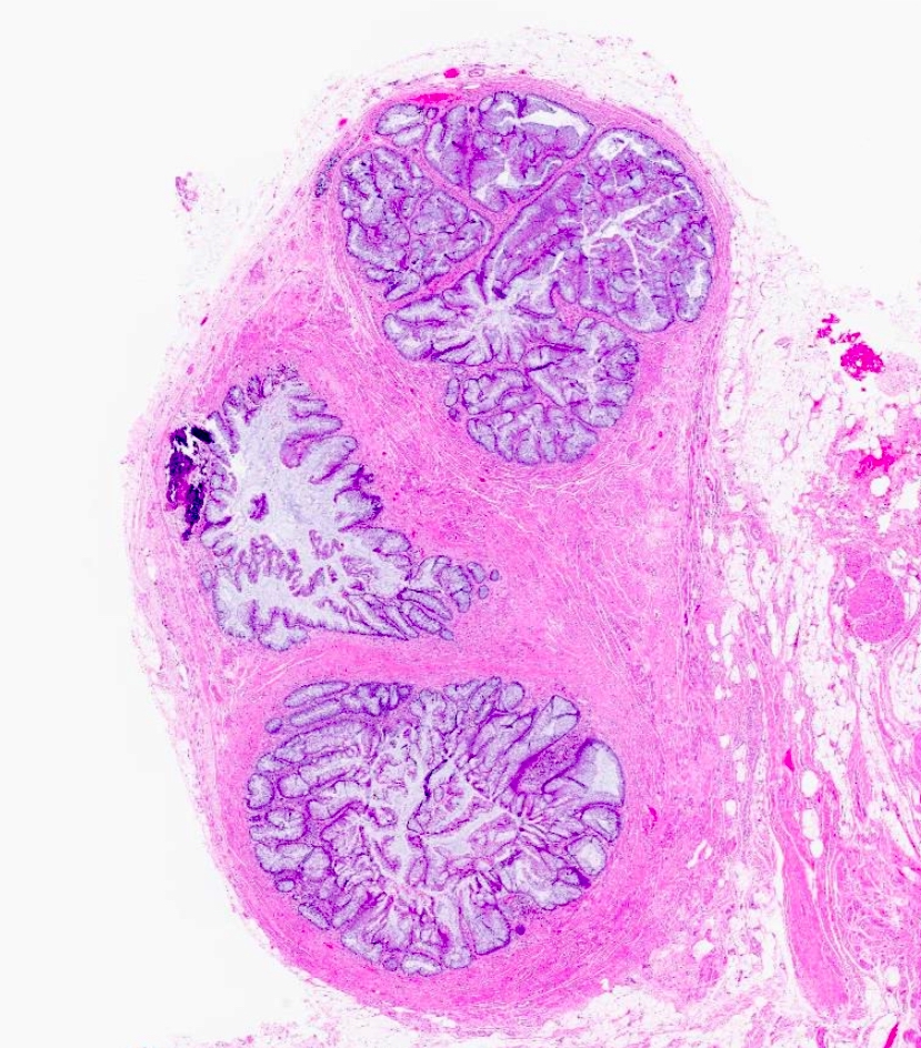
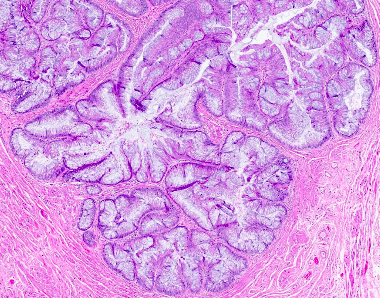
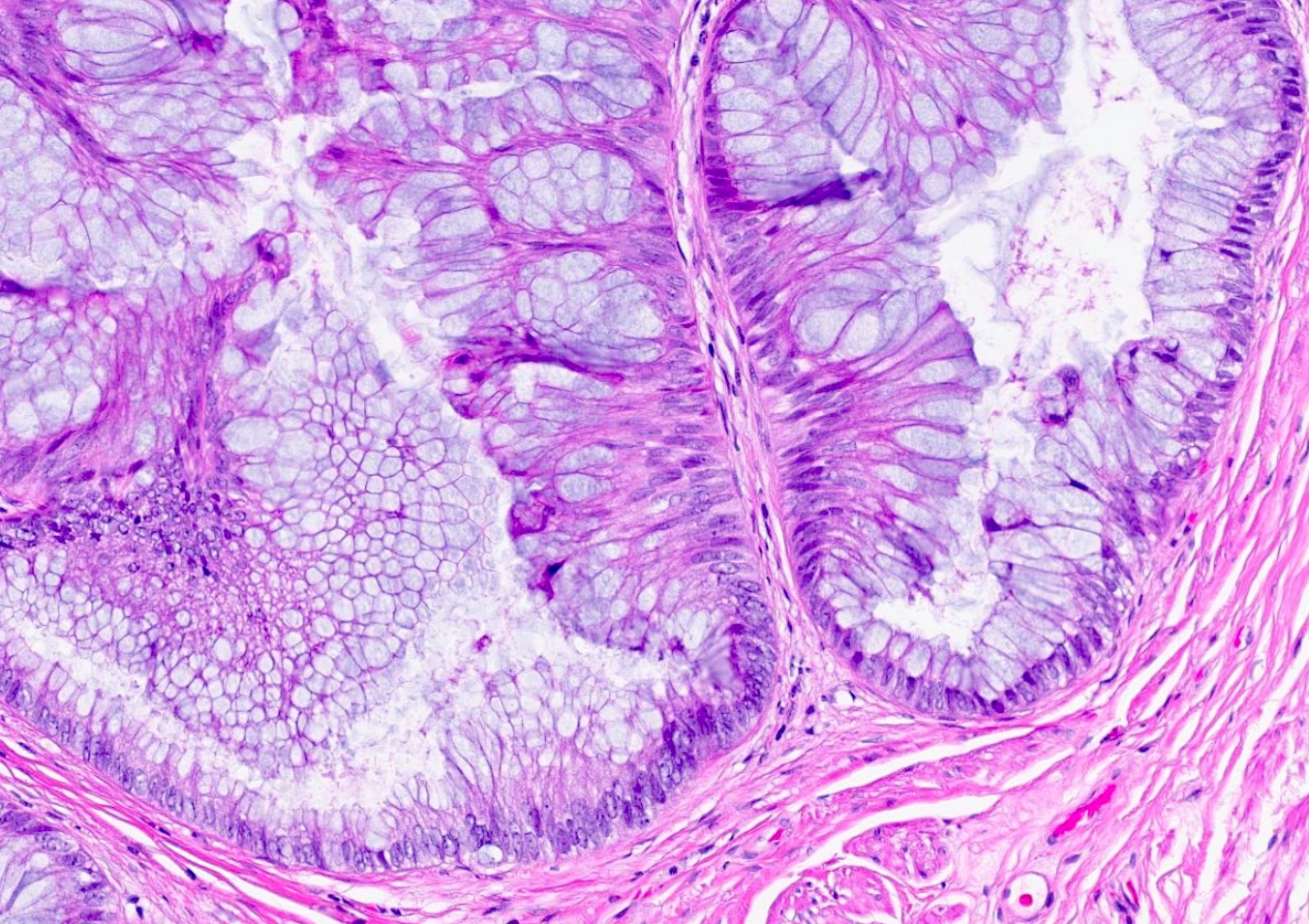
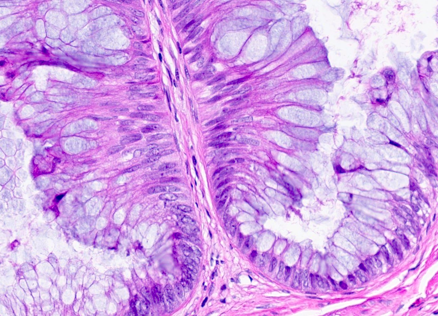
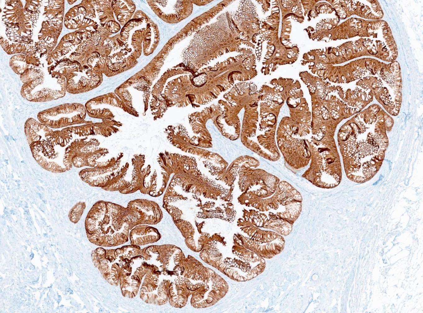
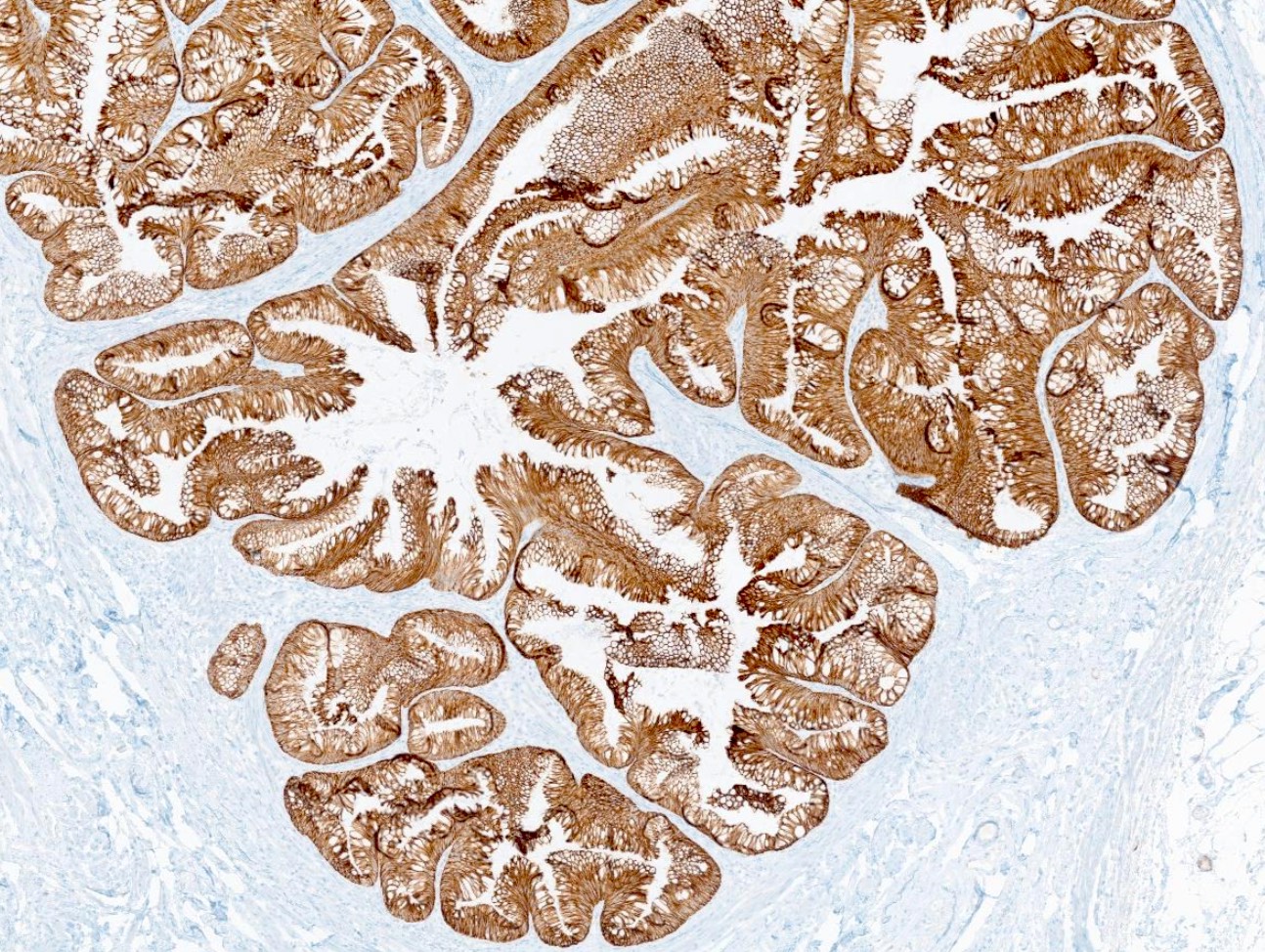
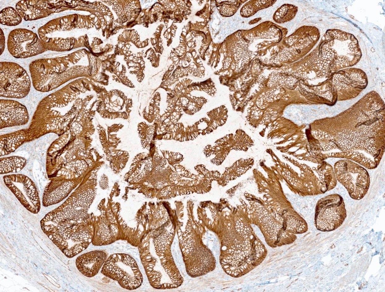
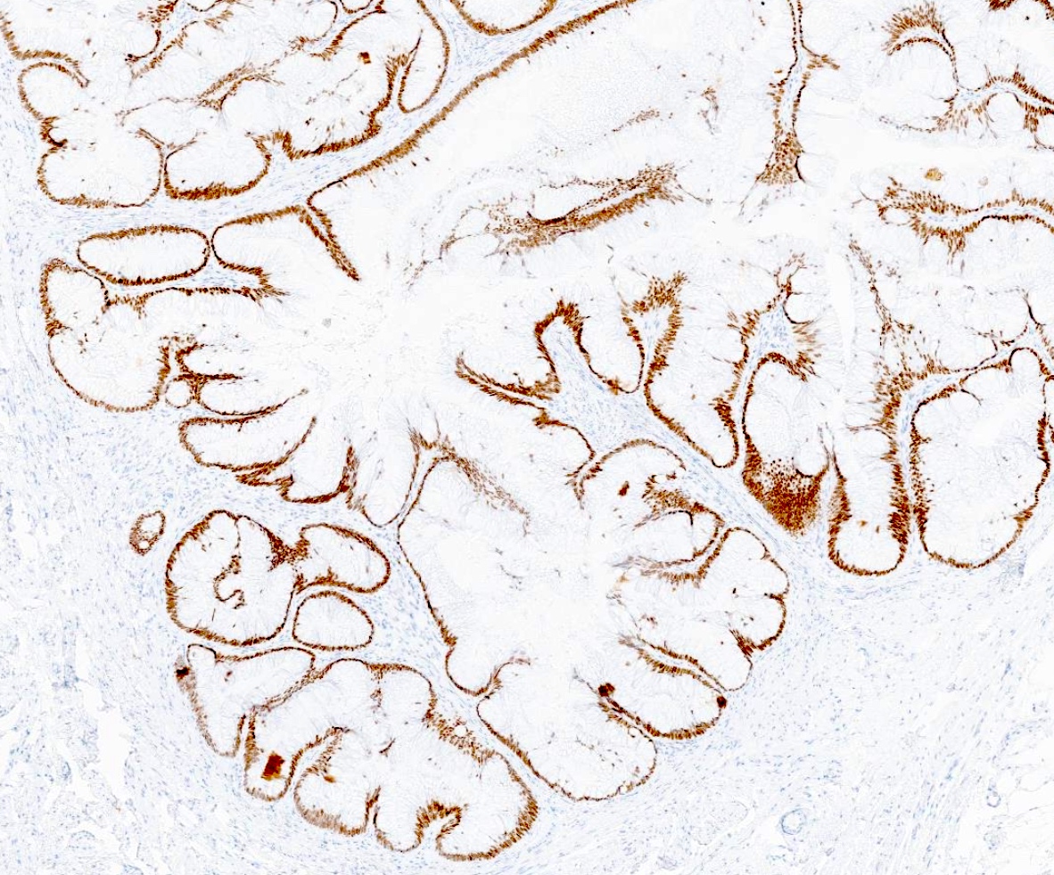
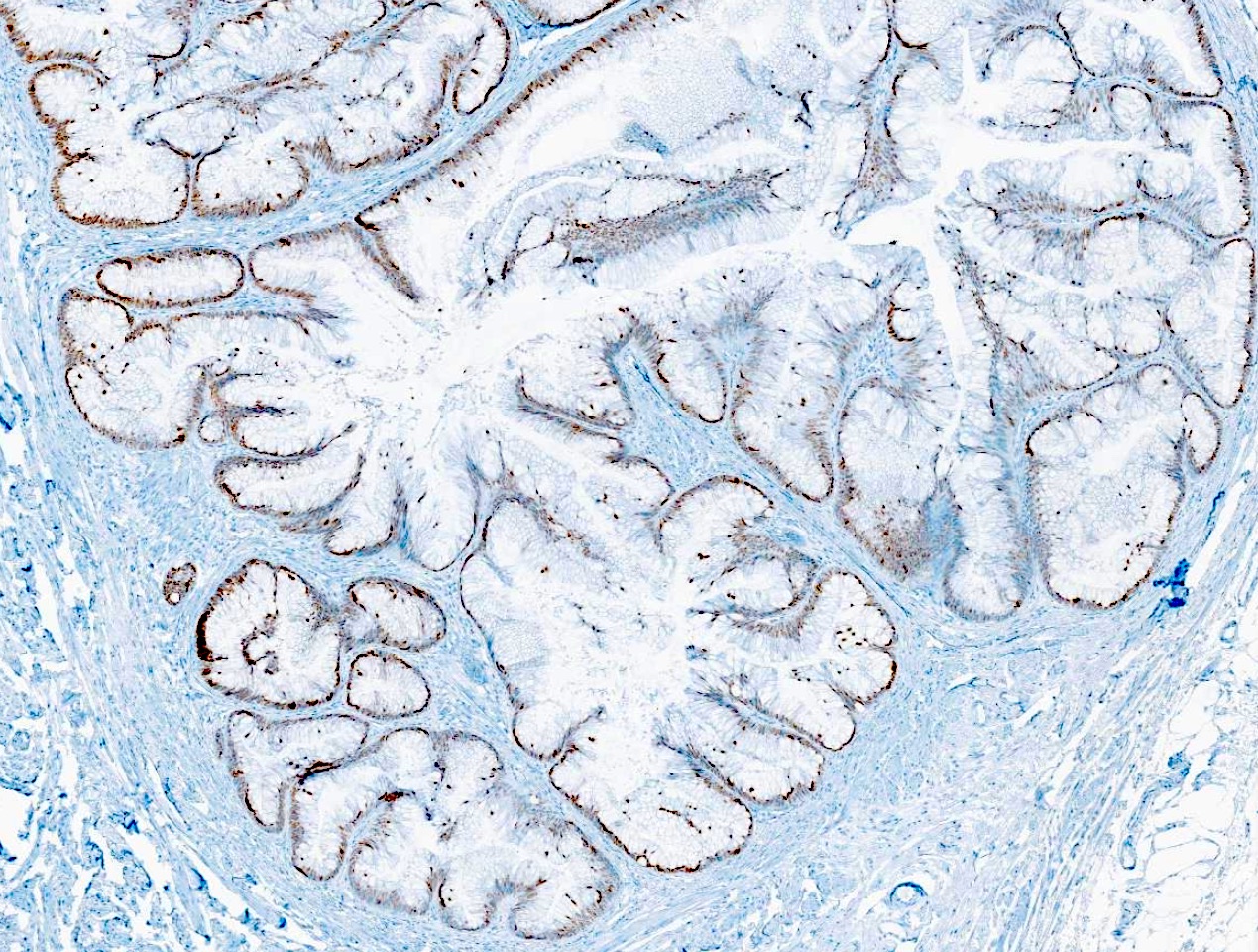
阳性染色
- CK20:弥漫阳性,支持肠型分化
- CDX2:弥漫阳性,常见
- SATB2:常呈阳性
- CEA:阳性情况不一 (Urol Case Rep 2025;61:103104)
- β catenin:胞膜或胞质染色模式
- MUC5AC 和 MUC2 可在部分区域表达 (Cureus 2025;17:e78562)
- CK7 可见于约 30% 的病例
- β catenin 和 CK7 可能有助于区分脐尿管腺癌与结直肠腺癌 (Am J Surg Pathol 2011;35:787)
- 参考文献: Arch Pathol Lab Med 2019;143:258, Case Rep Gastrointest Med 2019;2019:1434838
阴性染色
- 细胞核内的 β catenin
- GATA3
- ER / PR
- PSA
- AMACR
- p63 (Cureus 2023;15:e37036)
- 参考文献: Exp Ther Med 2023;26:555, Arch Pathol Lab Med 2019;143:258, Am J Surg Pathol 2011;35:787
分子 / 细胞遗传学描述
- TP53 和 KRAS 突变可见于脐尿管癌;但由于 MCTLMP 罕见,其分子数据仍然有限 (Oncotarget 2016;7:39293, Am J Clin Pathol 2022;158:263)
病理报告示例
- 膀胱,脐尿管憩室,切除标本:
- 脐尿管低度恶性潜能黏液性囊性肿瘤(MCTLMP)(见备注)
- 备注:组织学检查显示囊性腔隙衬覆分层的黏液性腺上皮,呈假乳头状结构,伴轻至中度细胞异型性,符合肠型分化。未见间质浸润。所提供的免疫组织化学染色显示病变细胞 CK7、CK20、SATB2 和 CDX2 阳性,GATA3 阴性。β catenin 呈胞膜染色模式。
鉴别诊断
- 诊断需要对囊壁进行完整的组织学评估
- 必须结合影像学及临床表现,以排除结直肠或阑尾黏液性肿瘤的继发累及 (Adv Anat Pathol 2016;23:71)
- 伴上皮内癌的 MCTLMP:
- 上皮可有明显分层及恶性细胞学特征
- 细胞异型性的程度与绒毛状腺瘤相似
- 间质稀少的乳头
- 结构较常规 MCTLMP 更为复杂(如乳头状或筛状生长)
- 需充分取材以排除浸润性癌灶
- 上皮可有明显分层及恶性细胞学特征
- 脐尿管黏液性囊腺癌:
- 浸润性间质侵犯
- 微浸润:浸润灶 < 2 mm,且占肿瘤的 < 5%
- 明确浸润:更广泛的破坏性间质浸润
- 破坏性生长模式
- 显著的细胞异型性
- 浸润性间质侵犯
- 脐尿管黏液性囊腺瘤:
- 由单层黏液性柱状上皮衬覆的囊性结构
- 无异型性
- 脐尿管绒毛状腺瘤:
- 外生性绒毛状结构
- 转移性结直肠腺癌:
- 常表现为弥漫性细胞核 β catenin 阳性
- 参考文献: Arch Pathol Lab Med 2019;143:258, Adv Anat Pathol 2016;23:71
练习题 #1
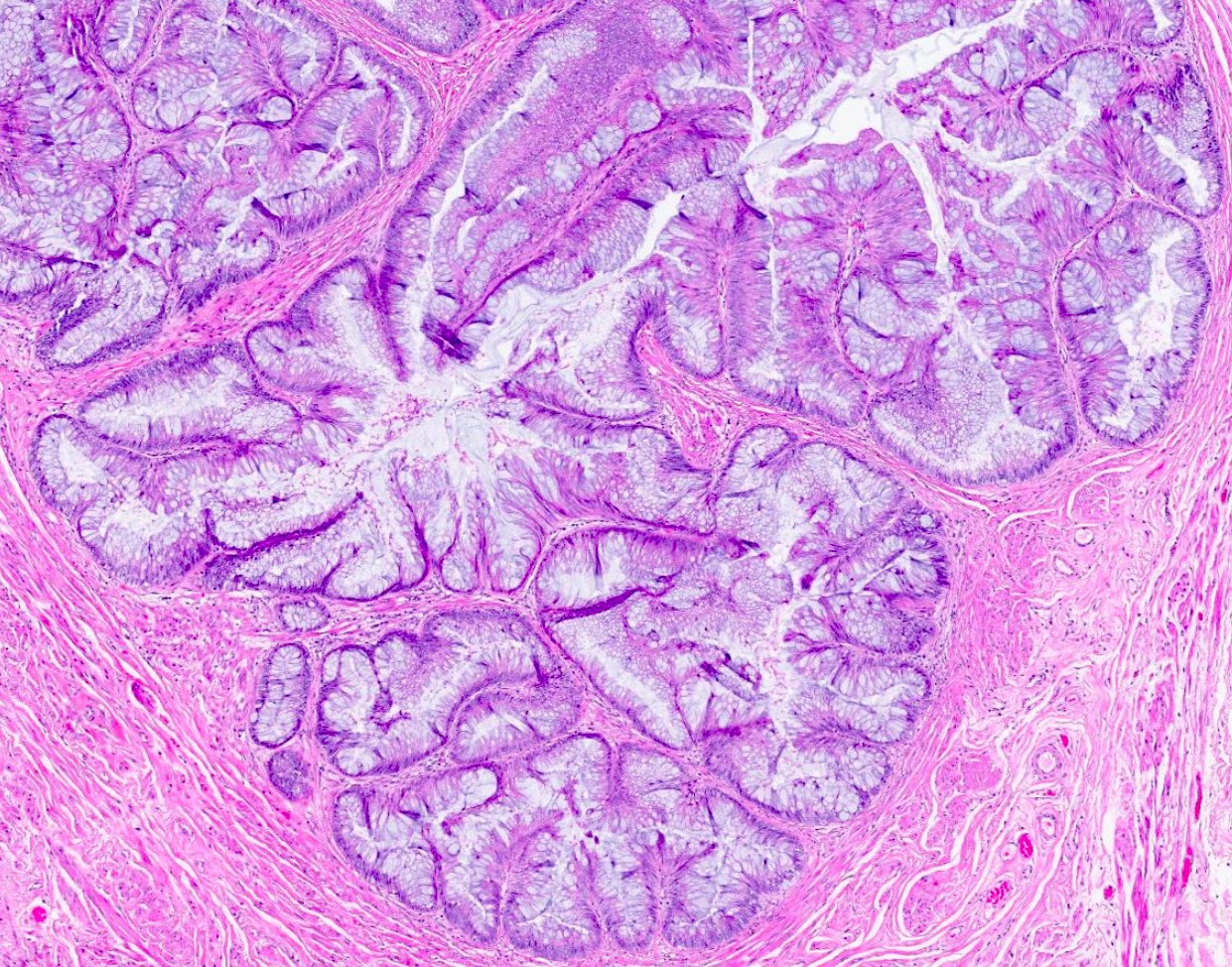
一名 43 岁女性接受了膀胱顶起源的囊性病变切除术。组织学检查显示多房性囊性腔隙,衬覆具有轻至中度异型性的黏液性柱状上皮。诊断是什么?
- 脐尿管浸润性黏液性囊腺癌
- 脐尿管黏液性囊腺瘤
- 脐尿管低度恶性潜能黏液性囊性肿瘤(MCTLMP)
- 绒毛状腺瘤
练习题答案 #1
C. 脐尿管低度恶性潜能黏液性囊性肿瘤(MCTLMP)。MCTLMP 表现为囊性黏液性上皮,伴轻至中度细胞异型性及有限的结构复杂性,如假乳头状生长或分层。选项 D 错误,因为绒毛状腺瘤通常呈具有纤维血管轴心的外生性绒毛状结构,而非多房性囊性腔隙。选项 B 错误,因为黏液性囊腺瘤缺乏明显异型性及上皮分层。选项 A 错误,因为浸润性黏液性囊腺癌必须存在间质浸润。
参考文献: 脐尿管低度恶性潜能黏液性囊性肿瘤
- PathologyOutlines Textbook Updates, 26 October 2026 (Sydney Rashid, Bushra Zafar Sayeed, Reima El Naili; reviewed by Rola M. Saleeb): a cystic mucinous epithelial neoplasm of the urachus or its remnants with low grade atypia and no invasion, resembling LAMN and ovarian mucinous borderline tumours.
- Diagnostic points: extravasated acellular stromal mucin is common and should not be read as invasion (rupture may cause pseudomyxoma peritonei); architectural complexity and invasion matter more than cytologic atypia alone, and extensive sampling is needed to exclude small foci of carcinoma or microinvasion.
- IHC: CK20 and CDX2 diffuse, SATB2 often, CK7 in ~30%, membranous/cytoplasmic but not nuclear β-catenin; GATA3, ER/PR, PSA, AMACR and p63 negative; correlate with imaging and clinical findings to exclude secondary colorectal or appendiceal mucinous neoplasms.
- Outcome and treatment: complete excision (partial cystectomy with en bloc excision of the urachal remnant) is typically curative, whereas invasive urachal adenocarcinoma has ~45% 5-year survival. Ten microscopic/IHC images, a sample report and a practice question.
Scope: the full PathologyOutlines topic “Urachal mucinous cystic tumor of low malignant potential” with all 10 images and captions. The same 26 October 2026 Textbook Updates also covered factor VIII deficiency (hemophilia A), appendiceal neoplasms metastatic to ovary, polymorphous adenocarcinoma of salivary gland and testicular epidermoid cyst, which are not included.
Definition / general
- Glandular epithelial neoplasm arising from the urachus or urachal remnants, characterized by cystic architecture and mucinous epithelial lining with low grade cytologic atypia (Am J Surg Pathol 2014;38:1033)
- Morphologically resembles appendiceal low grade mucinous neoplasms and ovarian mucinous borderline tumors (Case Rep Gastrointest Med 2019;2019:1434838)
Essential features
- Urachal glandular neoplasm with low grade intestinal type mucinous epithelium and lack of infiltrative invasion
- May display papillary / villous epithelial proliferation
- Extravasated acellular stromal mucin is very common and should not be misinterpreted as invasion
- Rupture may result in pseudomyxoma peritonei (Curr Oncol 2014;21:e155, Exp Ther Med 2023;26:555)
- Architectural complexity and presence or absence of invasion appears to be more biologically significant than cytologic atypia alone (Adv Anat Pathol 2016;23:71)
- Extensive sampling is critical to exclude small foci of carcinoma or focal microinvasion (Am J Surg Pathol 2014;38:1033)
Terminology
- Mucinous cystic tumor of low malignant potential (MCTLMP) (preferred)
- Designation of low malignant potential is preferred because these tumors demonstrate epithelial atypia exceeding that seen in benign cystadenomas but lack destructive stromal invasion required for a diagnosis of adenocarcinoma (Am J Surg Pathol 2014;38:1033)
- Borderline mucinous cystic tumor of urachus
- Low grade mucinous cystic tumor of urachus
- Urachal mucinous borderline tumor
- Historically grouped with urachal mucinous cystadenoma or mucinous adenocarcinoma before modern subclassification (Adv Anat Pathol 2016;23:71, Arch Pathol Lab Med 2019;143:258)
ICD coding
- Overlaps with urachal mucinous glandular neoplasms; coding may depend on institutional classification
- ICD-O: 8472/1 - mucinous cystic tumor of borderline malignancy
- ICD-10: Q64.4 - congenital anomaly of urachus; may be associated clinically
- ICD-11: 2F77 & XH2FF0 - neoplasm of uncertain behavior of male genital organs & mucinous cystic tumor of borderline malignancy
- No universally standardized dedicated code identified at this time for MCTLMP specifically
Epidemiology
- Adults with a wide age range: 26 - 80 years old (Am J Surg Pathol 2014;38:1033, Int Cancer Conf J 2022;11:104)
- Urachal tumors overall account for < 1% of bladder malignancies (J Urol 2012;188:1102)
- Tumor size varies widely, with reported sizes ranging from ~2 to 8 cm (Am J Surg Pathol 2014;38:1033, Int Cancer Conf J 2022;11:104)
- No clear association between gender and risk of MCTLMP has been identified (Int Cancer Conf J 2022;11:104)
Sites
- Urachus / urachal remnants
- Most commonly involves the bladder dome or supravesical midline region between the bladder and umbilicus
- May extend toward the umbilicus or anterior abdominal wall (Adv Anat Pathol 2016;23:71)
Pathophysiology
- Persistence of the urachus leaves epithelial lined remnants susceptible to neoplastic transformation
- Thought to arise from glandular / intestinal metaplasia within urachal remnants (Adv Anat Pathol 2016;23:71, Am J Clin Pathol 2022;158:263)
- Demonstrates morphologic progression analogous to ovarian mucinous epithelial neoplasms
- Mucinous cystadenoma → MCTLMP → MCTLMP with intraepithelial carcinoma → mucinous cystadenocarcinoma (with micro or frank invasion) (Adv Anat Pathol 2016;23:71, Am J Surg Pathol 2014;38:1033)
Etiology
- Associated with persistent urachus (embryologic remnant of the allantois)
- May arise secondary to chronic urine irritation of urachal remnant epithelium (Radiographics 2025;45:e240122, Adv Anat Pathol 2016;23:71)
- Chronic urinary stasis, inflammation and infection within persistent urachal remnants have been proposed as contributors to neoplastic transformation
Diagrams / tables
Images hosted on other servers:
Bladder and umbilical connection
Clinical features
- Most often discovered as an incidental finding on imaging
- May present with hematuria, mucosuria, abdominal pain and palpable mass (Am J Surg Pathol 2014;38:1033, Cureus 2025;17:e90905, Arch Pathol Lab Med 2019;143:258)
- Rare cases present with pseudomyxoma peritonei after rupture or mucin spillage (Int Cancer Conf J 2022;11:104, Exp Ther Med 2023;26:555, Curr Oncol 2014;21:e155)
- Current classification schemes divide urachal mucinous cystic neoplasms analogously to ovarian mucinous epithelial tumors into the following (Adv Anat Pathol 2016;23:71, Am J Surg Pathol 2014;38:1033)
- Mucinous cystadenoma
- Mucinous cystic tumor of low malignant potential (MCTLMP)
- MCTLMP with intraepithelial carcinoma
- Mucinous cystadenocarcinoma with microinvasion
- Mucinous cystadenocarcinoma with frank invasion
Diagnosis
- Diagnosis can be made on biopsy
- CT of abdomen / pelvis shows a cystic lesion extending from the umbilicus to the dome of the bladder (Int Cancer Conf J 2022;11:104, Medicine (Baltimore) 2017;96:e8740)
- Thorough histologic examination and extensive sampling are necessary to exclude infiltrative invasion or microinvasion (Am J Surg Pathol 2014;38:1033, Arch Pathol Lab Med 2019;143:258)
- Distinction from benign urachal cyst and invasive adenocarcinoma is critical
Laboratory
- Serum CEA and CA 19-9 levels have been elevated in some cases (Exp Ther Med 2023;26:555)
Radiology description
- Enhanced CT commonly demonstrates a unilocular or multiloculated supravesical cystic lesion without enhancement (Urol Case Rep 2025;61:103104, Exp Ther Med 2023;26:555)
- CT may demonstrate a stable cystic lesion adjacent to the bladder dome that can mimic a benign urachal cyst or diverticulum (Int Cancer Conf J 2022;11:104, Case Rep Gastrointest Med 2019;2019:1434838)
- Rim may be calcified (Case Rep Gastrointest Med 2019;2019:1434838)
Radiology images
Images hosted on other servers:
Prognostic factors
- Architectural complexity and presence or absence of stromal invasion are key factors in classification and risk stratification of urachal mucinous neoplasms
- Presence or absence of stromal invasion is one of the most important determinants of biologic behavior
- Reported MCTLMP cases are typically free of recurrence or metastasis on follow up after complete surgical resection
- In contrast to MCTLMP, invasive urachal adenocarcinoma demonstrates substantially worse prognosis with reported 5 year survival rates near 45%
- References: Am J Surg Pathol 2014;38:1033, Adv Anat Pathol 2016;23:71, Int Cancer Conf J 2022;11:104
Case reports
- 38 year old man with incidental finding on ultrasound (Cureus 2025;17:e90905)
- 47 year old man with asymptomatic incidental mass (Cureus 2023;15:e37036)
- 54 year old man with dysuria and suprapubic discomfort (Cureus 2025;17:e78562)
- 74 year old man with lower abdominal pain with peritoneal pseudomyxoma (Exp Ther Med 2023;26:555)
- 77 year old man hospitalized for pneumonia (Urol Case Rep 2025;61:103104)
Treatment
- Complete surgical excision is the primary treatment and prognosis is generally favorable for localized noninvasive lesions (Am J Surg Pathol 2014;38:1033, Adv Anat Pathol 2016;23:71)
- Surgical management most commonly consists of partial cystectomy with en bloc excision of the urachal remnant and associated cystic lesion (Cureus 2023;15:e37036, Cureus 2025;17:e78562)
Gross description
- Urachal mucinous cystic tumors are typically well circumscribed unilocular or multilocular cystic lesions arising from the bladder dome or urachal remnant (Arch Pathol Lab Med 2019;143:258, Adv Anat Pathol 2016;23:71)
- Usually filled with abundant viscous mucinous or gelatinous material (Cureus 2025;17:e78562)
Gross images
Images hosted on other servers:
Microscopic (histologic) description
- Epithelial stratification up to ~3 cell layers
- Lined by mucinous cuboidal to columnar epithelium
- Mild to moderate nuclear atypia and regions of nuclear pseudostratification
- Rare mitotic figures and low proliferative activity
- Mild to moderate cytologic atypia
- Pseudopapillary or villous architectural complexity
- Absence of stromal invasion
- Multiloculated or unilocular cystic lesion filled with mucinous material
- Thick fibromuscular cyst wall resembling urachal remnant
- May show goblet cells
- May demonstrate papillary, villous, tufted or pseudopapillary epithelial architecture
- References: Cureus 2025;17:e78562, Int Cancer Conf J 2022;11:104, Adv Anat Pathol 2016;23:71, Am J Surg Pathol 2014;38:1033, Am J Surg Pathol 2011;35:787, Arch Pathol Lab Med 2019;143:258
Microscopic (histologic) images
Contributed by Reima El Naili, M.D.
Positive stains
- CK20: diffuse, supporting intestinal type differentiation
- CDX2: diffuse, frequent
- SATB2: often
- CEA: variable (Urol Case Rep 2025;61:103104)
- β catenin: membranous or cytoplasmic staining pattern
- MUC5AC and MUC2 may be present in some areas (Cureus 2025;17:e78562)
- CK7 may be seen in ~30% of cases
- β catenin and CK7 may be of help in distinguishing urachal from colorectal adenocarcinoma (Am J Surg Pathol 2011;35:787)
- References: Arch Pathol Lab Med 2019;143:258, Case Rep Gastrointest Med 2019;2019:1434838
Negative stains
- Nuclear β catenin
- GATA3
- ER / PR
- PSA
- AMACR
- p63 (Cureus 2023;15:e37036)
- References: Exp Ther Med 2023;26:555, Arch Pathol Lab Med 2019;143:258, Am J Surg Pathol 2011;35:787
Molecular / cytogenetics description
- TP53 and KRAS mutations occur in urachal carcinoma; however, molecular data for MCTLMP remain limited because of the rarity of these tumors (Oncotarget 2016;7:39293, Am J Clin Pathol 2022;158:263)
Sample pathology report
- Bladder, urachal diverticulum, excision:
- Urachal mucinous cystic tumor of low malignant potential (MCTLMP) (see comment)
- Comment: Histologic examination shows cystic spaces lined by stratified mucinous glandular epithelium with pseudopapillary architecture and low to moderate cytologic atypia, consistent with intestinal differentiation. No stromal invasion is identified. Provided immunohistochemical stains show the lesional cells to be positive for CK7, CK20, SATB2 and CDX2 but negative for GATA3. β catenin shows membranous staining pattern.
Differential diagnosis
- Diagnosis requires complete histologic evaluation of the cyst wall
- Correlation with imaging and clinical findings is necessary to exclude secondary involvement by colorectal or appendiceal mucinous neoplasms (Adv Anat Pathol 2016;23:71)
- MCTLMP with intraepithelial carcinoma:
- Epithelium may have significant stratification and malignant cytologic features
- Degree of cytologic atypia present is similar to villous adenomas
- Stroma poor papillae
- Architecturally more complex than conventional MCTLMP (e.g., papillary or cribriform growth)
- Thorough sampling is required to rule out foci of invasive carcinoma
- Epithelium may have significant stratification and malignant cytologic features
- Mucinous cystadenocarcinoma of the urachus:
- Infiltrative stromal invasion
- Microinvasion: invasive focus < 2 mm and comprising < 5% of tumor
- Frank invasion: more extensive destructive stromal invasion
- Destructive growth pattern
- Marked cytologic atypia
- Infiltrative stromal invasion
- Mucinous cystadenoma of the urachus:
- Single mucinous columnar epithelium lined cystic architecture
- No atypia
- Villous adenoma of the urachus:
- Exophytic villiform architecture
- Metastatic colorectal adenocarcinoma:
- Often shows diffuse nuclear β catenin positivity
- References: Arch Pathol Lab Med 2019;143:258, Adv Anat Pathol 2016;23:71
Practice question #1
A cystic lesion arising from the dome of the bladder was resected from a 43 year old woman. Histologic examination shows multiloculated cystic spaces lined by mucinous columnar epithelium with mild to moderate atypia. What is the diagnosis?
- Invasive mucinous cystadenocarcinoma of the urachus
- Mucinous cystadenoma of the urachus
- Mucinous cystic tumor of low malignant potential (MCTLMP) of the urachus
- Villous adenoma
Practice answer #1
C. Mucinous cystic tumor of low malignant potential (MCTLMP) of the urachus. MCTLMP demonstrates cystic mucinous epithelium with mild to moderate cytologic atypia and limited architectural complexity such as pseudopapillary growth or stratification. Answer D is incorrect because villous adenoma typically shows exophytic villous architecture with fibrovascular cores rather than multiloculated cystic spaces. Answer B is incorrect because mucinous cystadenoma lacks significant atypia and epithelial stratification. Answer A is incorrect because invasive mucinous cystadenocarcinoma requires stromal invasion.
Reference: Urachal mucinous cystic tumor of low malignant potential