- Jerad Gardner 在 KiKo 发布的 AIP France 2021 讲座病例 4 讲义(2026-10-28 他在 X 上重新分享):45 岁男性,拇指结节缓慢增大,临床印象为囊肿,诊断指(趾)乳头状腺癌。
- 2022 年更新:HPV42 是该肿瘤的致癌驱动因素,见于 96% 的病例。
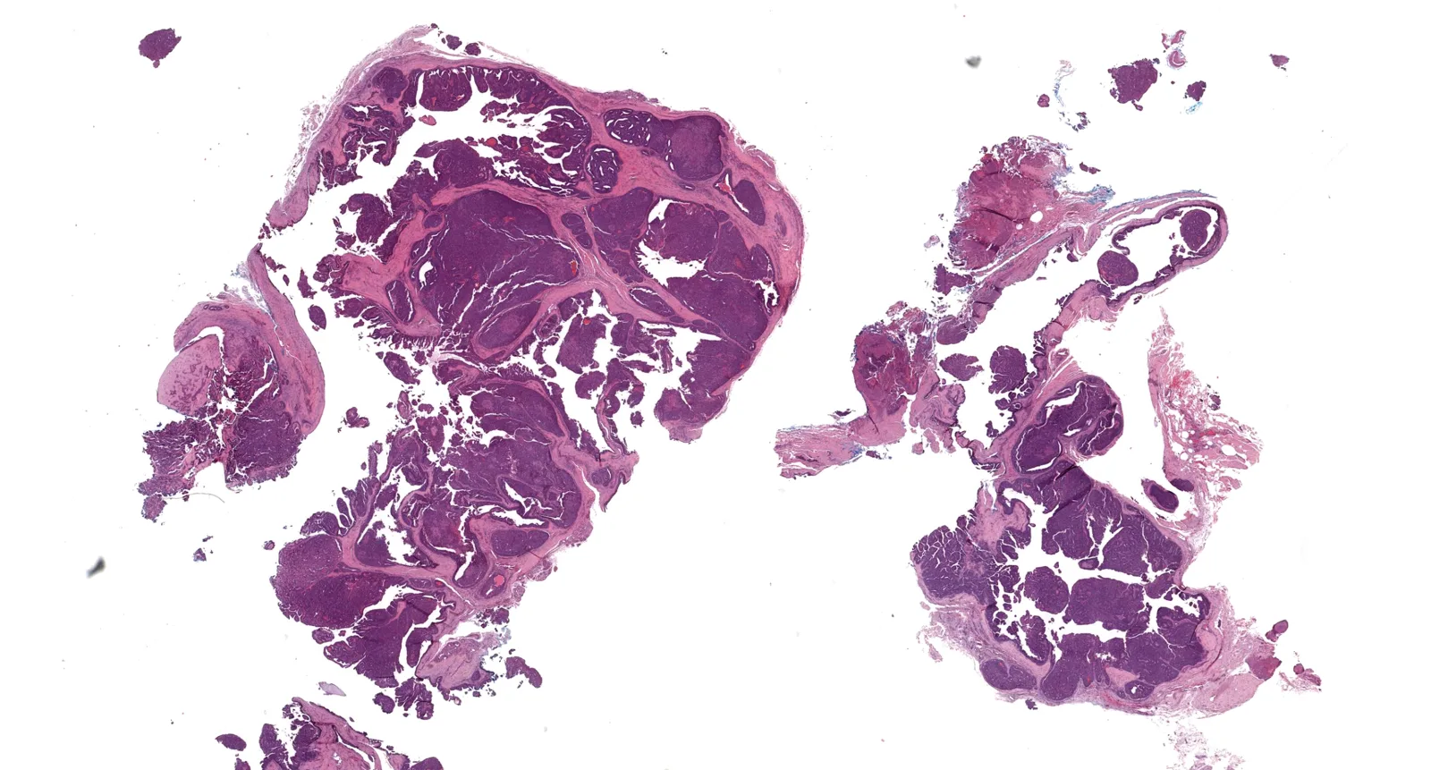
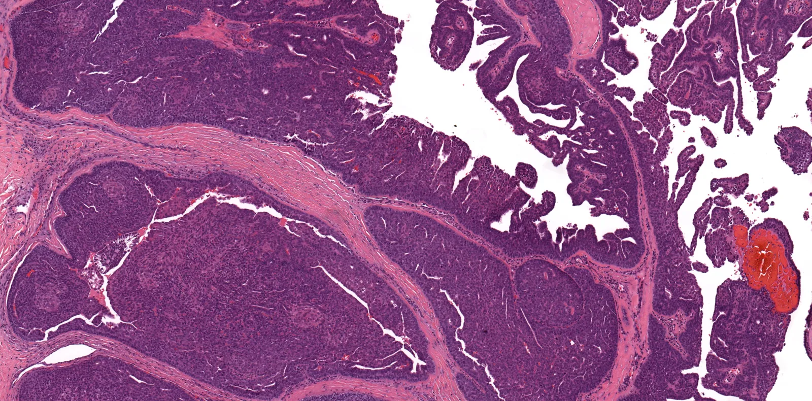
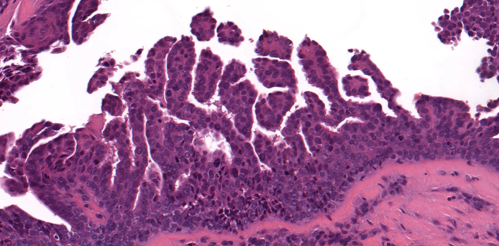
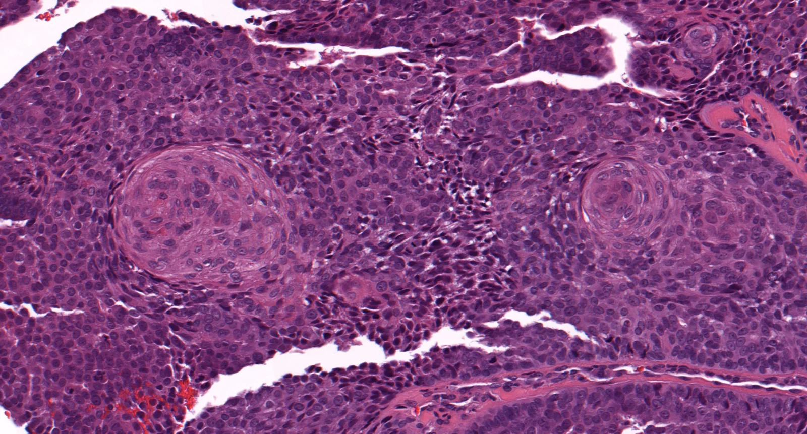
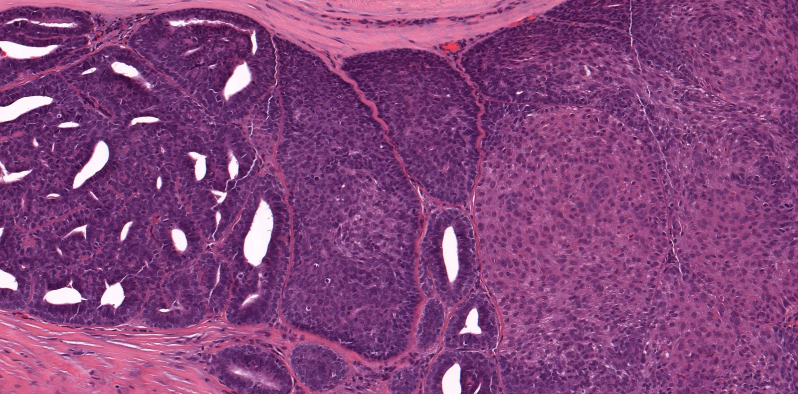
- 形态要点:真皮深层/皮下多结节实性兼囊性肿瘤,不连表皮;囊腔内乳头/微乳头,实性区散在衬覆柱状至立方上皮的小管,可见鳞样漩涡;核平淡、核分裂少,外观具迷惑性。
- 实践提示:指(趾)部任何附属器肿瘤在确切排除之前都应考虑本病,有疑问请专家会诊;报告模板建议切缘阴性切除,并请骨肿瘤外科/手外科会诊。
收录范围:KiKo 帖子全文(临床病史、诊断、2022 年更新、讨论节选、病理报告示例模板及注释、参考文献)与全部 5 幅图及图注,中英对照。数字切片与讲座视频请经链接观看。
病例 4
临床病史:45 岁男性,拇指结节缓慢增大。临床印象:囊肿。
数字切片:https://kikoxp.com/posts/6976
诊断:指(趾)乳头状腺癌
视频:https://kikoxp.com/posts/8379
2022 年 10 月更新:人乳头瘤病毒 42 型(HPV42)是指(趾)乳头状腺癌的致癌驱动因素,见于 96% 的病例。相关文章:https://pubmed.ncbi.nlm.nih.gov/35331965/ · Cancer Discovery:HPV42 驱动指(趾)乳头状腺癌
讨论
节选自我的著作《Survival Guide to Dermatopathology》(皮肤病理生存指南),经出版方 Innovative Science Press 许可使用:
指(趾)乳头状腺癌
这种罕见的汗腺肿瘤表现为手指远端(有时为足趾)的结节,通常见于中年成年男性。局部复发常见,部分病例会转移至区域淋巴结或肺,有时发生在初诊很久之后。治疗包括扩大局部切除,部分病例甚至需截指(趾)。肿瘤在真皮深层或皮下呈单个或多个结节,与表皮不相连。结节通常界限清楚,周围有致密纤维化,但部分病例周边可呈浸润性。结节特征性地见显著的囊性腔隙,数量不等的乳头和(或)微乳头突入腔内。通常存在实性区,由卵圆形、圆形乃至胖梭形细胞构成的富细胞片块组成。实性区内散在导管/小管,衬覆柱状至立方上皮。含小管的实性区呈双相外观,部分病例让我有点联想到双相型滑膜肉瘤。可见鳞样漩涡。多数指(趾)乳头状腺癌按常规细胞学和组织学标准看具有迷惑性的良性外观:细胞核通常平淡,核分裂活性相对较低。指(趾)部位和乳头结构是识别本瘤、避免误诊为良性肿瘤的关键。对发生在指(趾)的任何附属器肿瘤,在能确切排除之前,都应把指(趾)乳头状腺癌列入初始鉴别诊断。如有任何疑问,我强烈建议请专家会诊。这里对病理医生和患者来说都利害攸关。
病理报告示例模板
使用我的任何病理报告示例模板之前,请先阅读以下免责声明:https://kikoxp.com/posts/5105
指(趾)乳头状腺癌,切缘阳性(见备注)
备注:镜下特征符合指(趾)乳头状腺癌的特点,中年成年男性指部肿块这一临床情境也相符。指(趾)乳头状腺癌很容易局部复发,部分病例会转移至区域淋巴结或远处转移至肺。建议切除并达到切缘阴性。我认为由擅长处理这种罕见恶性附属器肿瘤的外科医生治疗这类患者十分重要。我发现,鉴于这些部位手术处理的复杂性,请骨肿瘤外科医生*和(或)手外科医生会诊,常常非常有助于优化指(趾)乳头状腺癌及其他手足恶性汗腺肿瘤的治疗方案。
镜下描述:病变为位于真皮深层和皮下的结节,兼有实性区和囊性区。实性区由圆形至卵圆形细胞构成的片块及密集排列的巢团组成。实性区内遍布小管结构,并可见鳞状化生桑葚样小体。囊性区可见乳头状及微乳头状内陷。
*注:orthopedic oncologist(骨肿瘤外科医生)是美国的叫法,指专门从事肉瘤及其他骨与软组织肿瘤外科治疗的骨科医生(而非肿瘤内科医生)。我不确定法国是否也用这个说法,还是只在美国使用。
参考文献
- Survival Guide to Dermatopathology. JM Gardner. Innovative Science Press. 2019.
- Fulton EH, Kaley J, Gardner JM. Skin adnexal tumors in plain language: a practical approach for the general surgical pathologist. Arch Pathol Lab Med. 2019 Jul;143(7):832-851.
- Scolyer RA, et al. Digital papillary adenocarcinoma: a tumour that should be considered in the differential diagnosis of neoplasms involving the digits. Pathology. 2013 Jan;45(1):55-61.
图示
- Jerad Gardner’s KiKo handout for Case 4 of his AIP France 2021 lecture (re-shared on X on 28 October 2026): a 45-year-old man with a slowly growing thumb nodule, clinically thought to be a cyst — digital papillary adenocarcinoma.
- Update 2022: HPV42 is the oncogenic driver, present in 96% of cases.
- Morphology: multinodular solid and cystic tumour in the deep dermis/subcutis without epidermal connection; papillae/micropapillae in cystic spaces, tubules lined by columnar to cuboidal epithelium in solid zones, squamoid whorls; bland nuclei and low mitotic activity give a deceptively benign look.
- Practice point: consider it for any adnexal tumour on the digits until confidently excluded and seek expert consultation if in doubt; the report template recommends excision with free margins and orthopedic oncology/hand surgery input.
Scope: the full KiKo post (clinical history, diagnosis, 2022 update, discussion excerpt, sample pathology report template and note, references) with all 5 figures and captions. The digital slide and lecture video are available via the links.
Case 4
Clinical history: 45 year old man with slowly growing thumb nodule. Clinical impression: cyst.
Digital slide: https://kikoxp.com/posts/6976
Diagnosis: Digital papillary adenocarcinoma
Video: https://kikoxp.com/posts/8379
Update 10/2022: Human papillomavirus 42 (HPV42) is the oncogenic driver of digital papillary adenocarcinoma and is present in 96% of cases. Articles here: https://pubmed.ncbi.nlm.nih.gov/35331965/ · Cancer Discovery: Human papillomavirus 42 drives digital papillary adenocarcinoma
Discussion
Excerpt from my book, Survival Guide to Dermatopathology, used with permission from the publisher (Innovative Science Press):
Digital papillary adenocarcinoma
This rare sweat gland tumor presents as a nodule on the distal finger (or sometimes toe), usually of middle-aged adult men. Local recurrence is common, and a subset of cases metastasize to regional lymph nodes or lung, sometimes long after initial diagnosis. Treatment involves wide local excision or even digit amputation in some cases. The tumor displays single or multiple nodules in the deep dermis or subcutis with no epidermal connection. The nodules are usually circumscribed and surrounded by dense fibrosis, although some cases may be infiltrative at the periphery. The nodules characteristically display prominent cystic spaces with variable numbers of papillae and/or micropapillae projecting into the spaces. Solid areas are usually present composed of cellular sheets of oval, round, or even plump spindle cells. Ducts/tubules punctuate the solid zones; these are lined by columnar to cuboidal epithelium. The solid zones with tubules have a biphasic appearance that in some cases reminds me a bit of biphasic synovial sarcoma. Squamoid whorls may be seen. Most cases of digital papillary adenocarcinoma have a deceptively benign appearance by conventional cytologic and histologic criteria; they usually have banal nuclei and relatively low mitotic activity. The digital location and presence of papillae are the crucial keys to recognizing this tumor and not misdiagnosing it as a benign tumor. Digital papillary adenocarcinoma should be in the initial differential diagnosis for any adnexal tumor on the digits until it can be confidently excluded. If there is any doubt, I strongly recommend expert consultation. A lot is at stake for both the pathologist and the patient here.
Sample pathology report template
Please read this disclaimer before using any of my sample pathology report templates: https://kikoxp.com/posts/5105
Digital papillary adenocarcinoma, margins positive (see comment)
Comment: The microscopic features are characteristic of digital papillary adenocarcinoma, as is the clinical scenario of a digital mass in a middle aged adult male. Digital papillary adenocarcinoma has a strong predilection for local recurrence, and a subset will metastasize either to regional lymph nodes or distantly to the lungs. Excision with free margins is recommended. I believe it is important for these patients to be treated by a surgeon skilled in handling this rare malignant adnexal tumor. I have found that consultation with an orthopedic oncologist* and/or hand surgeon is often very helpful in optimizing the treatment plan for digital papillary adenocarcinoma and other malignant sweat gland tumors of the hands and feet given the complexity of surgical management at these sites.
Microscopic Description: The lesion is nodule in the deep dermis and subcutis with both solid and cystic areas. The solid areas are composed of sheets and closely packed nests of round to oval cells. Tubular structures are present throughout these solid zones, and morules of squamous metaplasia are also seen. The cystic areas show papillary and micropapillary invaginations.
*Note: orthopedic oncologist is the USA terminology for an orthopedic surgeon (not a medical oncologist) who specializes in surgical treatment of sarcomas and other bone and soft tissue tumors. I was not sure if this term was used in France or if it is just used in the USA.
References
- Survival Guide to Dermatopathology. JM Gardner. Innovative Science Press. 2019.
- Fulton EH, Kaley J, Gardner JM. Skin adnexal tumors in plain language: a practical approach for the general surgical pathologist. Arch Pathol Lab Med. 2019 Jul;143(7):832-851.
- Scolyer RA, et al. Digital papillary adenocarcinoma: a tumour that should be considered in the differential diagnosis of neoplasms involving the digits. Pathology. 2013 Jan;45(1):55-61.
Illustration