- Jerad Gardner 在 KiKo 的软组织病理教学帖(2026-10-29 他在 X 上分享新生儿深部软组织肿物病例时再次推荐):人字形排列是束状排列的一种亚型,梭形细胞束彼此以锐角相交,形似鱼骨或「V」形折线纹。
- 过去人字形排列几乎等同于纤维肉瘤;借助免疫组化与分子检测,历史上大多数「纤维肉瘤」已可重新归类,真正的成人型纤维肉瘤极其罕见。
- 可见人字形排列的肿瘤:滑膜肉瘤、MPNST、去分化脂肪肉瘤、梭形细胞黑色素瘤、富于细胞性神经鞘瘤、富于细胞性皮肤纤维瘤、婴儿型纤维肉瘤、纤维肉瘤型 DFSP 等。
- 实践提示:「纤维肉瘤」一词仅限 3 种情形——纤维肉瘤型 DFSP、婴儿型纤维肉瘤(ETV6::NTRK3)、名称中含该词的特定肿瘤;作者建议非软组织专家不要在此之外诊断纤维肉瘤。
收录范围:KiKo 帖子全文与帖内图片(1 幅)及图注,中英对照。短视频与数字切片请经链接观看。
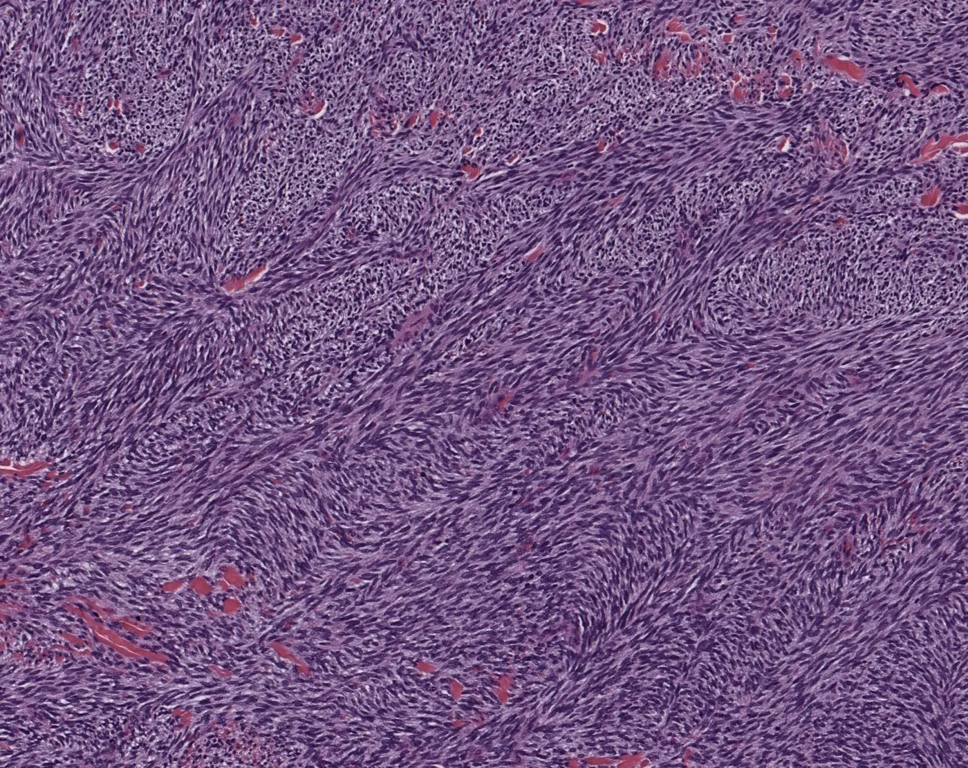
这张人字形排列的图片取自一例恶性外周神经鞘瘤(MPNST)。更多信息见作者的 MPNST 入门视频:https://kikoxp.com/posts/4092
人字形排列短视频:https://kikoxp.com/posts/22808。或继续往下读……
什么是束状与人字形排列
软组织肿瘤中的束状排列,是指梭形细胞排成细长的束,束内细胞彼此平行。人字形排列是束状排列的一种特殊亚型:细胞束彼此以锐角相交,在一些观察者看来像鱼骨或「V」形折线纹。我个人联想到的是 1990 年代流行的「魔眼」书(那种由无数彩色小点组成的图,看得恰到好处、两眼一对焦,就会浮现出 3D 图像……顺便说一句,它的学名叫自动立体图,详见:https://en.wikipedia.org/wiki/Autostereogram)。在显微镜下看到人字形排列时,我会觉得组织变成了立体的,而不是扁平的二维:有些细胞束像隆起的山脊,有些像下陷的山谷(也许我做病理太久了……)。
过去,人字形排列是纤维肉瘤的「关键词」。如今,纤维肉瘤已是极其罕见的实体。借助现代免疫组化和分子检测,历史上大多数「纤维肉瘤」都可以重新归入其他实体。这篇论文讲得很好:https://pubmed.ncbi.nlm.nih.gov/20829680/。
可见人字形排列的肿瘤
- 滑膜肉瘤
- 恶性外周神经鞘瘤(MPNST)
- 去分化脂肪肉瘤
- 梭形细胞黑色素瘤
- 富于细胞性神经鞘瘤
- 富于细胞性皮肤纤维瘤
- 婴儿型纤维肉瘤
- 纤维肉瘤型隆突性皮肤纤维肉瘤
- ……以及其他
现代软组织病理仍使用「纤维肉瘤」一词的 3 种情形
- 纤维肉瘤型隆突性皮肤纤维肉瘤(即发生于 DFSP 的纤维肉瘤)——DFSP 中出现的人字形富于细胞区。
- 婴儿型纤维肉瘤(发生于婴儿、具 ETV6::NTRK3 融合的独立肉瘤)。
- 纤维肉瘤是某个特定肿瘤名称的一部分(如黏液纤维肉瘤、硬化性上皮样纤维肉瘤等)。
即使真正的「成人型」纤维肉瘤仍然存在,也极其罕见。2020 年 WHO 骨与软组织肿瘤分类仍把成人型纤维肉瘤列为一个实体,但作为软组织病理医生,我执业 10 年从未做出过这一诊断。我强烈建议病理医生不要在上述情形之外使用「纤维肉瘤」一词(除非您是软组织病理专家),因为一旦这样诊断,很可能就是误诊。
其他可能有用的视频与病例
- MPNST 入门视频:https://kikoxp.com/posts/4092
- 全切片数字切片(发生于丛状神经纤维瘤的 MPNST):https://kikoxp.com/posts/12967
- 滑膜肉瘤入门视频:https://kikoxp.com/posts/4105
- 全切片数字切片(单相型滑膜肉瘤,典型人字形):https://kikoxp.com/posts/12966
- 脂肪肉瘤入门视频:https://kikoxp.com/posts/2634
- 全切片数字切片(含黏液样脂肪肉瘤样区及人字形排列):https://kikoxp.com/posts/6837
- 全切片数字切片(纤维肉瘤型 DFSP,兼有席纹状与人字形排列):https://kikoxp.com/posts/4603
作者全部视频、数字切片、图片及病理报告示例的分类索引:https://kikoxp.com/posts/5084(皮肤病理)与 https://kikoxp.com/posts/5083(骨/软组织肉瘤病理)。
- Jerad Gardner’s KiKo soft tissue teaching post (recommended again on X on 29 October 2026 alongside a neonatal deep soft tissue mass case): herringbone is a subtype of fascicular pattern in which spindle cell fascicles intersect at acute angles, resembling fish bones or chevrons.
- Herringbone used to be a buzzword for fibrosarcoma; with immunostains and molecular testing most historic “fibrosarcomas” can be reclassified, and true adult-type fibrosarcoma is vanishingly rare.
- Tumours showing herringbone pattern include synovial sarcoma, MPNST, dedifferentiated liposarcoma, spindle cell melanoma, cellular schwannoma, cellular dermatofibroma, infantile fibrosarcoma and fibrosarcomatous DFSP.
- Practice point: reserve “fibrosarcoma” for fibrosarcomatous DFSP, infantile fibrosarcoma (ETV6::NTRK3) and specific tumour names containing the word; the author advises non-experts not to diagnose fibrosarcoma otherwise.
Scope: the full KiKo post with its single figure and caption. The short video and digital slides are available via the links.
This picture of herringbone pattern was taken from a case of malignant peripheral nerve sheath tumor (MPNST). See my MPNST 101 video for more info: https://kikoxp.com/posts/4092
Short video about herringbone pattern: https://kikoxp.com/posts/22808. Or keep reading...
Fascicular and herringbone pattern
Fascicular pattern in soft tissue tumors means that the spindle cells are arranged in elongated bundles with the cells running in parallel to each other. Herringbone pattern is a distinct subtype of the fascicular pattern. In herringbone pattern, these fascicles intersect with one another at acute angles, forming a pattern that resembles fish bones or chevrons to some observers. What it makes me think of personally is those Magic Eye books that were popular in the 1990s (the ones with those multicolor pictures made of tiny dots that when you look at it just right and cross your eyes then a 3D image stands out...FYI the fancy word for that is autostereogram and you can read more about it here: https://en.wikipedia.org/wiki/Autostereogram). When I look at herringbone pattern under the microscope, it feels as if the tissue has become 3 dimensional rather than flat and 2D. It feels like some of the fascicles are raised ridges and others are depressed valleys (maybe I've just been doing pathology for too long...).
In the olden days, herringbone pattern was a buzzword for fibrosarcoma. In modern times, fibrosarcoma is a vanishingly rare entity. With modern immunostains and molecular, most historic examples of “fibrosarcoma” can now be reclassified as other entities. Here's an excellent paper about this: https://pubmed.ncbi.nlm.nih.gov/20829680/.
Herringbone pattern can be seen in these tumors
- Synovial sarcoma
- Malignant peripheral nerve sheath tumor (MPNST)
- Dedifferentiated liposarcoma
- Spindle cell melanoma
- Cellular schwannoma
- Cellular dermatofibroma
- Infantile fibrosarcoma
- Fibrosarcomatous dermatofibrosarcoma protuberans
- ...and others
There are 3 scenarios where "fibrosarcoma" is still used in modern soft tissue pathology
- Fibrosarcomatous dermatofibrosarcoma protuberans (aka fibrosarcoma arising in DFSP) – herringbone hypercellular area arising in DFSP.
- Infantile fibrosarcoma (a distinct sarcoma arising in infants that has an ETV6::NTRK3 fusion).
- If fibrosarcoma is part of a specific tumor name (e.g. myxofibrosarcoma, sclerosing epithelioid fibrosarcoma, etc).
If true “adult-type” fibrosarcoma still exists, it is vanishingly rare. Adult-type fibrosarcoma is still listed as an entity in the 2020 WHO bone and soft tissue tumor book, but it is a diagnosis that I have never made in 10 years of practice as a soft tissue pathologist. I strongly encourage pathologists not to use the term fibrosarcoma outside of the above scenarios (unless one is a soft tissue pathology expert), because if you do, you are very likely to be making an incorrect diagnosis.
Other videos and cases you might find useful
- MPNST 101 video: https://kikoxp.com/posts/4092
- WSI digital slides (MPNST arising in plexiform neurofibroma): https://kikoxp.com/posts/12967
- Synovial sarcoma 101 video: https://kikoxp.com/posts/4105
- WSI digital slide (monophasic synovial sarcoma perfect herringbone): https://kikoxp.com/posts/12966
- Liposarcoma 101 video: https://kikoxp.com/posts/2634
- WSI digital slides (with myxoid liposarcoma-like areas & herringbone): https://kikoxp.com/posts/6837
- WSI digital slides (fibrosarcomatous DFSP w storiform & herringbone): https://kikoxp.com/posts/4603
A complete organized library of all my videos, digital slides, pics, & sample pathology reports is available here: https://kikoxp.com/posts/5084 (dermpath) & https://kikoxp.com/posts/5083 (bone/soft tissue sarcoma pathology).