- Acta Neuropathologica 叙述性综述(Nay、Skripuletz、Isenmann 等,汉诺威医学院牵头,德国脑脊液诊断与临床神经化学学会 DGLN 共识作者组),CC BY 开放获取,本站收录全文与 7 幅图、表 1。
- 分析前因素决定诊断准确性:送检延迟会导致 pH 依赖的自溶和大量细胞丢失;快速细胞离心涂片可保存形态,推荐常规制备离心涂片染色并由专家镜检。
- 自动细胞计数仪在低细胞数及轻度升高范围精度有限、形态评估能力不足;计数池手工计数仍是定量金标准,专家镜检仍是形态学参考标准。
- 影像进步之下,CSF 细胞学在感染、肿瘤及蛛网膜下腔出血评估中仍有独特价值:吞红细胞、含铁血黄素巨噬细胞与橙色血质结晶提示出血及其时期;AI 与远程细胞学只能在经过验证的流程中使用。
收录范围:全文(摘要、正文各节、7 幅图及图注、表 1),中英对照;参考文献列表从略,正文引用链接指向出版社页面。
摘要
尽管神经影像学、分子诊断和数字技术不断进步,脑脊液细胞学仍是中枢神经系统感染性、自身免疫性、出血性和肿瘤性疾病诊断评估的基石。本篇叙述性综述总结了脑脊液细胞学的既定原则,强调了方法学局限和诊断陷阱,并整合了包括自动细胞计数、人工智能和远程细胞学在内的新兴进展。分析前因素是诊断准确性的关键决定因素。标本处理延迟会促进 pH 依赖性自溶并导致大量细胞丢失,而快速细胞离心能够保留细胞形态,有助于可靠评估。自动细胞计数仪在常规实验室中的应用日益增加,在技术人员有限的环境中具有优势。然而,现有系统在细胞计数低和轻度升高的标本中精密度有限,形态学评估能力也有限。手工计数池计数,尤其是在正常和低细胞计数范围内,仍是 CSF 细胞定量评估的金标准,而专家显微镜检查仍是定性细胞形态学评估的参考标准。虽然神经影像学的敏感性持续提高,脑脊液细胞学在感染性和肿瘤性疾病的检出以及蛛网膜下腔出血的评估中仍具有独特的诊断作用。通过细胞学检出吞红细胞巨噬细胞(噬红细胞)、含铁血黄素巨噬细胞(噬含铁血黄素细胞)和橙色血质结晶,可以揭示出血并为其时间阶段提供线索。总体而言,将脑脊液细胞学与临床、实验室、微生物学、免疫学和影像学结果相结合,仍是一种有价值的诊断工具。仔细识别常见陷阱至关重要。我们建议快速处理脑脊液,常规制备细胞离心涂片并染色,由专家进行显微镜复核,且仅在经过明确验证的工作流程中实施自动化或数字化方法。
引言:脑脊液细胞分类分析的基本原理
脑脊液(CSF)主要由侧脑室脉络丛产生,经脑室系统、蛛网膜下腔和脊髓中央管循环。它既为中枢神经系统(CNS)提供机械缓冲,也作为运输信号分子、代谢物和蛋白质的介质。每日 CSF 生成量约为 500 mL,而现存总量为 150–170 mL。因此,CSF 每五至七小时更新一次 [125]。出于诊断目的,通常在 L3/4 或 L4/5 椎间隙行腰椎穿刺采集 CSF。枕下穿刺或颈侧穿刺仅极少实施,已基本被腰椎和脑室 CSF 采集途径取代。在特定情况下,可获取脑室 CSF,通常通过外引流系统进行。生理条件下,CSF 肉眼呈清亮、无色,几乎不含细胞。充足的 CSF 量对于确保诊断准确性至关重要,尤其是在初次诊断检查时。除细胞计数和显微镜检查外,标准分析还应包括 CSF 蛋白质、葡萄糖和乳酸水平,以及白蛋白和免疫球蛋白商值 [128]。在某些地区,尤其是德语国家,常规使用 CSF/血清商值图解来解释这些参数 [102, 128]。虽然这篇以细胞学为重点的综述不会详细讨论这些参数,但应注意,将其采集并与其他结果和临床发现整合,对于形成累积性综合 CSF 报告至关重要。重要的是,至少对一张染色涂片进行细胞学评估是必不可少的 [128]。
表 1 总结了在 CNS 病理诊断检查中指导 CSF 细胞学的关键实践问题,本综述将逐一讨论这些问题。
关键问题 | 参考文献 |
|---|---|
1. CSF 细胞诊断的金标准是什么? 手工计数池计数仍是 CSF 细胞定量评估的金标准(尤其是在正常和低细胞计数范围内),而自动分析仪可用作一线方法,以确保检测的可及性和快速出具结果。如果尚未建立经本地验证的更低阈值,可将 <30 个细胞/µL 的阈值作为需要手工确认的实用标准 细胞分类计数和形态学评估必须由经验丰富的细胞学专家对细胞离心制备的涂片进行显微镜检查,理想情况下采用 Pappenheim 染色(May-Grünwald-Giemsa 染色)。自动分类可辅助筛查,但不能取代专门的细胞学评估 | [128, 134, 136] |
2. 对于 CSF 细胞计数正常的标本,制备细胞学涂片是否多余? 否。即使细胞总数正常,也可能存在相关病理发现(例如恶性细胞、活化淋巴细胞、巨噬细胞包涵体) 因此,细胞学能够提供超出单纯定量细胞计数的诊断信息 | [11, 118] |
3. 分析前因素是否与 CSF 细胞计数和细胞形态学结果的解读有关? 是。腰椎穿刺至分析之间的延迟、储存温度、血液污染以及标本量不足,都会显著影响细胞完整性和形态 在 1 至 2 h 内处理 CSF 标本被认为至关重要 尤其在初次检查时,应至少采集 5 mL,最好为 10–15 mL,以进行全面的 CSF 诊断 仅用于细胞学的标本量就应至少为 2–3 mL,而怀疑恶性肿瘤时建议采集更多标本 | [17, 29, 77, 97, 99, 115, 128] |
4. 血液污染是否影响 CSF 细胞学的诊断准确性? 是。血液混入可能引起假性细胞增多,并使细胞分类计数的解释复杂化 | [9, 56] |
5. 脑室脑脊液在细胞学上是否与腰椎 CSF 不同? 是。与腰椎 CSF 相比,脑室 CSF 的细胞计数通常较低 由于头尾方向的梯度和局部病变(例如脑室内出血、分流相关炎症),细胞组成可能有所不同 因此,源自腰椎 CSF 的参考值不能直接用于脑室标本 | [26, 54, 95] |
6. 在成人中建立的细胞学参考值和解释是否也适用于儿童 CSF 标本? 否。新生儿和小婴儿的 CSF 白细胞计数在生理状态下高于成人 必须采用年龄特异性参考范围,以避免将生理表现误判为病理表现 细胞形态学解读必须考虑发育因素 | [59, 81, 120, 144] |
7. 鞘内治疗能否改变 CSF 细胞学表现? 是。鞘内治疗(例如诺西那生、托夫生、糖皮质激素)和全身治疗(例如 IVIG)可能诱发无菌性细胞增多或特征性的巨噬细胞包涵体 这些变化通常无症状,必须识别为治疗相关改变,以免误诊 | [40, 58, 65, 119, 126, 137] |
文献检索与综述方法
本文设计为由专家主导的叙述性综述,而非系统性综述。在整个稿件撰写过程中,通过反复进行针对特定主题的数据库检索来确定文献,并辅以参考文献追溯,以及查阅相关教科书、专著和细胞学图谱。鉴于本综述旨在将 CSF 细胞学的历史发展与当前诊断实践及新兴技术相整合,未预先设定发表时间范围或固定检索式。历史和当代出版物均纳入考虑。文献依据其与综述范围的相关性,以及对当前诊断实践或历史背景的贡献进行选择。对于不同出版物中存在差异的研究结果或建议,保留并讨论这些差异,而非通过正式的证据加权予以解决,并使用现行指南、综述和原始研究为不确定领域提供背景。未设置地域限制,在相关情况下考虑了国际指南和原始研究中的证据。
关于附加表 1:为更新文献 [134] 所总结的手工与自动 CSF 细胞定量比较证据,于 2026 年 9 月 19 日检索 PubMed/MEDLINE 和 Cochrane 图书馆/CENTRAL,查找 2019 年 1 月 1 日至 2026 年 9 月 19 日发表的研究。这项针对性检索将脑脊液/CSF 的检索词与自动细胞计数或体液分析仪以及手工计数方法的相关词组合,包括基于计数池/血细胞计数板的方法。数据库检索获得 373 条记录(PubMed/MEDLINE,n = 341;Cochrane 图书馆/CENTRAL,n = 32)。去除四条重复记录后,在标题和摘要筛选阶段排除了 369 条记录中的 355 条。评估了十四篇全文文章,其中纳入 11 篇。引文筛选额外发现六篇全文文章,其中四篇符合条件。在数据库和引文筛选中,共排除五篇全文文章:一篇因已被 Waldrop 等纳入,两篇因主要定量比较在自动化平台之间进行,缺少合格的手工定量参照,一篇因手工显微镜检查仅限于分类评估,缺少适当的手工 WBC/TNC 参照比较,另一篇因无法单独提取 CSF 特异性定量结果。补充表 S1 共纳入 15 项研究。
CSF 细胞学——历史与基本原理
Quincke 于十九世纪末报告了首次经皮腰椎穿刺,并在随后数年继续研究 CSF,包括 CSF 压力、化学成分、细胞学以及各种病变中的表现 [98]。CSF 诊断学的早期发展大多发生在德语国家,例如 Fuchs-Rosenthal 计数池的开发 [35],以及所谓的全景 Pappenheim 染色(May-Grünwald-Giemsa 染色)[90]。早期细胞获取量很少,细胞形态也常遭破坏,使形态学诊断困难,最早的 CSF 图谱中即展示了这一点 [53, 101]。
正是 Sayk 在 1950 年代引入沉降室 [108],为更可靠的 CSF 细胞分析铺平了道路。因此,在 1960 年代和 1970 年代,德国耶拿和哈雷出现了具有高质量细胞形态图像的图文著作。Sayk 提供了第一部摄影图谱 [109],随后是 Schmidt [111],后者就炎症过程、组织再生、吞噬作用和异型细胞的 CSF 细胞学开展了广泛工作。随着采用细胞离心机的温和离心技术问世,CSF 细胞学变得更加可行,随后不同作者又发表了更多主要以英语出版的著作 [64, 85]。与此同时,美国的相关著作也独立出现 [44, 106],彼此引用有限。
与 CSF 生物化学和生物标志物研究取得的重大进展相比,CSF 中细胞的基本形态学特征和染色特性始终保持稳定。CSF 细胞学的历史争议主要涉及细胞群的解释和分类,而非形态变化,包括淋巴细胞亚型、巨噬细胞活化状态,以及反应性细胞与肿瘤细胞的区分。与此同时,不断演变的肿瘤命名和分子分类系统使得对较早的细胞学文献需要谨慎解读。
对于希望全面了解以实践为导向的 CSF 细胞学方法的读者,目前已有数部专门图谱和教科书可供参考 [74, 141]。Kluge 及合著者提供了一部当时最新的德文 CSF 细胞学图谱,在约 20 年间确立了标准 [61],而且独特的是,该书被译为英文 [62],并附有两种语言的 DVD,供教学和学习使用。较近时期,一部涵盖临床 CSF 诊断学各个方面的综合著作进行了更新,目前反映了最先进的实践 [141]。本文部分作者参与了该著作的编写,出于透明性考虑在此披露。其中的 CSF 细胞学章节 [48] 和综合病例图谱 [47] 展示了正常及病理标本中的典型 CSF 细胞学表现,并采用现行 WHO 分类中的肿瘤命名 [72],推荐进一步阅读。
分析前及方法学考量
为确保诊断准确性,尤其是在初次检查时,应至少采集 5 mL,最好为 10–15 mL [128]。仅用于细胞学检查的标本量就至少需要 2–3 mL;怀疑恶性肿瘤时,更多标本量至关重要,因为敏感性随标本量增加而提高 [97]。从分析前的角度看,采集较大量、最高达 30 mL 的 CSF 被认为是安全的 [78],当代数据表明,腰椎穿刺后头痛主要取决于穿刺针特征(针尖类型、针径),而非抽取的 CSF 量 [4, 67, 78]。
由于蛋白含量低且缓冲能力弱,CSF 极易受到分析前变异的影响。标本应采集于无菌聚丙烯管中。玻璃管可能使细胞黏附于管壁,导致细胞计数被低估。同样,普通 EDTA 管或氟化钠管由于可能引起细胞降解和化学干扰,也不适用于细胞学分析 [22]。然而,有关这些变化对 CSF 结果影响程度的可用数据仍然有限。出于诊断目的,通常将 CSF 依次采集到多个试管中,以便在创伤性腰椎穿刺时评估血液污染,并将标本分配用于常规和专门分析,包括送往外部实验室,而无需随后在容器之间转移。
处理前的时间至关重要。采样后,CSF 的 pH 在数小时内从约 7.3 [112] 升至碱性值(在实验条件下可达 pH 10)[20],促进细胞自溶,而环境温度升高会加速这些有害过程。因此,运输过程中应避免暴露于高环境温度。尤其是在炎热天气,温控运输可能有助于保留细胞形态完整性 [87],尽管关于 CSF 常规冷藏的现有数据并不一致,快速处理仍是分析前的首要任务。若不进行稳定处理,白细胞可能迅速丢失,因此在 1–2 h 内处理 CSF 标本被认为至关重要。新生儿和成人患者腰椎及脑室 CSF 的研究报告显示,延迟可导致淋巴细胞显著减少,5 h 内的变化范围为 −10 至 −53%,24 h 内可达 −60% [17, 29, 77, 99, 121]。对于更长时间,Schwenkenbecher 等的研究显示,72 h 后,血液污染标本中 40 至 78% 的细胞发生溶解 [115]。这可能造成误诊,尤其是在轻度细胞增多的标本中。
中性粒细胞和肿瘤细胞被认为更加脆弱。如果完整的细胞学检查延迟,至少应在 2 h 内及时完成自动或手工细胞计数,随后尽快制备涂片。关于更长时间内自溶程度的数据有限。在解读细胞计数时,必须仔细考虑这些分析中和分析前因素。
计数池,例如广泛使用的 Fuchs-Rosenthal 计数池,常用于手工 CSF 细胞计数。将新鲜采集的原始 CSF(检查前不足两小时采集)用移液器加入计数池(总体积 3.2 µL)。由于即使短时间运输也可能发生沉降,移液前应轻轻旋动试管数次。移液后应静置沉降数分钟。无核红细胞即使在未染色标本中通常也容易区分,应将其与其余有核细胞分别计数。由于在未染色标本中通常无法可靠区分白细胞与其他有核细胞(例如骨髓细胞),应注意,可能会报告细胞计数“假性”升高,而这并不反映真正的 CNS 病变。细胞学专家在评估后续细胞学结果时应注意这一点,这也是支持 CSF 细胞学诊断意义的重要依据,因为需要通过显微镜评估来确定有核细胞的性质和来源,避免误解定量结果。
对于细胞丰富或受到血液污染的 CSF,可用 0.9% NaCl 稀释(例如 1:10)后进行细胞计数。随后在细胞计数结果中考虑稀释倍数,出于易于理解和可比性的考虑,结果单位始终以“细胞数/微升”表示。
越来越多的实验室采用自动血液分析仪进行 CSF 细胞计数。自动化改善了结果周转时间和标准化水平,在细胞计数较高时以及红细胞和有核细胞计数均较高的严重血性神经外科标本中表现良好,已有报道显示其具有可接受的精密度和明确的工作流程优势 [136]。
然而,尽管技术有所改进,如果未调整并验证设备特异性参考限值,CSF 白细胞可靠的定量下限仍在 20 个细胞/µL 左右(取决于设备),较旧系统的限值可能在 50 个细胞/µL 左右 [134, 136, 143]。因此,专家评估得出结论:低于 30 个细胞/µL 的细胞计数必须通过手工计数池计数进行确认。Waldrop 等的系统性综述和荟萃分析 [134] 再次强调,在接近参考区间的极低计数范围内,关于自动计数准确性的数据稀少,且亚组分析显示,计数 <20 个细胞/µL 时相关性明显较弱,尽管其发现各研究中自动分析仪与手工显微镜检查的总体一致性良好,而且绝对计数较高时一致性更强。我们的针对性更新额外识别出 15 项比较研究,涵盖成熟的血液分析仪以及较新的基于图像和半自动化平台,补充表 S1(附加文件 1)。总体而言,这些研究报告的白细胞总数与手工计数池计数具有良好一致性,尤其是在细胞浓度较高时。虽然某些较新平台在低细胞计数时表现令人鼓舞,但现有证据仍依赖于具体设备,且常基于小规模、单中心队列。在 5 个细胞/µL 的决策阈值附近,仍观察到具有临床意义的差异。自动细胞分类计数的一致性通常较弱,且若无形态学复核,无法可靠识别异型或恶性细胞。因此,更新后的证据扩大了所评估平台的范围,但并未实质性改变 Waldrop 等的结论 [134]。
综上,可提出一项兼顾各方面的建议:由受过培训的人员进行手工计数池计数,仍是 CSF 白细胞定量评估的金标准;自动分析仪可作为一线方法,以确保检测的可及性和快速出具结果,前提是对低和轻度升高的细胞计数进行手工确认,并认识到方法学局限。
只要有原始 CSF 可用,就应获得白细胞定量计数,不应以细胞离心涂片上每高倍视野细胞数的近似估计来替代 [128]。细胞离心涂片上的细胞回收量很大程度上取决于制备技术、离心条件、标本量和沉积面积,而一个高倍视野所代表的面积还会因显微镜系统而异 [63, 104]。因此,基于每高倍视野细胞数的通用数值阈值尚未标准化,不应将其用于把 CSF 归类为细胞数正常或细胞增多 [63, 104]。只有在无法获得原始 CSF 进行定量计数的特殊情况下,例如复核外部制备的涂片时,才可对细胞丰富程度作定性描述,并明确注明未获得定量细胞计数。
除自动细胞计数外,常规分析仪提供的自动细胞分类充其量也只是粗略分类(例如粒细胞、淋巴细胞和单核细胞),应仅考虑用于筛查。关键的是,自动化系统无法可靠识别对 CSF 鉴别诊断起决定作用的细胞形态学类型(例如浆细胞、活化/反应性淋巴细胞、原始细胞、含铁血黄素巨噬细胞/吞红细胞巨噬细胞、散在肿瘤细胞或细胞团)[113, 136, 142]。因此,只要定性解释具有重要意义,就仍需要显微镜检查。
正常成人腰椎 CSF 白细胞计数参考范围并非由单项里程碑式试验界定,而是在历史上由多个“正常”患者的描述性系列研究逐渐形成,随后被整合为实验室和教科书中的参考范围 [2, 7]。这些范围本质上似乎是基于人群的统计学参考区间,源自无 CNS 疾病临床或实验室证据的队列,然后取整为常规实践中便于应用的截断值。
血液混入是 CSF 解读中最常见的挑战之一。它可能发生于腰椎穿刺过程中,并可显著影响细胞学和生物化学参数。近似而言,每混入 1000 个红细胞约带入 1 个白细胞。对于血液混入严重的标本(每 µL 超过 100,000 个红细胞(RBCs)),通过线性回归确定的白细胞(WBC)计数校正公式似乎不准确,往往高估混入的白细胞数,从而可能低估真实 WBC 计数 [56]。此外,对于血细胞计数显著变化的患者,例如白血病、活动性全身感染、贫血或白细胞减少症患者,这一比例可能显著改变,使上述校正因子失准。在此类情况下,我们建议考虑该患者实际血细胞计数中红细胞与白细胞的个体比例。
为尽量减少血液污染,应使用最后一管标本进行 CSF 细胞计数,并检查连续各管中血液污染是否消退。
细胞分类
由于 CSF 通常是一种几乎无细胞的液体,应将细胞浓集于载玻片上,以提高诊断准确性。目前,依据现行标准,主要采用细胞离心方法(细胞离心涂片)实现这一目的 [124]。细胞离心涂片风干后即可进行染色。Pappenheim 染色(又称 May-Grünwald-Giemsa 染色)已成为 CSF 实验室的标准细胞学染色方法 [22, 128]。针对特定检查还可采用其他染色方法(例如革兰染色、普鲁士蓝染色、印度墨汁染色、Ziehl–Neelsen 染色),但鉴于精细的分子生物学和成像技术已可应用,这些方法使用较少。尽管具有主观性,专家对染色细胞离心标本进行的显微镜检查仍是定性细胞分类的参考标准 [128]。受过培训的细胞学专家能够发现细微的形态学线索,例如反应性淋巴细胞、浆细胞、活化巨噬细胞、粒细胞、异型和恶性细胞、核分裂象以及细菌,这些发现在肿瘤性或炎症性疾病中至关重要。此外,细胞学检查能够区分真正的细胞增多与非白细胞或伪影细胞所致的细胞计数假性升高,而后者无法通过自动化方法或未染色标本可靠识别。
正常 CSF 细胞学
生理条件下在 CSF 中检出的细胞几乎完全是免疫细胞,并非源自神经组织。血液-CSF 屏障和血脑屏障有效阻止红细胞和粒细胞被动通过,同时允许淋巴细胞和单核细胞在严格调控下进行有限迁移,以实施免疫监视 [31, 98]。这一概念构成了 CSF 细胞计数诊断价值的基础,因为数量和性质上的偏离可为中枢神经系统内的炎症过程提供敏感指标。正常成人 CSF(图 1)含有 ≤4 个细胞/µL,主要由淋巴细胞和单核细胞组成,比例约为 70:30。
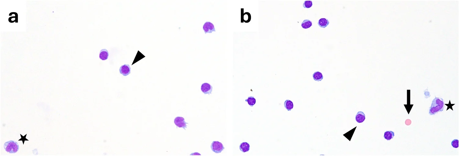
正常脑脊液细胞学(Pappenheim 染色(May-Grünwald-Giemsa 染色))。采用 Pappenheim 染色(May-Grünwald-Giemsa 染色)的代表性脑脊液细胞离心涂片,显示细胞稀少,以小的成熟淋巴细胞(箭头头部)为主,偶见单核细胞(星号)。淋巴细胞的细胞核圆形、深染,胞质少,而单核细胞较大,胞质更丰富、淡嗜碱性,染色质较疏松。在(b)中可见单个红细胞(箭头),符合无诊断意义的极少量血液污染。总体未见炎症或恶性肿瘤的细胞学征象(替代文字:代表性 CSF 细胞离心涂片,显示以小的成熟淋巴细胞为主,偶见单核细胞,无病理异常。)
应注意,常规手工 CSF 细胞学中的细胞分类计数通常基于对随机识别的 100 个细胞进行分类。因此,所得百分比是受二项分布变异影响的点估计。例如,测得比例为 30% 时,对应的近似 95% 置信区间为 21–40%,其他比例也适用类似的误差范围 [107]。因此,与预期分布的轻微偏离可能反映抽样变异,而非真正的生物学差异。
在 Pappenheim 染色(May-Grünwald-Giemsa 染色)的细胞离心涂片上,细胞核呈紫至紫罗兰色嗜碱性,胞质呈淡色至灰蓝色,可据此可靠区分白细胞类别。淋巴细胞为小的圆形细胞(≈7–9 µm),染色质致密、均匀,周围仅有一圈很少的嗜碱性胞质。活化淋巴细胞较静息淋巴细胞大,在正常 CSF 中不常见。重要的是,区分静息与活化淋巴细胞需要谨慎解读,因为即使在非病理标本中,细胞大小或胞质体积也可能存在轻微变化。因此,过早将淋巴细胞归为“活化”是常规 CSF 分类细胞学中过度解读的常见原因。单核细胞大小约为淋巴细胞的两倍(≈15–20 µm),在非活化状态下形态更为多样,通常具有凹陷的肾形核、细致的染色质和灰蓝色胞质,即使无明显活化,胞质也可能含有小空泡。
伪影
伪影(图 2),即涂片上可见但不代表患者体内 CSF 实际状况的细胞和现象,可通过两种不同方式产生:首先,细胞或颗粒可能在穿刺过程中进入标本。其次,伪影可能在制备和染色过程中被引入或产生。
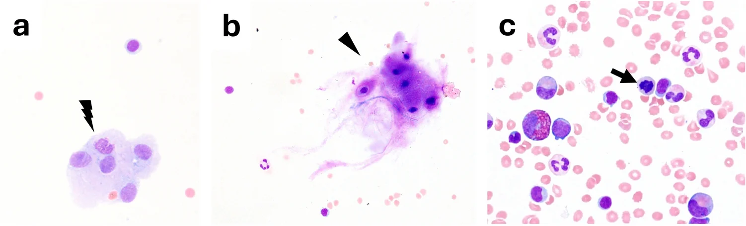
脑脊液中的细胞学伪影(Pappenheim 染色(May-Grünwald-Giemsa 染色))。代表性细胞离心涂片,展示脑脊液分析中遇到的常见细胞学伪影。a 内皮细胞(闪电符号),特征为大型多边形细胞形成黏附性细胞团,胞质丰富、淡染,细胞核呈卵圆形,通常由穿刺操作引入;b 软骨细胞(箭头头部),表现为大型细胞,胞质丰富、嗜碱性,细胞核圆形,代表穿刺过程中椎间盘物质造成的污染;c 骨髓污染,特征为红细胞背景中存在不同成熟阶段的造血前体细胞(有核红细胞;箭头)(替代文字:CSF 中具有代表性的细胞学伪影,包括内皮细胞、软骨污染和骨髓混入。)
除非在穿刺过程中被人为带入,或源自此前的(蛛网膜下腔)出血,否则不应存在红细胞。在所采集的全部 CSF 标本中,多达 70% 可检出不同程度的人为血液污染,而婴儿和新生儿中的发生率似乎更高 [116]。
在腰椎穿刺过程中,来自穿刺途经组织的细胞类型(例如皮肤、肌肉、软骨、骨髓、上皮细胞)可能作为伪影出现,而其余分类细胞学表现并无异常。内皮细胞(图 2a)可表现为大型多边形细胞形成的黏附性细胞团,胞质丰富、淡染,细胞核呈卵圆形。软骨(图 2b)表现为陷窝内的软骨细胞,嵌于嗜碱性的软骨样基质中(常呈异染性)。来自脑室系统的 CSF 标本也可能含有脉络丛上皮细胞、室管膜细胞以及(变性)脑实质碎片。室管膜或脉络丛细胞为立方形细胞,形成黏附性片状或蜂窝状细胞团,有时有纤毛,细胞核均匀、圆形,染色质细腻:应将其识别为与取样部位相关的污染物,而非癌。来自穿刺通道的骨骼肌表现为细长纤维或多边形碎片,胞质致密、纤维状,细胞核位于周边(Pappenheim 染色(May-Grünwald-Giemsa 染色)下横纹可能不明显)。平滑肌细胞呈梭形,细胞核为“雪茄状”。骨髓带入(罕见)可产生三系混合细胞(图 2c):幼红细胞、中幼粒细胞,以及偶见的巨核细胞碎片。值得注意的是,骨髓混入会伴有红细胞。
如前所述,根据所采用的计数方法,污染可导致细胞计数人为升高,必须在报告中记录,并通过细胞学确认其性质。
最后,人为混入的细菌(皮肤菌群或试剂沉淀)可能模拟感染。细菌通常存在于细胞间的“空隙”中,且出现时不伴细胞增多或中性粒细胞,应怀疑人为污染,并结合临床进行判断。
除人为混入细胞或微生物外,细胞丢失和染色伪影也可能影响诊断准确性。由于 CSF 蛋白含量低且缓冲能力弱,自溶在数小时内开始,首先影响粒细胞 [88]。有人假设肿瘤细胞降解得更快 [41]。典型自溶征象包括核溶解或核碎裂、细胞核模糊、胞质颗粒消失、空泡形成及“影”细胞(图 3a)。自溶可人为降低表观细胞计数,并使鉴别诊断模糊。重度自溶可显著降低细胞分类计数的诊断准确性,因此细胞学专家应予注明。
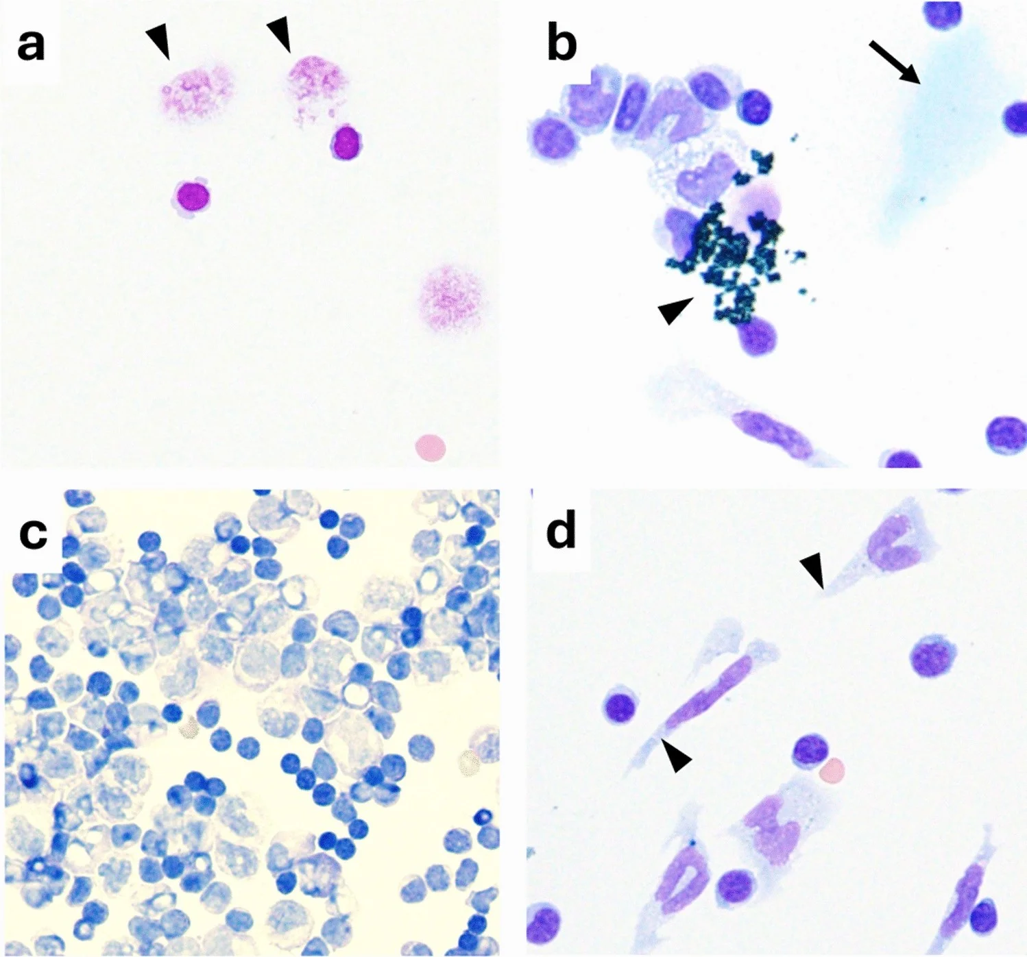
脑脊液细胞学中具有代表性的制备和染色伪影(Pappenheim 染色(May-Grünwald-Giemsa 染色))。a 自溶改变伴影细胞(箭头头部),特征为细胞和细胞核细节丢失。b 染色伪影,可见符合染料沉淀的颗粒状嗜碱性物质(箭头头部),以及局部背景染色不均/染液积聚(箭头)。c 细胞过载、细胞丰富的细胞离心涂片,显示染色不均,核质对比度降低。d 细胞离心涂片相关的机械性变形,细胞被拉长,形态改变(箭头头部)(替代文字:CSF 细胞学中具有代表性的制备和染色伪影,包括细胞离心涂片相关的细胞变形、细胞过载且染色不均的涂片、自溶影细胞,以及颗粒状/嗜碱性染色伪影。)
Pappenheim 染色(May-Grünwald-Giemsa 染色)中最常见的染色伪影包括大小不一、均匀深紫色的颗粒,其所在焦平面与细胞不同,可模拟细菌(图 3b)。这些颗粒通常在试剂陈旧或冲洗不充分时出现。此外,准确染色需要 pH 约为 6.8 的磷酸盐缓冲液。pH 漂移会使胞质浑浊、核细节不清(染色不足)。细胞丰富的标本必须在染色前稀释,因为细胞过载的细胞离心涂片形成的是多层而非单层细胞:染料和缓冲液渗透不均,因此中央表现为染色不足(细胞核淡染、胞质褪色),而边缘则呈染色过度,背景浑浊(图 3c)。另一种典型制备伪影由细胞离心涂片制备过程产生。在细胞离心涂片上,较高的 g 力可使脆弱细胞的细胞核变形,夸大细胞大小或改变细胞形状(图 3d)。
炎症性 CNS 疾病中的细胞学模式
在 CSF 中,炎症体现为细胞分类计数的改变和免疫细胞系的活化。了解活化模式可为潜在病理过程提供关键线索。然而,炎症性细胞学模式并无疾病特异性,不应解读为可单独确定病因。其表现可能受疾病阶段、抗微生物或免疫抑制治疗、鞘内治疗、血液污染和 CSF 采样部位影响,因此始终应结合临床和实验室背景进行解释。
反应性淋巴细胞增大(≈10–20 µm),染色质疏松,嗜碱性胞质更丰富,核轮廓多变。核凹陷、核外形不规则和偶见核仁可作为反应性活化的一部分出现,本身不应被解读为恶性特征。然而,有报道称,肿瘤性淋巴细胞出现显著胞质不规则和深核切迹的频率,明显高于炎症性淋巴细胞 [92]。重要的是,在炎症性 CSF 中观察到的大多数活化淋巴细胞是活化 T 淋巴细胞,而非 B 细胞谱系分化的表现 [49]。在高度活化的 CSF 中(例如急性神经莱姆病),偶尔可检出淋巴细胞核分裂象。浆细胞代表终末分化的 B 细胞 [94],因此反映体液免疫活化的较晚阶段,而非仅仅是一种活化状态。细胞学上,其特征为偏位、扁平的细胞核,染色质粗糙,核周有与高尔基体对应的淡染区,胞质深嗜碱性。CSF 中浆细胞的出现可早于鞘内免疫球蛋白合成的定量检出,凸显了细胞学检查在炎症性 CNS 疾病中的额外诊断价值。
单核细胞逐渐转变为活化巨噬细胞,伴有胞质增大和空泡形成。在病毒性脑膜炎或消退期细菌性脑膜炎中,吞噬细胞碎屑很常见。活化单核细胞可因大量空泡而显著增大,胞质可呈泡沫状(“泡沫细胞”),或在存在单个大空泡时几乎呈空白(“印戒细胞”)。
中性粒细胞呈圆形,具有分叶核。在暴发性细菌性炎症等急性期,可见细胞核呈“杆状”的未成熟中性粒细胞。在炎症过程中,核分叶增多,通常达到 4–6 叶。有报道称,散在中性粒细胞虽然也可出现在无细胞增多的“正常”CSF 中,但与 CSF 感染无关 [13, 24],尽管文献中支持这一假设的证据很少。
嗜酸性粒细胞的胞质通常具有明显的嗜酸性颗粒,多数情况下可清楚辨认出两叶核。以嗜酸性粒细胞为主的细胞增多很罕见,通常提示 CNS 寄生虫或球孢子菌感染,或者更罕见地,提示肿瘤性脑膜炎中的伴随反应,尤其见于霍奇金病累及 CNS 的报道,或提示分流相关反应 [36, 43, 46]。在某些自身免疫性炎症性 CNS 疾病中,最突出的是 AQP4-IgG 阳性的视神经脊髓炎谱系疾病(NMOSD),曾报道含有较大比例嗜酸性粒细胞的混合性细胞增多 [50]。在 CNS 血管炎性疾病中,也曾罕见报道 CSF 嗜酸性粒细胞增多,包括嗜酸性肉芽肿性多血管炎和原发性中枢神经系统血管炎 [45, 86]。CSF 嗜酸性粒细胞增多也可发生于药物诱导的无菌性脑膜炎,包括与 NSAIDs 和抗生素相关的病例 [80]。
嗜碱性粒细胞的胞质呈明显深蓝色或含嗜碱性颗粒,使日常实践中难以清楚区分细胞核与胞质。它们在 CSF 中很少出现,其诊断意义仍知之甚少。在无血液污染且其他方面无异常的 CSF 中,不应见到嗜碱性粒细胞。因此,一旦发现,应结合血液污染、血液系统肿瘤或软脑膜肿瘤累及的背景进行解释,极少数情况下也应考虑神经结节病或病毒性脑膜炎等炎症性 CNS 疾病 [18, 19, 25]。
感染性 CNS 炎症
急性化脓性细菌性脑膜炎是一种神经系统急症,病死率较高,存活者长期患病负担较重 [5]。就 CSF 细胞学表现而言,通常可见较高的细胞计数(通常每 µL 超过数个 1000 个细胞),以中性粒细胞为主(图 4a)。在成人的典型表现中,中性粒细胞通常占白细胞的 >90% [128, 129]。
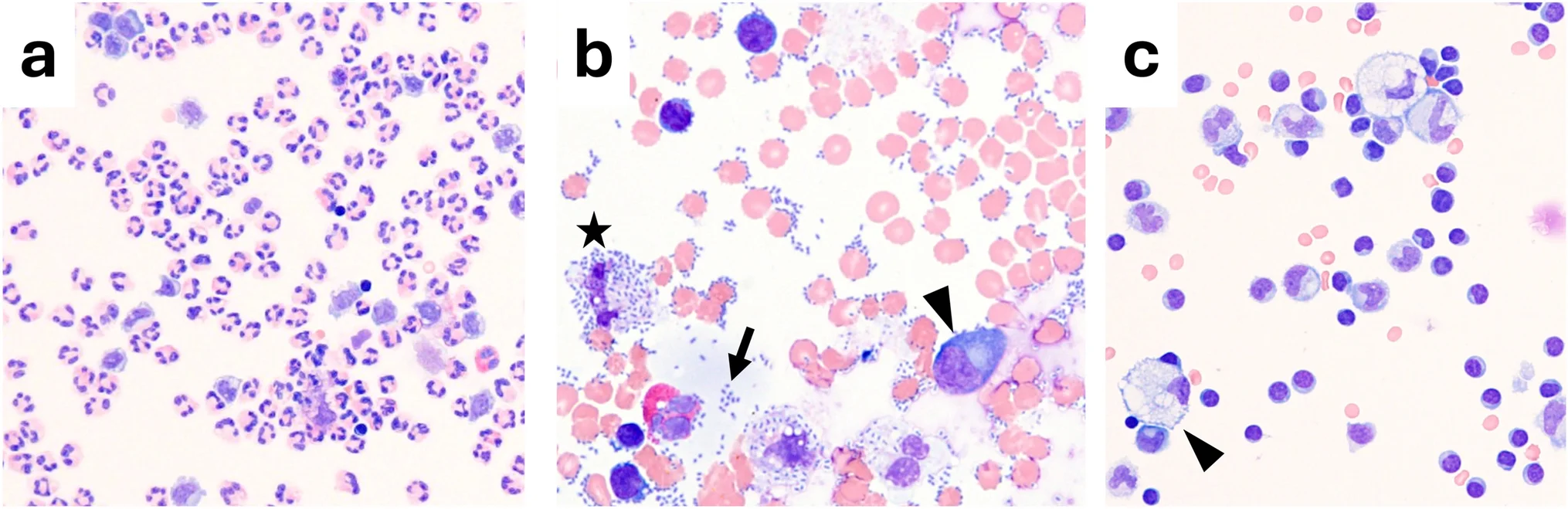
感染性炎症性 CNS 疾病的细胞学模式(Pappenheim 染色(May-Grünwald-Giemsa 染色))。代表性脑脊液细胞离心涂片,展示不同的感染相关炎症模式。a 细菌性(化脓性)脑膜炎中的显著中性粒细胞增多,未检出微生物。b 肺炎链球菌感染中的早期非化脓性脑膜脑炎,特征为在缺乏以中性粒细胞为主的浸润时,可见细胞外(箭头)和细胞内(星号)双球菌,并伴有一个大浆细胞(箭头头部)。c 病毒性 CNS 炎症较晚阶段伴吸收性改变,特征为巨噬细胞具有泡沫状胞质(“泡沫细胞”,箭头头部),反映通过吞噬清除细胞碎屑(替代文字:三幅 CSF 细胞离心涂片显微图,显示(a)显著中性粒细胞增多,(b)尽管中性粒细胞炎症有限,仍可见细胞内外双球菌及一个浆细胞,以及(c)炎症消退过程中的泡沫状巨噬细胞)
然而,应强调,感染自然呈阶段性进展,在细菌性 CNS 感染的早期或晚期,预期表现不同。在细菌性脑膜炎的早期阶段(约 12 h 或更短)[32],尤其如脑膜炎球菌和肺炎球菌疾病病例中所描述的,标本中可能直接检出细菌,同时炎症反应较弱;在此期间,细胞计数可能正常或接近正常,仅有轻微炎症征象,有时甚至无中性粒细胞浸润证据(图 4b)。这种特殊的细菌性脑膜炎也称为“非化脓性”脑膜炎,被认为尤其危险。它更常见于老年患者和免疫功能受损的情况下 [118]。
随着炎症进展,持续的免疫反应导致降解过程(图 4c),使中性粒细胞增多减轻,巨噬细胞、活化单核细胞和淋巴细胞相对增加。在腰椎穿刺前开始抗生素治疗(按照指南流程),可在数小时内使 CSF 无菌化,但在最初数小时内不会实质性改变白细胞计数 [55]。
另一种可导致结果偏离预期的情况是免疫抑制。在严重免疫功能受损的患者中,由于宿主炎症反应显著减弱,更常出现程度较低的细胞增多 [132],甚至白细胞计数正常 [118]。因此,白细胞计数正常不能排除感染。在此类情况下,诊断更依赖于进一步的直接或间接病原体诊断,以及患者病史和临床病程。
神经莱姆病通常可见中度细胞增多,介于每 µL 50 至 500 个细胞之间 [128]。可能存在一定比例的中性粒细胞,尤其在感染早期及治疗前 [89]。由于存在强烈的淋巴细胞活化,浆细胞比例往往显著增加,甚至出现核分裂象,初看时仅凭形态可能难以与肿瘤性脑膜炎区分。然而,淋巴瘤中通常缺乏轻度至中度活化的(“中间型”)淋巴细胞。对于诊断不明确的病例,需要进一步扩大检查。这些检查包括流式细胞术、整合鞘内免疫球蛋白合成以及通过 Reiber 图解评估的血液-CSF 屏障功能的综合 CSF 报告、CXCL-13、疏螺旋体特异性抗体指数计算,以及疏螺旋体特异性血清学检测 [103, 127, 138]。
理论上,病毒感染也被认为存在类似的阶段性病程,起初伴有中性粒细胞浸润 [15, 70]。然而,这些情况下向淋巴细胞为主的表现转变,似乎比大多数细菌病原体感染更快:细菌性脑膜炎的间隔 CSF 分析显示,即使接受有效抗微生物治疗,细胞增多和中性粒细胞占优势也常持续超过 48–72 h [10, 70]。然而,直接比较不同感染病因下 CSF 细胞变化时间动态的系统性数据仍然有限。病毒性 CNS 感染通常在诊断性腰椎穿刺时表现为淋巴细胞增多(通常每 µL 50–500 个细胞)[70]。不过,对于肠道病毒、西尼罗病毒、FSME 或 HSV 等若干病毒类型,CSF 可短暂不出现细胞增多,或显示不同程度的中性粒细胞偏多 [15]。白细胞计数通常轻度至中度升高。中枢神经系统机会性感染(例如人多瘤病毒 2 型(原称 JCV)、CMV)可引起不明显的细胞学表现,因此常只能通过进阶诊断手段检出。
虽然病毒性 CNS 感染中早期中性粒细胞反应、随后迅速转为淋巴细胞占优势的过程已得到充分确立 [70],但结核或真菌感染存在类似阶段性病程的证据有限,这些疾病中的 CSF 细胞学常表现为异质性或混合性细胞模式。
结核性脑膜炎通常表现为 CSF 白细胞计数中度至显著升高,范围约为每 µL 50 至 1000 个白细胞,特征为混合性细胞组成,淋巴细胞和中性粒细胞比例不一,尤其在疾病早期;随着病程进展或接受治疗,转为以淋巴细胞为主 [122]。结核性脑膜炎中偶有嗜酸性粒细胞的报道,可能使细胞学表现更具异质性 [66]。在 CNS 结核中,即使是通过其他方式确诊的病例,Ziehl–Neelsen 染色也仅能在少数病例中显示抗酸杆菌 [122]。因此,核酸扩增已成为诊断金标准,应常规抽取更大量的 CSF(7–10 mL)以提高诊断敏感性,尤其是在结核性脑膜炎中,因为结核分枝杆菌在 CSF 中通常浓度很低 [27]。
真菌性 CNS 感染的 CSF 炎症表现具有高度异质性,随病原体、疾病阶段和宿主免疫状态而异 [69]。与结核性脑膜炎类似,真菌病原体在 CSF 中的浓度往往较低,使微生物学和分子诊断高度依赖充足的标本量。
自身免疫性炎症
非感染性自身免疫疾病的 CSF 细胞学通常以淋巴细胞和单核细胞为主,程度为轻至中度,但某些疾病具有独特特征。解读应整合临床发现和进一步诊断检查。细胞学通常可支持这些诊断,但不足以确诊。
多发性硬化是最常见的需要详细 CSF 诊断检查的免疫性 CNS 疾病。常规 CSF 参数往往正常,或至多表现为轻度淋巴细胞和单核细胞增多,通常低于 50 个细胞/µL [2]。虽然有报道称,在复发性病程中,轻度细胞增多和包括产生免疫球蛋白的浆细胞在内的淋巴细胞活化征象更常见 [6],但 CSF 细胞计数与临床活动度之间相关性的强度仍有争议。
除多发性硬化外,还有其他自身免疫疾病可引起 CSF 炎症。值得注意的是,自身免疫过程中 CSF 白细胞计数和分类细胞学在形态上可模拟感染性疾病等情况,因此,结合额外诊断检查和随访观察所得的累积结果往往具有重要意义。
与多发性硬化不同,水通道蛋白-4-IgG 阳性 NMOSD 和 MOG 抗体相关疾病(MOGAD)在发作期间均表现为变化更大、且往往更明显的细胞增多(最高达每 µL 500 个细胞),常见中性粒细胞,偶见嗜酸性粒细胞,尤其是在 NMOSD 和儿童队列中——当临床表现与多发性硬化不相符时,这些模式应引起怀疑——并在最初最终模拟细菌感染 [50, 51]。
自身免疫性脑炎(AIE)并不一定表现为 CSF 细胞增多。尤其在发生于老年患者的 AIE 亚型中,CSF 白细胞计数往往正常 [8]。然而,若出现细胞增多,则常为轻度至中度淋巴细胞增多,而某些 AIE 的极急性期可出现中性粒细胞 [57],可能模拟感染性疾病。
CSF 炎症的其他原因
鞘内治疗可引起独特的细胞学变化。在脊髓性肌萎缩症患者接受诺西那生治疗期间,可出现含有大量边界清晰的紫色/蓝色包涵体的巨噬细胞(“诺西那生噬细胞”;图 5a),而无持续性炎症变化 [40]。接受鞘内托夫生治疗的患者中也描述了类似的巨噬细胞包涵体(图 5b)[119]。用于 SOD1 突变型肌萎缩侧索硬化的托夫生,常伴有 CSF 白细胞计数升高、鞘内免疫球蛋白合成增加及 CSF 蛋白升高,偶有无菌性/化学性脑膜炎或脊髓炎。这些实验室变化往往无症状,但必须识别为治疗相关改变 [137]。鞘内类固醇 [126] 和其他药物也可引起无菌性中性粒细胞增多(“化学性脑膜炎”)。此外,静脉注射免疫球蛋白(IVIG)治疗是公认的无菌性脑膜炎病因,通常在输注后数小时至数天内出现,特征为显著细胞增多,早期常以中性粒细胞为主,伴 CSF 蛋白升高,但微生物学检查阴性 [58, 65]。最后,短时间内反复腰椎穿刺后可出现轻度“反应性细胞增多”[110]。
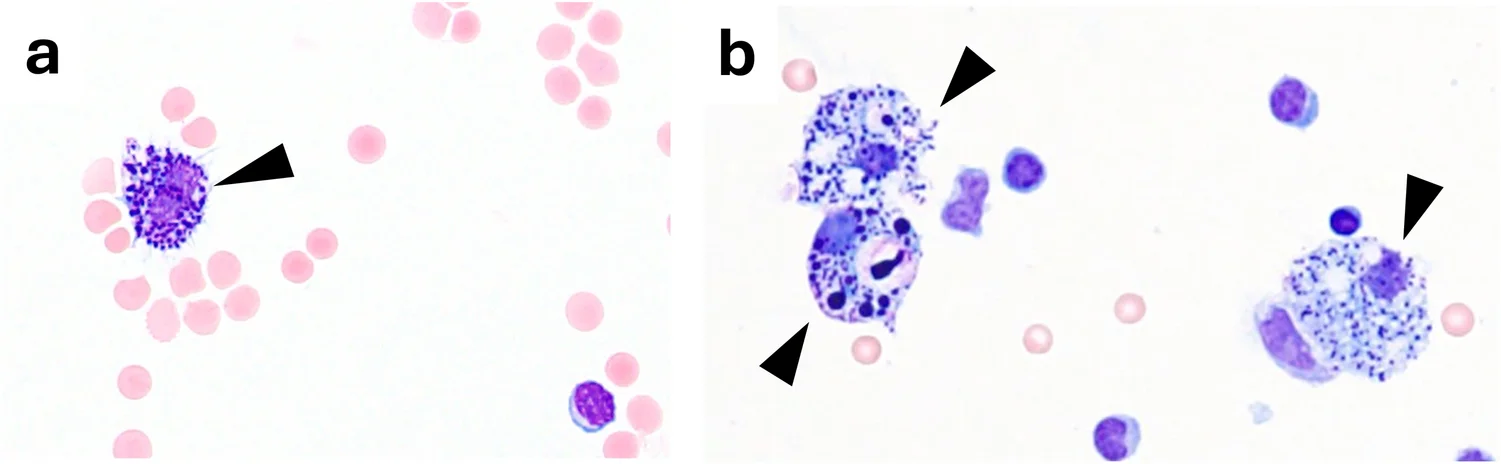
脑脊液中反义寡核苷酸相关的巨噬细胞包涵体(Pappenheim 染色(May-Grünwald-Giemsa 染色))。代表性脑脊液细胞离心涂片,显示鞘内反义寡核苷酸治疗后,巨噬细胞内出现大量边界鲜明、浓缩致密、嗜碱性至紫色的包涵体。a 单个巨噬细胞内胞质包涵体密集,符合诺西那生相关巨噬细胞包涵体细胞(“诺西那生噬细胞”;箭头头部),其余涂片细胞稀少,伴轻度人为血液污染。b 托夫生相关巨噬细胞包涵体(“托夫生噬细胞”;箭头头部),大量大小不一的嗜碱性至紫色包涵体遍布胞质(替代文字:两幅 CSF 细胞离心涂片显微图,显示鞘内反义寡核苷酸治疗后,巨噬细胞内出现紫蓝色胞质包涵体:(a)诺西那生相关包涵体和(b)托夫生相关包涵体。)
在无感染、自身免疫疾病或近期腰椎穿刺的情况下,偶尔也可观察到轻度炎症性 CSF 改变。在此类情况下,中枢神经系统组织损伤,例如手术、创伤或缺血性卒中后,可诱发能够通过 CSF 细胞学检出的反应性炎症改变 [100]。
中枢神经系统出血
众所周知,相当一部分蛛网膜下腔出血(SAH)患者的头颅计算机断层扫描(CT)影像学表现可能无明显异常,尤其是在亚急性情况下 [82, 135]。在这些情况下,肉眼观察脑脊液颜色、离心后上清液持续黄变、检测铁蛋白等化学指标,以及最重要的针对性脑脊液细胞病理学分析,可提供决定性证据 [82]。黄变和红细胞计数显著升高均不是 SAH 的特异性表现。脑脊液细胞学可显示既往出血随时间变化的标志性特征。出血后,可在脑脊液中检出吞噬血液成分的巨噬细胞。
通常在出血事件后 4–12 h 内可观察到的第一种巨噬细胞亚型是吞红细胞巨噬细胞(噬红细胞)[82, 133]。在随后的数日内,吞红细胞巨噬细胞(图 6a)逐渐演变为含铁血黄素巨噬细胞(噬含铁血黄素细胞)(图 6b),后者通常在 24–48 h 后出现 [82, 133]。这些细胞含有颗粒状深色色素(含铁血黄素),普鲁士蓝染色呈阳性(可在有疑问时使用)。在血红蛋白降解过程中,可出现中间型的混合性噬红细胞/噬铁细胞(噬红细胞/噬含铁血黄素细胞),同时含有部分保存的红细胞和细胞内含铁血黄素颗粒。它们代表吞红细胞巨噬细胞与含铁血黄素巨噬细胞之间的过渡形式,因此不应将其解释为具有明确时间窗的独立阶段 [61, 62, 124]。在连续采集的标本中,新近的红细胞吞噬现象与处于更晚阶段的含铁血黄素巨噬细胞同时存在,可能提示反复出血,应结合临床及影像学表现进行解释 [61, 62]。随着降解进展,被吞噬的红细胞可能失去染色特性,表现为光学上空亮的胞质内空泡,可能类似泡沫状巨噬细胞。这种空泡化巨噬细胞可提示此前发生过红细胞吞噬,但并无特异性,单独存在时不能构成可靠的出血细胞形态学证据。识别保存的红细胞、含铁血黄素或橙色血质可提供更具特异性的证据,而在难以确定的病例中,普鲁士蓝染色可帮助确认含铁色素 [82, 124]。
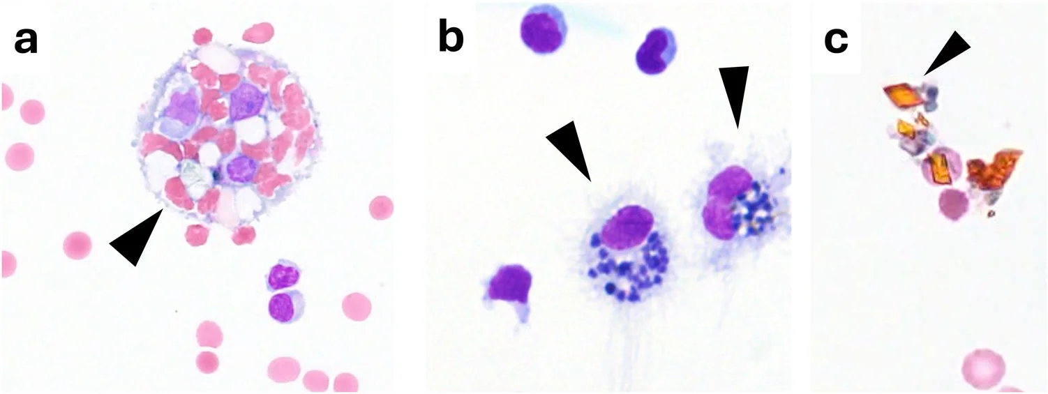
脑脊液中不同出血阶段的细胞学标志(Pappenheim 染色(May-Grünwald-Giemsa 染色))。a 吞红(白)细胞巨噬细胞(箭头),提示近期出血,内含完整的红细胞及偶见的白细胞残余。b 含铁血黄素巨噬细胞(箭头),反映亚急性出血,细胞内可见血红蛋白降解形成的含铁颗粒。c 细胞外橙色血质结晶(箭头),代表晚期出血,表现为细胞外间隙中的黄橙色、具有双折射性的结晶结构(替代文本:与颅内出血相关的代表性细胞学表现,包括吞红细胞巨噬细胞、含铁血黄素巨噬细胞和橙色血质结晶。)
随后,可在含铁血黄素巨噬细胞内检出特征性的黄橙色菱形或星状结晶结构,即橙色血质(胆红素)结晶(图 6c)。巨噬细胞凋亡后数月,仍可检出这些漂浮于脑脊液中的结晶。使用偏振光显微镜也可清晰显示这些晶样结构。对吞噬细胞进行精确的形态学分类,可用于推断出血的时间阶段或急性程度。然而,若结合连续脑脊液标本中吞红细胞巨噬细胞的再次出现进行考虑,这些表现会引起对反复或持续出血的担忧,应结合临床病程和神经影像学进行评估。
在鞘内治疗背景下,异物巨噬细胞可能构成一种罕见的诊断难题:由于它们在 Pappenheim 染色(May-Grünwald-Giemsa 染色)中也呈泡沫状、空泡化外观,并含有嗜碱性包涵体(通常显得较为清晰),例如托夫生或诺西那生等药物的残留,因此不能将其误认为含铁血黄素巨噬细胞。仔细结合患者病史与临床病程,并在有疑问时进一步染色(如普鲁士蓝染色),有助于避免误分类。
对于运输时间延长的情况,尚无证据表明脑脊液中人为混入血液可导致体外出现吞红细胞巨噬细胞 [23, 115, 133]。
脑脊液细胞学中的肿瘤性疾病
尽管神经影像学和脑脊液分子检测取得了重大进展,但只要怀疑软脑膜受累或中枢神经系统肿瘤性病变,常规脑脊液细胞学仍是常规检查中具有决定意义的组成部分。然而,在评估这些疑似诊断的细胞学表现时,仍有几个重要陷阱必须仔细考虑。肿瘤性脑膜炎等中枢神经系统肿瘤性疾病可导致显著的细胞增多。然而,脑脊液白细胞计数正常并不能排除中枢神经系统肿瘤 [11]。因此,指南制定小组建议,当临床/MRI 表现使验前概率保持较高水平时,应进行脑脊液细胞学检查 [105],必要时重复细胞学分析。能否从脑脊液中检出恶性细胞,似乎取决于组织学亚型、与脑脊液腔在解剖上是否相通(与蛛网膜下腔/脑室接触)、疾病负荷以及分析前因素(标本量、快速处理)[97]。软脑膜或室管膜边界完整的实质性肿块,如脑膜瘤,通常细胞学检查为阴性,除非非典型/间变性组织学改变或明显的软脑膜或脑室内播散形成了通向脑脊液的通道。
脑脊液中恶性细胞(图 7)的形态可有很大差异,取决于基础肿瘤性疾病。有经验的检查者依据恶性细胞通常呈现的形态学标准作出诊断 [21, 47, 48, 92, 124]:
-
细胞大小或形状异常:细胞增大,单个肿瘤细胞之间大小不一(直径通常为 20–25 µm,可达 200 µm)
-
细胞核异常:细胞核增大和/或呈多形性、形态不规则,可见核仁或假性包涵体,核塑形,多核,染色质结构和分布改变
-
非典型核分裂或凋亡
-
细胞质异常:例如腺癌细胞中的囊泡、胞质的伪足样突起、核质比高,以及胞质强嗜碱性
-
可能成簇(见于某些病种)
-
肿瘤细胞被吞噬
-
伴随炎症反应:淋巴细胞(可能为主要细胞类型)、单核细胞、中性粒细胞和红细胞
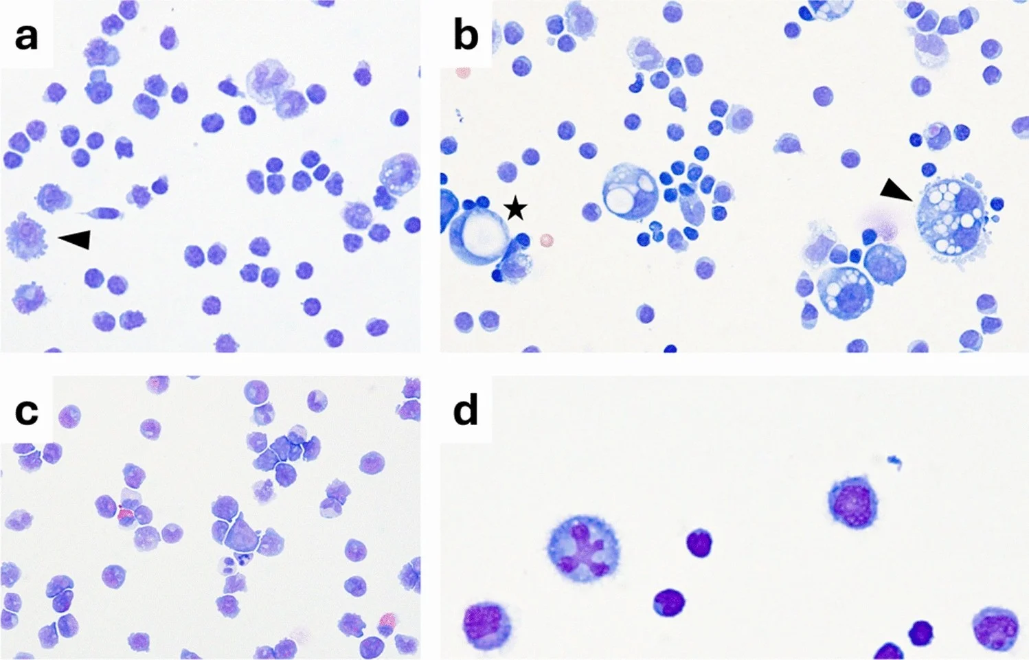
中枢神经系统肿瘤受累的细胞学特征(Pappenheim 染色(May-Grünwald-Giemsa 染色))。代表性脑脊液细胞离心涂片,显示不同形式的肿瘤性脑膜和脑实质受累。a 淋巴瘤性脑膜炎(Meningeosis lymphomatosa),显示非典型淋巴样细胞,其细胞核增大、染色质粗糙,并有明显核仁(箭头)。该肿瘤细胞还显示明显的胞质突起。b 癌性脑膜炎(Meningeosis carcinomatosa),特征为体积较大的恶性上皮细胞,胞质明显空泡化,包括一个印戒样细胞(星号),以及产生黏液的多核肿瘤细胞(箭头)。c 白血病性脑膜炎(Meningeosis leucaemica),可见未成熟造血细胞浸润,细胞核呈异型性,细胞大小不一。d 播散性脑生殖细胞瘤,显示非典型细胞,细胞核增大、形态不规则,并有明显的核异常(替代文本:代表性恶性脑脊液细胞学表现,显示来自不同血液系统及实体肿瘤病种的肿瘤细胞。)
没有任何单一细胞学参数可作为脑脊液中恶性肿瘤的特征性诊断依据 [92]。发现的恶性标准越多,肿瘤性疾病的诊断就越可靠。分析前标本劣化本身可产生误导性的细胞形态学特征。在自溶标本中,逐渐发生的细胞核褪染可形成表观上的“假核仁”,可能类似恶性细胞中明显的真核仁。因此,在难以确定的病例中,应谨慎解释,并可考虑重新采集标本、及时处理后再次评估。在黑色素瘤中,细胞内或细胞外黑色素可提供额外诊断线索,但应与含铁血黄素等血红蛋白来源的色素相鉴别。如果仍有疑问,可适当使用“非典型”细胞的表述,仅诊断为“疑似肿瘤性脑膜炎”,并建议重复穿刺和分析,必要时使用更大体积的脑脊液。还常可见其他活化细胞(如淋巴细胞、单核细胞和粒细胞),提示伴随炎症反应,因此使用“肿瘤性脑膜炎”这一术语。发生组织损伤时,也可能出现红细胞。另一方面,仅有少数散在的细胞体积增大、核异型性和胞质囊泡,也可能确立癌性脑膜炎的诊断,例如在腺癌中。
当细胞学检查为阳性时,仅凭形态学很少能够明确肿瘤分类(例外包括腺癌、急性白血病、某些淋巴瘤和黑色素瘤),因此需要采用免疫细胞化学、流式细胞术或活检等辅助技术。在血液系统恶性肿瘤中,尤其是淋巴系统肿瘤和白血病,多参数流式细胞术的敏感性可能高于常规细胞形态学,应将其视为重要的补充方法 [96, 117]。然而,阳性判定标准以及流式细胞术阳性/细胞学阴性结果的临床意义,仍未完全标准化 [96]。在细胞材料充足时,选用细胞块制备还可有助于免疫细胞化学或分子特征分析 [139]。在软脑膜转移中,新型循环肿瘤细胞检测的敏感性可能高于常规细胞学,但检测平台和阳性判定标准仍存在异质性。此外,脑脊液液体活检方法正越来越多地被用作神经肿瘤学的补充诊断工具。通过靶向测序、下一代测序、数字 PCR 或基于纳米孔的方法分析循环肿瘤细胞和游离肿瘤 DNA(cfDNA/ctDNA),可能提高诊断敏感性,并提供肿瘤特异性分子改变的信息,尤其是在软脑膜病变中 [1, 75, 83]。除突变和拷贝数分析外,脑脊液来源 cfDNA 的甲基化谱分析正在成为一种有前景的中枢神经系统肿瘤分子分类方法 [1]。目前,这些技术应被视为常规细胞学的补充,而非替代,应结合细胞形态学、影像学和临床背景进行解释。为进行潜在的后续分子分析,尤其是对于原发灶不明或存在多个可能原发肿瘤的癌症患者,可考虑保留无细胞脑脊液上清液。一些实验室将同一份脑脊液标本同时用于细胞学和分子分析:先离心,移取并保存无细胞上清液,再在制备细胞离心涂片前,用生理盐水或磷酸盐缓冲盐水补足移去的体积。然而,这类联合工作流程对细胞回收率、细胞形态保存和诊断性能的影响,尚未得到系统验证。日常临床实践建议如下:采集较大体积的脑脊液,及时处理,如果首次标本为阴性但仍有怀疑,则重复腰椎穿刺。对于各类癌症,首次穿刺的敏感性(~ 50–60%)可通过重复采样提高至 ~ 80% [42]。
婴幼儿和儿童的脑脊液细胞学:年龄特异性脑脊液白细胞计数及细胞分类模式
年龄相关参考范围:各年龄的“正常”是什么?
成人中被认为正常的脑脊液细胞学表现,不能直接应用于生命早期的脑脊液诊断。在新生儿中,可靠的队列研究和荟萃分析数据表明,脑脊液白细胞计数的参考上限较低,但略高于较大月龄婴儿和儿童。在一个纳入 318 名婴儿的队列中,早产新生儿脑脊液白细胞计数的第 95 百分位数为 12 个细胞/µL,足月新生儿为 14 个细胞/µL [120]。尽管针对早产儿和足月儿脑脊液细胞计数的大规模研究较少,但现有数据提示新生儿期的细胞计数较高 [144]。新生儿期之后,这些数值在出生后前 3 个月内逐渐接近 ≤ 4 个细胞/µL 的范围 [59, 81]。曾有人讨论,产程及分娩时头部受到挤压或机械应力是否可能导致新生儿脑脊液白细胞计数较高,但这一可能性未能得到证实 [131]。其他假说包括血液–脑脊液屏障不成熟或脑脊液循环不成熟,由于流动动力学显著减弱,可能使免疫细胞有更多时间迁入脑脊液。
然而,似乎尚无可靠的临床或实验研究。在健康新生儿中,脑脊液细胞分类计数以单个核细胞为主,由淋巴细胞和单核细胞组成,但在其他方面正常的脑脊液中,也可能出现少量中性粒细胞。尤其是在早产儿中,较高比例的中性粒细胞似乎更常见 [79, 144]。约 2 月龄后,脑脊液细胞比例应与成人参考值相仿。
生命早期特有的诊断陷阱
(i) 无脓性脑膜炎:与免疫抑制的成人患者相似,在新生儿和小婴儿中,无细胞增多证据的感染性脑膜炎或脑炎似乎比在免疫功能正常的健康成人中更常见 [145]。在一个纳入 40 名新生儿和小婴儿(年龄 < 90 天)的队列中,共发生 44 次经 PCR 或培养证实的细菌性脑膜炎,其中 44 次中的 13 次(30%)的脑脊液白细胞计数处于正常范围 [14]。一个规模更大的培养证实脑膜炎新生儿队列报告,6.5%(12/184)的病例白细胞计数无显著升高 [39]。相比之下,成人细菌性脑膜炎病例中仅约 2% 表现为无脓性 [118]。
(ii) 血液污染与校正公式:新生儿中创伤性穿刺很常见 [116]。然而,采用固定的 1000:1 红细胞(RBC):白细胞(WBC)校正可能导致病例误分类,并掩盖细菌性脑膜炎中的细胞增多。一项针对 ≤ 60 日龄婴儿的回顾性分析显示,采用 1000:1 比值进行校正,会将部分细菌性脑膜炎病例重新归类为“无细胞增多”[34]。两项大型队列研究表明,采用其他校正计算方法似乎比 1:1000 更准确:在新生儿的 432 次创伤性穿刺中,建议采用 1:400 的校正比值 [38];而在一个纳入 2880 名 < 60 日龄新生儿和小婴儿的队列中,1:877 可获得最准确的校正 [73]。因此,对于儿科脑脊液总体而言,多数作者建议同时报告未经校正的脑脊液白细胞计数和红细胞污染程度,并结合临床背景进行解释,而不是将细胞“校正掉”。
脑脊液细胞学的部位差异:脑室脑脊液的解读
脑室脑脊液中的细胞分类计数
脑室脑脊液(vCSF)通常在特定临床背景下采集,典型情况是在出血、神经外科手术、创伤或脑积水后通过脑室外引流(EVD)采样。组织颗粒的存在以及对合并脑室炎干扰的担忧,使仔细的显微镜检查成为必要。
由于细胞和较大颗粒在各脑脊液腔之间的分布平衡不均匀,与腰椎脑脊液相比,脑室标本可能低估细胞性炎症,因此“正常”的 vCSF 白细胞计数未必能反映远端腔室的情况。平行采样提示,即使存在感染,同一患者的腰椎白细胞计数通常也高于脑室计数。这很可能反映了脑脊液腔内的分隔、头尾方向梯度以及细胞成分沉降。在脑膜炎病例的比较系列研究中,同一次病程内的腰椎白细胞计数始终高于 vCSF 白细胞计数 [26, 54, 95]。尽管既往研究结果强调了采样部位对解释结果的重要性,但对于是否应为 vCSF 规定不同于腰椎脑脊液的正常范围,目前尚无共识。
从细胞学工作者的角度诊断脑室炎
尽管通常预期脑室炎会出现细胞增多,但似乎仍有相当数量患者的脑脊液白细胞计数不升高。一项综合荟萃分析描述,93% 的细菌性脑室炎患者白细胞计数升高,而另一方面,32% 无脑室炎的患者也出现升高 [28]。因此,在医疗相关脑室炎中,常规指标并不完善:脑脊液白细胞计数、葡萄糖或蛋白正常,均不能可靠地排除感染,尤其是在使用抗生素后,或疾病负荷较低、病变局限于某一腔室时。因此,指南优先强调脑脊液培养(以及仍有怀疑时重复检测),而非任何单一白细胞计数阈值 [130]。在实践中,vCSF 细胞增多——如果存在——通常以中性粒细胞为主,但可因既往手术、脑室内出血或鞘内治疗而减弱,而这些因素也都会升高红细胞计数,干扰结果解释 [130]。
考虑脑室脑脊液中的血液污染
在采集 vCSF 的病例中,经常可观察到血液污染。这种现象可归因于多种因素,包括近期置入 EVD、术后并发症、SAH 等基础病变,以及脑室炎等并发症。已有报告指出,采用 1:666 或 1:1000 等固定校正公式时,过度校正或校正不足的比例较高 [9]。一些作者建议,通常不应对脑脊液白细胞计数进行血液污染校正,因此 vCSF 应报告未经校正的白细胞计数。对于疑似中枢神经系统感染(如脑室炎)的诊断,一种更复杂但可能更精确的方法是采用患者特异性的校正指数(CI):
在一个观察性队列中,CI 相对于血液比值升高(≥ 4–5 倍)与脑室炎相关,并改善了与“血性但无菌”脑脊液的鉴别 [93]。近期一项 2024 年多中心分析支持将 CI 作为有用的辅助指标,同时指出截断值存在差异,且不能替代培养 [37]。此外,连续分析(每日或隔日)可及时发现 vCSF 中脑脊液白细胞计数及随后 CI 的骤然升高。
器械相关嗜酸性粒细胞增多和非典型细胞分类结果
已有充分文献描述,接受分流或引流的患者 vCSF 中可出现嗜酸性粒细胞,并与器械故障、脑脊液外渗以及有时发生的感染(如痤疮皮肤杆菌感染)相关 [36]。嗜酸性粒细胞增多可十分显著,但并无特异性;应据此评估机械性问题并进行针对性微生物学检查,而不应简单归为“过敏性”或“反应性”。以中性粒细胞为主但培养阴性的细胞增多,可反映神经外科术后的化学性脑膜炎或脑室内药物刺激;这类模式必须密切结合手术过程及治疗情况进行判断。
总体而言,与腰椎脑脊液相比,vCSF 细胞学更易受血液污染和器械相关异物反应的干扰,也更容易因空间梯度而出现“假性平淡”表现。确保正确解读的最稳妥方法包括明确标注采样部位、依据变化趋势进行解释,以及与临床表现、微生物学和影像学紧密整合。
与时俱进:数字化和人工智能(AI)时代脑脊液细胞病理学的新兴工具
目前,液体活检和高维免疫表型分析等新方法正以越来越快的速度发展。因此,提出经典脑脊液细胞病理学,乃至总体上的常规脑脊液诊断是否很快会变得不再那么重要这一问题,或许是合理的。然而,应注意到,细胞病理学诊断领域同样存在重大且有前景的发展,这些发展很可能会巩固传统细胞病理学作为强大新技术的基础及补充所持续具有的重要地位。
人工智能在脑脊液和体液细胞病理学中的应用:现状与前景
过去 5 年间,深度学习(DL)系统已从早期概念验证方法发展到小规模临床可行性研究。在脑脊液中,利用数字化细胞离心涂片训练的卷积模型能够对白细胞和肿瘤细胞进行五分类,甚至可根据推测的原发部位对癌细胞进行亚型分类 [52, 114, 140]。2022 年报道的一套五分类细胞分类模型流程,在来自 90 名软脑膜转移患者的 > 50k 张单细胞裁剪图像中,实现了稳健的癌细胞检出,甚至实现了稳健的癌症亚型分类;与此同时,其他研究团队展示了可解释的 DL,能够对具有诊断意义的脑脊液细胞类型进行分类,并在全切片图像上提供病例级标签 [140]。早期神经网络方法也已处理细胞计数和粗略的细胞谱系设门等预筛查任务,以确定优先供人工评估的视野,这对于脑脊液中常见的低细胞量标本似乎尤其有前景 [52]。尽管结果令人鼓舞,但这些研究仍为单中心研究,通常使用经过筛选整理的数据集,外部验证有限,因此尚不能保证其在不同实验室、染色方法和扫描仪之间的泛化能力。
目前,其他体液细胞学中的证据比脑脊液更充分,尤其是胸腔积液;在回顾性队列中,将 AI 应用于全切片图像,在恶性肿瘤检出和类别区分方面已达到诊断级性能 [91]。近期的叙述性综述和系统综述认为,AI 可以增强但不能替代专家评估,并强调多中心验证和工作流程整合的必要性 [16, 60]。
重要的是,受监管的 AI 分诊在细胞学领域已成为现实:Hologic Genius™ 数字诊断平台已获 FDA 许可(DEN210035)用于巴氏涂片,AI 可在病理医师签发报告前预筛查并呈现感兴趣区域。这提供了一个监管先例,表明细胞学中 AI 辅助的图像分诊是可行且可接受的;随着数字图像采集、数据标准化和模型验证不断成熟,这一框架可能推广至体液细胞学。早期转化研究还探索了成像流式细胞术结合 AI 应用,以富集积液中的恶性细胞,提示可建立混合流程,将 AI 标记的候选细胞交由免疫细胞化学或液体活检进行确认和分型 [71, 76, 84]。因此,脑脊液实验室近期可行的应用场景可能包括:AI 辅助预筛查,对疑有肿瘤细胞或异型性的切片/区域进行排序,以便尽早由人工复核;质量控制支持(标记分布外的染色、对焦情况或不合常理的细胞分类结果);以及在疑难病例中,通过可视化解释指出促成“恶性可疑”评分的细胞,为决策提供支持;在当前阶段,这些应用始终仍须处于受过培训的细胞病理医师的监督之下。下一步需要开展前瞻性、多中心研究,使用不依赖特定厂商的扫描仪、标准化染色和外部测试集,并报告校准情况、类别不平衡的处理方法以及人因结果(节省的人工时间、观察者间一致性)。
脑脊液远程细胞学:依据、验证、工作流程与局限
远程医疗方法,尤其是涉及(细胞)病理学的方法,已经发展起来,旨在维持广泛且及时的专家参与,同时尽量减少转运和报告周转延迟。
就细胞病理学总体而言,当前数字病理学指导文件支持对细胞学应用进行验证,包括最低病例数、与玻璃切片一致性的记录,以及持续质量检查 [3, 33]。远程细胞学可通过远程控制的实时显微镜检查(配备摄像头的显微镜,采用安全、低延迟的流式传输),或采用全切片成像(WSI)进行存储转发式会诊。然而,脑脊液细胞离心涂片的常规全切片数字化可能在技术上存在挑战,尤其是细胞稀少的标本,因为自动细胞检测和自动对焦算法可能无法识别稀疏分布的细胞材料或最佳焦平面 [30, 68]。从一般细胞病理学借鉴的实用方法包括手动界定细胞沉积区域并增加对焦点,以及多平面或 Z 轴堆叠扫描,或在条件允许时采用扩展景深采集 [30, 68]。这些方法可改善不同焦平面上细胞材料的显示,但代价是扫描时间更长、文件体积更大。重要的是,这些策略主要源于一般细胞学标本的经验,尚未专门针对脑脊液细胞离心涂片进行系统验证。细胞病理学领域的前瞻性研究(包括体液制片和快速现场评价(ROSE)工作流程)表明,在阅片者受过培训且基础设施稳定时,标本充分性判定和初步诊断具有较高一致性 [12, 123]。
在脑脊液细胞病理学中,远程细胞学虽然尚未广泛采用,但具有相当大的潜力:在经济压力日益增大、医院与实验室服务不断整合的背景下,它提供了一种切实可行的方法,可集中专业力量,促进获得专家细胞病理学评估的机会,尤其适用于缺乏本地专业人员的基层机构。建立区域远程网络,需要中心与基层机构共同协作,并就联合诊断的适当标准达成一致(例如,将按标准制备并固定的细胞离心涂片送至专家中心,或进行现场显微镜检查并配合远程会诊)。在分散的机构中,如果本地无法直接获得质量方面的反馈,则更难在分析前、制片和染色环节维持高标准。
总结
总体而言,脑脊液细胞学已经发展成为、并将继续作为脑脊液分析的有力工具。将细胞学发现与临床资料相联系,并在综合报告中将结果与蛋白检测结果一并整合,至关重要。专家显微镜评估仍是定性脑脊液细胞学的参考标准。然而,计算机辅助技术正迅速发展,具有相当大的潜力。未来十年,随着脑脊液诊断领域技术和组织层面的变化逐步展开,这些技术的实施应伴随审慎评价和验证,以确保诊断准确性。
数据可得性
本综述未生成或分析任何新数据。因此,数据共享不适用于本文。
- A narrative review in Acta Neuropathologica (Nay, Skripuletz, Isenmann and colleagues, led from Hannover Medical School on behalf of the German Society for CSF Diagnostics and Clinical Neurochemistry, DGLN); CC BY open access — full text with 7 figures and Table 1.
- Pre-analytical factors determine accuracy: delayed processing causes pH-dependent autolysis and substantial cell loss; rapid cytocentrifugation preserves morphology, and stained cytospin slides with expert review are recommended routinely.
- Automated counters show limited precision at low and slightly elevated counts and limited morphology; manual chamber counting remains the quantitative gold standard and expert microscopy the reference for cytomorphology.
- CSF cytology keeps a unique role in infection, neoplasia and subarachnoid haemorrhage: erythrophages, siderophages and haematoidin crystals reveal bleeding and its stage; AI and telecytology belong only in validated workflows.
Scope: the full article (abstract, all sections, 7 figures with legends and Table 1). The reference list is omitted; in-text citation links point to the publisher page.
Abstract
Cerebrospinal fluid cytology remains a cornerstone of diagnostic evaluation in infectious, autoimmune, haemorrhagic, and neoplastic disorders of the central nervous system despite advances in neuroimaging, molecular diagnostics, and digital technologies. This narrative review summarises established principles of cerebrospinal fluid cytology, highlights methodological limitations and diagnostic pitfalls, and integrates emerging developments, including automated cell counting, artificial intelligence, and telecytology. Pre-analytical factors are critical determinants of diagnostic accuracy. Delayed sample processing promotes pH-dependent autolysis and substantial cell loss, whereas rapid cytocentrifugation preserves cellular morphology and facilitates reliable assessment. Automated cell counters are increasingly implemented in routine laboratories and offer advantages in settings with limited technical staffing. However, current systems show limited precision in samples with low and slightly elevated cell counts and limited morphological evaluation. Manual chamber counting, particularly in the normal and low-cell-count range, remains the gold standard for quantitative CSF cell assessment, whereas expert microscopic examination remains the reference standard for qualitative cytomorphological assessment. Although the sensitivity of neuroimaging continues to improve, cerebrospinal fluid cytology retains a unique diagnostic role in the detection of infectious and neoplastic conditions and in the evaluation of subarachnoid haemorrhage. Cytological detection of erythrophages, siderophages, and haematoidin crystals can reveal haemorrhage and provide clues to its temporal stage. Overall, cerebrospinal fluid cytology remains a valuable diagnostic tool when integrated with clinical, laboratory, microbiological, immunological, and imaging findings. Careful recognition of common pitfalls is essential. We recommend rapid cerebrospinal fluid processing, routine preparation of cytospin-based stained slides with expert microscopic review, and the implementation of automated or digital methods only within clearly validated workflows.
Introduction: fundamentals of cerebrospinal-fluid differential cell analysis
Cerebrospinal fluid (CSF) is primarily produced by the choroid plexus of the lateral ventricles and circulates through the ventricular system, subarachnoid space, and the central canal of the spinal cord. It acts both as a mechanical cushion for the central nervous system (CNS) and a medium for transporting signalling molecules, metabolites, and proteins. Daily CSF production amounts to approximately 500 mL, while the total volume present is 150–170 mL. Therefore, the CSF is replaced every five to seven hours [125]. For diagnostic purposes, CSF is typically collected via lumbar puncture at the L3/4 or L4/5 intervertebral space. Suboccipital or lateral cervical puncture is performed only rarely and has largely been replaced by lumbar and ventricular CSF access. In selected scenarios, ventricular CSF may be obtained, usually via external drainage systems. Under physiological conditions, CSF is macroscopically clear, colourless, and nearly acellular. Adequate CSF volumes are essential to ensure diagnostic accuracy, particularly during the initial diagnostic work-up. In addition to cell counting and microscopy, standard analysis should include CSF protein, glucose, and lactate levels, as well as albumin and immunoglobulin quotients [128]. In some regions, particularly in German-speaking countries, these parameters are routinely interpreted using CSF/serum quotient diagrams [102, 128]. While this cytology-focused review will not elaborate on these parameters, it should be noted that collecting and integrating them alongside the other results and clinical findings is essential for preparing a cumulative integrated CSF report. Importantly, cytological assessment of at least one stained slide is crucial [128].
Table 1 summarises key practical questions that guide CSF cytology in the diagnostic work-up of CNS pathology which are addressed throughout this review.
Key questions | References |
|---|---|
1. What is the gold standard for CSF cell diagnostics? Manual chamber counting remains the gold standard for quantitative CSF cell assessment (particularly in the normal and low-cell-count range), while automated analysers may be used as a first-line approach to ensure availability and rapid turnaround. A threshold of <30 cells/µL may be used as a pragmatic criterion for manual confirmation when no lower, locally validated threshold has been established Differential cell count and morphological assessment must be performed by an experienced cytologist using microscopy of cytocentrifuge-prepared slides, ideally stained with Pappenheim (May-Grünwald-Giemsa). Automated differentiation may support screening but does not replace dedicated cytological evaluation | [128, 134, 136] |
2. Is preparation of cytological slides redundant in samples with normal CSF cell count? No. Relevant pathological findings (e.g. malignant cells, activated lymphocytes, macrophage inclusions) may occur despite normal total cell counts Cytology therefore provides diagnostic information beyond quantitative cell counts alone | [11, 118] |
3. Are pre-analytical factors relevant for interpretation of CSF cell count and cytomorphology findings? Yes. Delay between lumbar puncture and analysis, storage temperature, blood contamination, and insufficient sample volume significantly affect cell integrity and morphology Processing CSF samples within 1 to 2 h is considered critical Particularly on first work-up, a minimum of 5 mL—preferably 10–15 mL—should be collected for comprehensive CSF diagnostics At least 2–3 mL should be available for cytology alone, while higher volumes are recommended when malignancy is suspected | [17, 29, 77, 97, 99, 115, 128] |
4. Does blood contamination affect the diagnostic accuracy of CSF cytology? Yes. Blood admixture may cause pseudopleocytosis and complicates interpretation of differential cell counts | [9, 56] |
5. Does ventricular cerebrospinal fluid differ cytologically from lumbar CSF? Yes. Ventricular CSF typically shows lower cell counts compared with lumbar CSF Cellular composition may differ due to rostrocaudal gradients and local pathology (e.g. intraventricular haemorrhage, shunt-associated inflammation) Reference values derived from lumbar CSF are therefore not directly applicable to ventricular samples | [26, 54, 95] |
6. Do cytological reference values and interpretations established in adults also apply for paediatric CSF samples? No. Neonates and young infants physiologically exhibit higher CSF white blood cell counts compared to adults Age-specific reference ranges are mandatory to avoid misclassification of physiological findings as pathological Cytomorphological interpretation must take developmental aspects into account | [59, 81, 120, 144] |
7. Can CSF cytology be altered by intrathecal therapies? Yes. Intrathecal therapies (e.g. nusinersen, tofersen, corticosteroids) and systemic treatments (e.g. IVIG) may induce sterile pleocytosis or characteristic macrophage inclusions These changes are often asymptomatic and must be recognized as treatment-related to avoid misdiagnosis | [40, 58, 65, 119, 126, 137] |
Literature search and review approach
This article was conceived as a narrative, expert-driven review rather than a systematic review. Literature was identified through iterative, topic-specific database searches throughout manuscript preparation, supplemented by reference tracing and consultation of relevant textbooks, monographs, and cytology atlases. Given the aim of integrating the historical development of CSF cytology with current diagnostic practice and emerging technologies, no predefined publication period or fixed search string was applied. Both historical and contemporary publications were considered. Literature was selected according to its relevance to the scope of the review and its contribution to current diagnostic practice or historical context. Where findings or recommendations differed between publications, these discrepancies were retained and discussed rather than resolved by formal evidence weighting, with current guidelines, reviews, and primary studies used to contextualize areas of uncertainty. No geographical restrictions were applied, and evidence from international guidelines and primary studies was considered where relevant.
Concerning Additional Table 1: To update the evidence concerning manual vs. automated CSF cell quantification summarised by [134] PubMed/MEDLINE and the Cochrane Library/CENTRAL were searched on 19 September 2026 for studies published between 1 January 2019 and 19 September 2026. The targeted search combined terms for cerebrospinal fluid/CSF with terms relating to automated cell counting or body-fluid analysers and manual counting methods, including counting chamber/haemocytometer-based approaches. The database search identified 373 records (PubMed/MEDLINE, n = 341; Cochrane Library/CENTRAL, n = 32). After removal of four duplicates, 355 of 369 records were excluded during title and abstract screening. Fourteen full-text articles were assessed, of which 11 were included. Citation screening identified six additional full-text articles, of which four were eligible. Across database and citation screening, five full-text articles were excluded: one because it had already been included in Waldrop et al., two because the primary quantitative comparison was between automated platforms without an eligible manual quantitative reference, one because manual microscopy was restricted to differential assessment without an appropriate manual WBC/TNC reference comparison, and one because CSF-specific quantitative results were not separately extractable. In total, 15 studies were included in Supplementary Table S1.
CSF cytology—history and fundamentals
Quincke reported on the first transcutaneous lumbar puncture in the late nineteenth century and continued to study CSF in the ensuing years, including CSF pressure, chemical components, cytology, and findings in various pathologies [98]. Much of the early development in CSF diagnostics took place in German-speaking countries, such as development of the Fuchs-Rosenthal chamber [35] and the so-called panoptic Pappenheim staining [90]. In the early days, cell yield was sparse and cellular appearance often disrupted, making morphological diagnosis difficult, as illustrated in the first CSF atlases [53, 101].
It was the introduction of the sedimentation chamber by Sayk [108] in the 1950 s that paved the way to more reliable analysis of CSF cells. Thus, illustrated works with good-quality cell appearance appeared in the 1960 s and 1970 s from Jena and Halle, Germany. Sayk provided a first photographic atlas [109], followed by Schmidt [111], who conducted extensive work on CSF cytology of inflammatory processes, tissue regeneration, phagocytosis, and atypical cells. With the arrival of the gentle centrifugation technique using a cytocentrifuge, CSF cytology became more feasible, and further works followed by independent authors that were primarily published in English [64, 85]. In parallel, American works appeared independently [44, 106], with limited cross-referencing.
In contrast to the substantial advances in CSF biochemistry and biomarker research, the fundamental morphological features and staining characteristics of cells in the CSF have remained stable over time. Rather than changes in morphology, historical controversies in CSF cytology mainly concerned the interpretation and classification of cell populations, including lymphocyte subtypes, macrophage activation states, and the distinction between reactive and neoplastic cells. In parallel, evolving tumour nomenclature and molecular classification systems necessitate cautious interpretation of older cytological literature.
For readers interested in a comprehensive and practice-oriented approach to CSF cytology, several dedicated atlases and textbooks are currently available [74, 141]. Kluge and co-authors provided an up-to-date CSF-cytology atlas in German that set the standards for approximately 20 years [61], and—uniquely—was translated into English [62] and supplemented by DVDs for teaching and learning purposes in both languages. More recently, a comprehensive work on all aspects of clinical CSF diagnostics was updated and now reflects the state-of-the-art practice [141]. Some of the authors of the present article contributed to this work, which is disclosed here for reasons of transparency. The chapter on CSF cytology [48], and an integrated case atlas [47] illustrate typical CSF cytology findings, both in normal and pathological specimens, and use the nomenclature of tumours according to the current WHO classification [72], and are recommended for further reading.
Pre-analytical and methodological considerations
To ensure diagnostic accuracy, particularly on the first work-up, a minimum of 5 mL—preferably 10–15 mL—should be collected [128]. At least 2–3 mL are required for cytology alone, and higher volumes are essential when malignancy is suspected, as sensitivity increases with sample size [97]. From a pre-analytical perspective, the collection of larger CSF volumes up to 30 mL [78] is considered safe, with contemporary data demonstrating that post-lumbar puncture headache is mainly determined by needle characteristics (tip type, gauge) rather than by the amount of CSF removed [4, 67, 78].
Because of its low protein content and weak buffering capacity, CSF is highly susceptible to pre-analytical variations. Samples should be collected in sterile polypropylene tubes. Glass tubes may cause adherence of cells to the tube wall, leading to underestimation of cell counts. Similarly, regular EDTA or sodium fluoride tubes are unsuitable for cytological analysis due to potential cellular degradation and chemical interference [22]. However, the data available on the extent of changes in relation to CSF findings remain limited. For diagnostic purposes, CSF is typically collected sequentially into multiple tubes, allowing assessment of blood contamination in cases of traumatic lumbar puncture and enabling distribution of samples for routine and specialised analyses, including shipping to external laboratories, without the need for subsequent transfer between containers.
Time to processing is critical. After sampling, CSF pH rises from about 7.3 [112] to alkaline values (up to pH 10 in experimental settings) within a few hours [20], promoting cellular autolysis, and these detrimental processes are accelerated by increased ambient temperatures. Exposure to high ambient temperatures should therefore be avoided during transport. Particularly on hot days, temperature-controlled transport may help preserve cytomorphological integrity [87], although available data on routine refrigeration of CSF are heterogeneous and rapid processing remains the primary pre-analytical priority. Without stabilization, leukocyte loss can be rapid: processing CSF samples within 1–2 h is therefore considered critical. Delays may result in significant reduction of lymphocytes ranging from − 10 to − 53% within 5 h and up to − 60% within 24 h as reported for lumbar and ventricular CSF by studies in neonatal and adult patients [17, 29, 77, 99, 121]. For longer periods, a study by Schwenkenbecher et al. showed that after 72 h 40 to 78% of cells were lytic in samples with blood contamination [115]. This can result in misdiagnosis, particularly in samples with mild pleocytosis.
Neutrophils and tumour cells are considered even more vulnerable. If full cytological workup is delayed, at least a prompt automated or manual cell count should be performed within 2 h followed by slide preparation as soon as possible. There is limited data on the degree of autolysis over longer time periods. These analytical and pre-analytical factors must be carefully considered when interpreting cell counts.
Counting chambers, such as the widely used Fuchs-Rosenthal chamber, are commonly employed for manual CSF cell counting. Freshly collected native CSF (collected less than two hours prior to examination) is pipetted into the chamber (total volume 3.2 µL). As sedimentation can occur even during short transport times, the tube should be gently swirled several times before pipetting. After pipetting, sedimentation should be allowed to proceed for a few minutes. Both anuclear erythrocytes, which can usually be easily distinguished even in an unstained preparation, and remaining nucleated cells are counted individually. As it is usually not possible to distinguish reliably between leukocytes and other nucleated cells (e.g., bone marrow cells) in an unstained preparation, it should be noted that a “false” increase in cell count may be reported, which does not reflect genuine CNS pathology. This should be noted by the cytologist when evaluating the subsequent cytological findings and represents a key argument for the diagnostic relevance of CSF cytology, as microscopic evaluation is required to identify the nature and origin of nucleated cells and to avoid misinterpretation of quantitative results.
In the case of cell-rich or blood-contaminated CSF, dilution with 0.9% NaCl (e.g., 1:10) can be performed for cell counting. The dilution factor is then considered in the cell-count output, as the output unit is always given as “cells per microliter” for reasons of comprehensibility and comparability.
A growing number of laboratories implement automated haematology analysers for CSF cell counting. Automation improves turnaround time and standardization and can perform well at higher cell counts, and in heavily haemorrhagic neurosurgical samples with high erythrocyte and nucleated cell counts, where acceptable precision and clear workflow advantages have been reported [136].
However, despite technical improvements, a reliable lower limit of quantification for CSF leukocytes is still around 20 cells/µL (device dependent) and older systems may have limits around 50 cells/µL [134, 136, 143], if device-specific reference limits are not adjusted and validated. Consequently, expert appraisal concludes that cell counts below 30 cells/µL require mandatory manual confirmation by chamber counting. The systematic review and meta-analysis by Waldrop et al. [134] again highlighted that data on the accuracy of automated counting in the very low range near the reference interval are sparse, and subgroup analyses show markedly weaker correlations for counts <20 cells/µL, although it found good overall agreement between automated analysers and manual microscopy across studies, with stronger agreement at higher absolute counts. Our targeted update identified 15 additional comparative studies covering established haematology analysers as well as newer image-based and semi-automated platforms Supplementary Table S1 (Additional file 1). Overall, these studies reported good agreement with manual chamber counting for total leukocyte counts, particularly at higher cell concentrations. Although some newer platforms showed encouraging performance at low cell counts, the available evidence remained device-dependent and was frequently based on small, single-centre cohorts. Clinically relevant discrepancies around the 5 cells/µL decision threshold were still observed. Automated differential counts generally showed weaker agreement, and atypical or malignant cells could not be reliably identified without morphological review. Thus, the updated evidence broadens the range of evaluated platforms but does not materially alter the conclusions of Waldrop et al. [134].
In summary, a balanced recommendation emerges: manual chamber counting by trained personnel remains the gold standard for quantitative CSF leukocyte assessment, while automated analysers may be used as a first-line approach to ensure availability and rapid turnaround, provided that low and slightly elevated cell counts are confirmed manually and methodological limitations are acknowledged.
A quantitative leukocyte count should be obtained whenever native CSF is available and should not be replaced by an approximate estimate of cells per high-power field on cytospin preparations [128]. Cell recovery on cytospin slides depends substantially on the preparation technique, centrifugation conditions, sample volume, and deposition area, while the area represented by a high-power field additionally varies between microscope systems [63, 104]. Accordingly, universal numerical thresholds based on cells per high-power field are not standardized and should not be used to classify CSF as normocellular or pleocytic [63, 104]. Only in exceptional circumstances in which no native CSF is available for quantitative counting, for example when reviewing an externally prepared slide, may cellularity be described qualitatively, with explicit indication that no quantitative cell count was available.
Apart from automated cell counting, automated cell differentiation provided by routine analysers is—at best—coarse (e.g., granulocytes vs lymphocytes vs monocytes) and should be considered for screening only. Critically, automated systems will not reliably recognize cytomorphological entities that drive CSF differential diagnosis (e.g., plasma cells, activated/reactive lymphocytes, blasts, siderophages/erythrophages, scattered tumour cells, or cell clusters) [113, 136, 142]. Therefore, microscopy remains required whenever qualitative interpretation matters.
Normal adult lumbar CSF leukocyte count reference ranges were not defined in a single landmark trial, but emerged historically from multiple descriptive series of “normal” patients and were later consolidated in laboratory and textbook reference ranges [2, 7]. These ranges appear to be essentially population‑based statistical reference intervals derived from cohorts without clinical or laboratory evidence of CNS disease, then rounded to pragmatic cutoffs for routine.
Blood admixture is one of the most frequent challenges in CSF interpretation. It can occur during lumbar puncture and may significantly affect cytological and biochemical parameters. As an approximation, about 1 leukocyte is introduced per 1000 erythrocytes. Correction formulas for white blood cell (WBC) count determined by linear regression for samples with strong blood admixture (more than 100,000 red blood cells (RBCs) per µL) seem to be inaccurate and tend to overestimate the number of leukocytes added, potentially leading to underestimation of the true WBC count [56]. Moreover, in patients with significant blood-count changes—e.g., in the context of leukaemia, active systemic infections, anaemia or leukopenia—the ratio may be significantly shifted, leading to inaccuracy in the aforementioned correction factor. In such cases, we recommend considering the individual ratio between erythrocytes and leukocytes in the actual patient's blood count.
To minimize blood contamination, CSF cell count should be performed from the last tube, and serial tubes should be inspected for clearance of blood contamination.
Cell differentiation
Since CSF is usually a nearly acellular fluid, cells should be concentrated onto slides to enhance diagnostic accuracy. Today, cyto-centrifugation methods (cytospin) are primarily used for this purpose in accordance with current standards [124]. After air drying the cytocentrifuge slide, staining can be performed. The Pappenheim (also known as May-Grünwald-Giemsa) stain has become the standard cytological staining method in CSF laboratories [22, 128]. Other staining methods are available for specific investigations (e.g., Gram, Prussian blue, India Ink, Ziehl–Neelsen), but are used less often given the availability of refined molecular biology and imaging techniques. Despite its subjective nature, expert microscopic examination of stained cytocentrifuge preparation remains the reference standard for qualitative cell differentiation [128]. Trained cytologists can detect subtle morphological clues such as reactive lymphocytes, plasma cells, activated macrophages, granulocytes, atypical and malignant cells, mitotic figures, as well as bacteria—findings critical in neoplastic or inflammatory disorders. Moreover, cytological examination enables the distinction between genuine pleocytosis and falsely elevated cell counts caused by non-leukocytic or artefactual cells that cannot be reliably identified by automated methods or unstained preparations.
Normal CSF cytology
Cells detected in CSF under physiological conditions are almost exclusively immune cells and do not originate from neural tissue. The blood-CSF and blood–brain barriers effectively prevent the passive passage of erythrocytes and granulocytes, while allowing limited, tightly regulated migration of lymphocytes and monocytes for immune surveillance [31, 98]. This concept underlies the diagnostic value of CSF cell counts, as quantitative and qualitative deviations provide sensitive indicators of inflammatory processes within the central nervous system. Normal adult CSF (Fig. 1) contains ≤ 4 cells/µL, composed predominantly of lymphocytes and monocytes in a roughly 70:30 ratio.
Normal cerebrospinal fluid cytology (Pappenheim stain). Representative cytospin preparations of cerebrospinal fluid stained with Pappenheim stain, showing low cellularity with predominance of small mature lymphocytes (arrowheads) and occasional monocytes (asterisks). Lymphocytes display a round, densely stained nucleus with scant cytoplasm, whereas monocytes are larger with more abundant, lightly basophilic cytoplasm and less condensed chromatin. In (b), a single erythrocyte (arrow) is visible, consistent with minimal blood contamination without diagnostic relevance. Overall, no cytological signs of inflammation or malignancy are present (Alt text: representative CSF cytospin showing predominantly small mature lymphocytes and occasional monocytes without pathological abnormalities.)
It should be noted that differential cell counts in manual routine CSF cytology are commonly based on the classification of 100 randomly identified cells. Consequently, the resulting percentages represent point estimates subject to binomial variation. For example, a measured proportion of 30% corresponds to an approximate 95% confidence interval of 21–40%, and similar margins apply to other proportions [107]. Minor deviations from the expected distribution may therefore reflect sampling variability rather than true biological differences.
On Pappenheim stained cytospins, nuclei show purple-violet basophilia and cytoplasm appears pale to grey-blue, which allows reliable distinction of leukocyte classes. Lymphocytes are small (≈ 7–9 µm), round cells with dense, homogeneous chromatin and a scant rim of basophilic cytoplasm. Activated lymphocytes are larger than quiescent lymphocytes and uncommon in normal CSF. Importantly, the distinction between resting and activated lymphocytes requires cautious interpretation, as minor variations in cell size or cytoplasmic volume may occur even in non-pathological samples. Premature classification of lymphocytes as “activated” is therefore a common source of over-interpretation in routine CSF differential cytology. Monocytes are about twice the size (≈ 15–20 µm) compared to lymphocytes and morphologically more heterogeneous in non-activated states, typically with indented, kidney-shaped nuclei, fine chromatin, and grey-blue cytoplasm that may contain small vacuoles even without overt activation.
Artefacts
Artefacts (Fig. 2), i.e., cells and phenomena seen on the slide that do not represent the situation in the patient’s CSF in vivo, may arise in two distinct ways: firstly, cells or particles may enter the sample during the puncture procedure. Second, artefacts may be added, or emerge, during the preparation and staining procedure.
Cytological artefacts in cerebrospinal fluid (Pappenheim stain). Representative cytospin preparations illustrating common cytological artefacts encountered in cerebrospinal fluid analysis. a Endothelial cells (lightning bolt), characterized by cohesive clusters of large polygonal cells with abundant pale cytoplasm and oval nuclei, typically introduced by the puncture procedure; b cartilage cells (arrowhead), showing large cells with abundant basophilic cytoplasm and round nuclei, representing contamination from intervertebral disc material during puncture; c bone marrow contamination, characterized by the presence of haematopoietic precursor cells (normoblast; arrow) of varying maturation stages within a background of erythrocytes (Alt text: representative cytological artefacts in CSF including endothelial cells, cartilage contamination and bone marrow admixture.)
Erythrocytes are absent unless introduced during the tap procedure (artificially) or via a preceding (subarachnoid) haemorrhage. Artificial blood contamination of varying intensity has been detected in up to 70% of all CSF samples taken, with the incidence appearing to be even higher in infants and neonates [116].
During lumbar puncture, cell types from traversed tissues (e.g., skin, muscle, cartilage, bone marrow, epithelial cells) may appear as artefacts in addition to the otherwise unremarkable differential cytology. Endothelial cells (Fig. 2a) may appear as cohesive clusters of large polygonal cells with abundant pale cytoplasm and oval nuclei. Cartilage (Fig. 2b) presents as chondrocytes in lacunae embedded in a basophilic, chondroid matrix (often metachromatic). CSF samples from the ventricular system may also contain plexus epithelial cells, ependymal cells, and fragments of (degenerating) brain parenchyma. Ependymal or choroid-plexus cells form cohesive sheets or honeycomb clusters of cuboidal cells, sometimes ciliated with uniform round nuclei and delicate chromatin: these should be recognized as site-related contaminants, not carcinoma. Skeletal muscle from the puncture tract appears as elongated fibres or polygonal fragments with dense, fibrillar cytoplasm and peripheral nuclei (striations may be faint on Pappenheim). Smooth muscle cells show spindle cells with “cigar-shaped” nuclei. Bone-marrow carry-over (rare) produces a trilineage mix (Fig. 2c): erythroblasts, myelocytes, and occasional megakaryocytic fragments. Notably, bone marrow admixture will be accompanied by the presence of erythrocytes.
As stated before, contamination can lead to artificially increased cell counts depending on the counting method applied, and must be documented in the report, with cytology used to confirm their nature.
Finally, artificial bacterial admixture (skin flora or reagent precipitates) may mimic infection. The presence of bacteria usually found in “empty voids” between cells, and occurrence without pleocytosis or neutrophils is suspicious for artificial contamination and should prompt clinical correlation.
Apart from artificial admixture of cells or microorganisms, cell-loss and staining artefacts can impact diagnostic accuracy. Because CSF is protein-poor and weakly buffered, autolysis begins within hours and first affects granulocytes [88]. It has been hypothesized that tumour cells undergo an even more rapid degradation [41]. Typical signs of autolysis include karyolysis or karyorrhexis, smudged nuclei, loss of cytoplasmic granularity, vacuolation, and “shadow” cells (Fig. 3a). Autolysis can artificially lower apparent cell counts and blur differential diagnoses. Advanced autolysis can significantly reduce the diagnostic accuracy of a differential cell count, and therefore should be annotated by the cytologist.
Representative preparation and staining artefacts in cerebrospinal fluid cytology (Pappenheim stain). a Autolytic changes with shadow cells (arrowheads), characterized by loss of cellular and nuclear detail. b Staining artefacts with granular basophilic material compatible with stain precipitate (arrowhead) and localized uneven background staining/stain pooling (arrow). c Overloaded, cell-rich cytospin preparation showing uneven staining with reduced nuclear and cytoplasmic contrast. d Cytospin-related mechanical distortion with elongated and altered cellular morphology (arrowheads) (Alt text: representative preparation and staining artefacts in CSF cytology, including cytospin-related cell distortion, an overloaded unevenly stained preparation, autolytic shadow cells, and granular/basophilic staining artefacts.)
The most common staining artefact in Pappenheim staining includes homogeneously deep-purple granules of variable size that sit in a different focal plane from cells and can mimic bacteria (Fig. 3b). Those granules usually occur when reagents are old or rinsing is inadequate. Furthermore, accurate staining requires phosphate buffer at ~ pH 6.8. pH drift yields muddy cytoplasm and poor nuclear detail (under-stained). Cell-rich samples have to be diluted before staining, because overloaded cytospins produce a multilayer instead of a monolayer: dyes and buffer penetrate unevenly, so the centre appears under-stained (pale nuclei, washed-out cytoplasm), while edges look over-stained with a muddy background (Fig. 3c). Another typical preparation artefact is produced by cytospin preparation. On cytospins, high g-forces can distort nuclei in fragile cells and exaggerate size or alter cell shape (Fig. 3d).
Cytological patterns in inflammatory CNS disorders
In the CSF, inflammation is reflected by shifts in the differential cell count and by activation of immune cell lines. Understanding activation patterns can provide key insights into the underlying pathology. However, inflammatory cytological patterns are not disease-specific and should not be interpreted as providing aetiological assignment on their own. Their appearance may be influenced by disease phase, antimicrobial or immunosuppressive treatment, intrathecal therapy, blood contamination, and the site of CSF sampling, and should therefore always be interpreted in the clinical and laboratory context.
Reactive lymphocytes enlarge (≈ 10–20 µm), with open chromatin and more abundant basophilic cytoplasm, and variable nuclear contours. Nuclear indentations, irregular nuclear outlines, and occasional nucleoli may occur as part of reactive activation and should not per se be interpreted as malignant features. However, pronounced cytoplasmic irregularity and deep nuclear notches have been reported significantly more frequently in neoplastic than in inflammatory lymphocytes [92]. Importantly, the majority of activated lymphocytes observed in inflammatory CSF conditions represent activated T-lymphocytes, rather than B-cell lineage differentiation [49]. In highly activated CSF (e.g., acute neuroborreliosis), mitoses of lymphocytes can occasionally be detected. Plasma cells represent terminally differentiated B-cells [94] and therefore reflect advanced humoral immune activation rather than a mere activation state. Cytologically, they are characterized by an eccentrically flattened nucleus with coarse chromatin, a perinuclear pale zone corresponding to the Golgi apparatus, and deeply basophilic cytoplasm. The presence of plasma cells in CSF may precede the quantitative detection of intrathecal immunoglobulin synthesis, underscoring the added diagnostic value of cytological examination in inflammatory CNS disorders.
Monocytes gradually transform to activated macrophages with enlargement and vacuolation of the cytoplasm. Phagocytosis of cellular debris is common in viral or resolving bacterial meningitis. Activated monocytes can become remarkably enlarged due to numerous vacuoles, and the cytoplasm can appear foamy (“foam cells”) or—in the case of individual, large vacuoles—almost empty (“signet ring cell”).
Neutrophil granulocytes appear round and have segmented nuclei. During acute phases, such as fulminant bacterial inflammation, immature neutrophils with “band-shaped” nuclei may be observed. During inflammation, an increase in hyper-segmentation to usually 4–6 nuclear segments, occurs. It has been reported that scattered neutrophils, which can nevertheless occur in “normal” CSF without pleocytosis, are not associated with CSF infection [13, 24], although there is little evidence for this assumption in the literature.
Eosinophil granulocytes usually have distinct eosinophilic granulated cytoplasm and, in most cases, two clearly recognizable nuclear segments. Eosinophil-dominated pleocytosis is rare and usually indicates the presence of a parasitic or coccidioidal CNS infection, or, more rarely, a concomitant reaction in neoplastic meningitis, especially reported in CNS involvement of Hodgkin’s disease, or a shunt-related reaction [36, 43, 46]. In certain autoimmune inflammatory CNS disorders, most notably AQP4-IgG-positive neuromyelitis optica spectrum disorder (NMOSD), a mixed pleocytosis with a substantial proportion of eosinophilic granulocytes has been reported [50]. Rarely, CSF eosinophilia has also been reported in vasculitic CNS disorders, including eosinophilic granulomatosis with polyangiitis and primary angiitis of the central nervous system [45, 86]. CSF eosinophilia may also occur in drug-induced aseptic meningitis, including cases associated with NSAIDs and antibiotics [80].
Basophilic granulocytes are distinctly deep blue or basophilic granulated in the cytoplasm, making it difficult to clearly distinguish between the nucleus and plasma in everyday practice. They are rarely present in CSF, and their diagnostic significance remains poorly understood. In non-blood-contaminated and otherwise unremarkable CSF, basophils are not expected. When identified, their presence should therefore be interpreted in the context of blood contamination, haematological or leptomeningeal neoplastic involvement, and, exceptionally, inflammatory CNS disorders such as neurosarcoidosis or viral meningitis [18, 19, 25].
Infectious CNS inflammation
Acute purulent bacterial meningitis is a neurological emergency with substantial case fatality and long-term morbidity among survivors [5]. Regarding the cytological CSF findings, high cell counts (usually exceeding several 1000 cells per µL) with neutrophil predominance (Fig. 4a) are typically found. In adults, neutrophils typically constitute > 90% of leukocytes in classic presentations [128, 129].
Cytological patterns of infectious inflammatory CNS disease (Pappenheim stain). Representative cytospin preparations of cerebrospinal fluid illustrating different infection-associated inflammatory patterns. a Pronounced neutrophilic pleocytosis in bacterial (purulent) meningitis without detectable microorganisms. b Early apurulent meningoencephalitis in Streptococcus pneumoniae infection, characterized by the presence of extra- (arrow) and intracellular (asterisks) diplococci in the absence of a predominant neutrophilic infiltrate, accompanied by a large plasma cell (arrowhead). c Later stage of viral CNS inflammation with resorptive changes, characterized by macrophages with foamy cytoplasm (“foam cells”, arrowhead) reflecting phagocytic clearance of cellular debris (Alt text: three CSF cytospin micrographs showing (a) marked neutrophilic pleocytosis, (b) intra- and extracellular diplococci with a plasma cell despite limited neutrophilic inflammation, and (c) foamy macrophages during resolving inflammation)
Nevertheless, it should be emphasized, that infections naturally progress in phases, with different findings expected in the early or late stages of bacterial CNS infections. In the early phase of bacterial meningitis (approximately 12 h or less) [32]—as particularly described in cases of meningococcal and pneumococcal disease—bacteria may be directly detectable in the specimen alongside a lower inflammatory response and, during this period, the cell count may appear normal or near to normal, with only mild signs of inflammation present, sometimes even without evidence of neutrophilic infiltration (Fig. 4b). This particular case of bacterial meningitis is also known as “apurulent” meningitis and is considered particularly threatening. It occurs more frequently in elderly patients and in the context of immunocompromising conditions [118].
As inflammation progresses, the ongoing immune response leads to degradation processes (Fig. 4c), resulting in a decrease in neutrophilia and a relative increase in macrophages, activated monocytes and lymphocytes. Initiation of antibiotic treatment before lumbar puncture (according to guideline procedures) can sterilize CSF within a few hours, but does not substantially change leukocyte counts within the first hours [55].
Another scenario that can lead to deviations from expected findings is immunosuppression. In severely immunocompromised patients, lower pleocytosis [132] or even normal leukocyte counts [118] may occur more frequently due to a significantly reduced inflammatory host response. Therefore, normal leukocyte count does not rule out infection. In such cases, diagnosis relies more readily on further direct or indirect pathogen diagnostics, as well as the patient history and clinical course.
In neuroborreliosis, usually moderate pleocytosis between 50 and 500 cells per µL is seen [128]. A relevant proportion of neutrophilic granulocytes might be present, especially in early infection and before treatment [89]. Due to the presence of strong lymphocytic activation and often remarkably increased plasma cell frequency and even the presence of mitotic figures, it may be difficult to distinguish from neoplastic meningitis at first glance based on morphology alone. However, in lymphoma slightly to moderately activated (“intermediate”) lymphocytes will usually be absent. In diagnostically ambiguous cases, extended investigations are required. These include flow cytometry, an integrated CSF report incorporating intrathecal immunoglobulin synthesis and blood-CSF barrier function assessed by Reiber diagrams, CXCL-13, Borrelia-specific antibody index calculation, and Borrelia-specific serological testing [103, 127, 138].
In theory, a similar phasic course involving initial neutrophil infiltration is also assumed for viral infections [15, 70]. However, the transition to lymphocyte-dominated findings appears to occur more rapidly in these cases than with most bacterial pathogens: interval CSF analyses in bacterial meningitis have shown that pleocytosis and neutrophil predominance frequently persist beyond 48–72 h, even under effective antimicrobial therapy [10, 70]. However, systematic data directly comparing the temporal dynamics of CSF cellular changes across different infectious etiologies remain limited. Viral CNS infections usually show lymphocytic pleocytosis (typically 50–500 cells per µL) by the time of diagnostic lumbar puncture [70]. Nevertheless, for several virus types like enterovirus, West Nile, FSME, or HSV, CSF findings may transiently lack pleocytosis or show a variable neutrophilic skew [15]. Leukocyte counts are usually slightly to moderately elevated. Opportunistic infections of the central nervous system (e.g., Human polyomavirus 2 (formerly JCV), CMV) can cause inconspicuous cytological findings, so they often can only be detected through advanced diagnostics.
While an early neutrophilic response with subsequent rapid lymphocytic predominance is well established in viral CNS infections [70], evidence for a comparable phasic course in tuberculous or fungal infections is limited, and CSF cytology in these conditions often shows heterogeneous or mixed cellular patterns.
Tuberculous meningitis typically presents with a moderately to markedly elevated CSF white blood cell count ranging from approximately 50 to 1000 leukocytes per µL, characterized by a mixed cellular composition with variable proportions of lymphocytes and neutrophilic granulocytes, particularly in early disease stages, and a shift toward lymphocytic predominance with disease progression or under therapy [122]. Occasionally, eosinophils have been reported in tuberculous meningitis and may contribute to a heterogeneous cytological appearance [66]. In CNS tuberculosis, Ziehl–Neelsen staining reveals acid-fast bacilli in only a minority even of otherwise confirmed cases [122]. Thus, nucleic acid amplification has become the gold standard for diagnosis, and larger CSF volumes (7–10 mL) should be drawn routinely to enhance diagnostic sensitivity, particularly in tuberculous meningitis, because Mycobacterium tuberculosis is often present in very low concentrations in CSF [27].
Fungal CNS infections are associated with a highly heterogeneous CSF inflammatory profile that varies with pathogen, disease stage, and host immune status [69]. Similar to tuberculous meningitis, fungal pathogens are often present at low concentrations in CSF, rendering microbiological and molecular diagnostics strongly dependent on adequate sample volume.
Autoimmune inflammation
CSF cytology in non-infectious autoimmune disorders is typically lympho-monocytic and of low-to-moderate extent, but certain entities show distinctive features. Interpretation should integrate clinical findings and further diagnostic testing. Cytology usually supports but is not sufficient to establish these diagnoses.
Multiple sclerosis is the most common immune CNS disease that requires detailed CSF diagnostics. Routine CSF parameters are often normal or show at most a mild lymphomonocytic pleocytosis, typically below 50 cells/µL [2]. While mild pleocytosis and signs of lymphocytic activation including immunoglobulin-producing plasma cells have been reported more frequently in relapsing disease courses [6], the strength of correlation between CSF cell count and clinical activity remains debated.
In addition to multiple sclerosis, there are additional autoimmune diseases that can cause CSF inflammation. It is noteworthy that CSF leukocyte count and differential cell cytology in autoimmune processes can morphologically mimic conditions, such as infectious diseases, so that the cumulative findings, considering additional diagnostic tests and follow-up observations, are often of great relevance.
In contrast to multiple sclerosis, both aquaporin-4-IgG-positive NMOSD and MOG-antibody-associated disease (MOGAD) display more variable and often more pronounced pleocytosis (up to 500 cells per µL) during attacks, with frequent neutrophils and occasional eosinophils, particularly in NMOSD and in paediatric cohorts—patterns that should raise suspicion when the clinical picture is not compatible with multiple sclerosis—and initially eventually mimic bacterial infection [50, 51].
Autoimmune encephalitis (AIE) does not necessarily present with CSF pleocytosis. Especially in AIE subtypes that occur in elderly patients, CSF leukocyte count is often normal [8]. However, if present, mild to moderate lymphocytic pleocytosis is common, and neutrophils [57] can appear in the very acute phase of some AIEs, which may mimic infectious disease.
Other causes of CSF inflammation
Intrathecal therapies may provoke distinctive cytological changes. During nusinersen treatment in patients with spinal muscular atrophy, macrophages with numerous sharply defined purple/blue inclusions (“nusinophages”; Fig. 5a) appear without sustained inflammatory shifts [40]. Similar macrophage inclusions have been described in patients receiving intrathecal Tofersen (Fig. 5b) [119]. Tofersen for SOD1-mutant amyotrophic lateral sclerosis is associated with frequent elevations of CSF leukocyte count, intrathecal immunoglobulin synthesis and CSF protein, and occasional aseptic/chemical meningitis or myelitis. These laboratory changes are often asymptomatic but have to be recognized as treatment-related [137]. Intrathecal steroids [126] and other agents can also cause sterile neutrophilic pleocytosis (“chemical meningitis”). In addition, intravenous immunoglobulin (IVIG) therapy is a well-recognized cause of aseptic meningitis, typically presenting within hours to days after infusion and characterized by a marked pleocytosis, often neutrophil-predominant in the early phase, with elevated CSF protein but negative microbiological studies [58, 65]. Finally, mild “reactive pleocytosis” can follow repeated lumbar punctures within short intervals [110].
Antisense oligonucleotide-associated macrophage inclusions in cerebrospinal fluid (Pappenheim stain). Representative cytospin preparations of cerebrospinal fluid showing macrophages with numerous sharply condensed, basophilic to purple intracellular inclusions after intrathecal antisense oligonucleotide therapy. a Single macrophage with densely packed cytoplasmic inclusions consistent with a nusinersen-associated macrophage inclusion cell (“nusinophage”; arrowhead) in an otherwise paucicellular preparation with mild artificial blood contamination. b Tofersen-associated macrophage inclusions (“tofersenophages”; arrowheads) with numerous variably sized basophilic to purple inclusions distributed throughout the cytoplasm (Alt text: two CSF cytospin micrographs showing macrophages with purple-blue cytoplasmic inclusions following intrathecal antisense oligonucleotide therapy: (a) nusinersen-associated and (b) tofersen-associated inclusions.)
Mild inflammatory CSF changes may occasionally be observed in the absence of infection, autoimmune disease, or recent lumbar puncture. In such cases, tissue damage of the central nervous system, for example following surgery, trauma, or ischemic stroke, can induce reactive inflammatory changes detectable in CSF cytology [100].
CNS haemorrhage
It is well known that the imaging findings obtained by cranial computed tomography (CT) may be unremarkable in a relevant proportion of patients with subarachnoid haemorrhage (SAH), especially in subacute scenarios [82, 135]. In these cases, visual inspection of CSF colour, persisting xanthochromia of the supernatant after centrifugation, testing chemical parameters such as ferritin, and most importantly, targeted CSF cytopathological analysis provides decisive evidence [82]. Neither xanthochromia nor a remarkably elevated erythrocyte count are specific for SAH. CSF cytology yields time-dependent hallmarks of prior haemorrhage. After bleeding, macrophages that phagocytose blood components can be detected in CSF.
The first subtype of macrophages, which can usually be observed within 4–12 h after the event, are erythrophages [82, 133]. Within the next days, erythrophages (Fig. 6a) gradually evolve to siderophages (Fig. 6b), which usually occur after 24–48 h [82, 133]. They contain granular dark pigment (hemosiderin) and stain positively with Prussian Blue (which may be used in cases of doubt). Intermediate mixed erythro-/siderophages (erythro-/haemosiderophages) may occur during haemoglobin degradation and contain both partially preserved erythrocytes and intracellular haemosiderin granules. They represent transitional forms between erythrophages and siderophages and should therefore not be interpreted as a separate stage with a sharply defined time window [61, 62, 124]. In sequential samples, the concomitant presence of fresh erythrophagocytosis and more advanced haemosiderin-containing macrophages may raise suspicion of recurrent bleeding and should be interpreted together with the clinical and imaging findings [61, 62]. During progressive degradation, phagocytosed erythrocytes may lose their staining properties and appear as optically empty intracytoplasmic vacuoles, potentially mimicking foamy macrophages. Such vacuolated macrophages may raise suspicion of preceding erythrocyte phagocytosis but are non-specific and, in isolation, do not constitute reliable cytomorphological evidence of haemorrhage. Identification of preserved erythrocytes, haemosiderin, or haematoidin provides more specific evidence, and Prussian blue staining may help confirm iron-containing pigment in equivocal cases [82, 124].
Cytological markers of different stages of haemorrhage in the CSF (Pappenheim stain). a Erythro(leuko)phage (arrowhead), indicating recent haemorrhage, containing intact erythrocytes and occasional leukocytic remnants. b Siderophages (arrowhead), reflecting subacute haemorrhage, with intracellular iron-containing granules resulting from haemoglobin degradation. c Extracellular haematoidin crystals (arrowhead), representing late-stage haemorrhage, appearing as yellow-orange, birefringent crystalline structures within the extracellular space (Alt text: representative cytological findings associated with intracranial haemorrhage including erythrophages, siderophages and haematoidin-crystals.)
Later, characteristic yellow-orange rhomboid or star-like crystalline structures can be detected within the siderophages: the haematoidin (bilirubin) crystals (Fig. 6c). These may be detected floating in CSF for months after macrophage apoptosis. By using polarized-light microscopy, the crystalloid structures can also be made clearly visible. The precise morphological classification of phagocytic cells can be used to draw conclusions about the temporal stage or acuity of bleeding. However, when considered alongside the reappearance of erythrophages in sequential CSF samples, they raise concerns for repeated or persistent bleeding and should prompt correlation with clinical course and neuroimaging.
In the context of intrathecal therapies, a rare diagnostic challenge may be foreign-body macrophages: Since they share a foamy, vacuolated appearance with basophilic inclusions (which often appear less blurred) e.g., of drug remnants such as tofersen or nusinersen on Pappenheim stains, they must not be mistaken for siderophages. Careful correlation with patient history and clinical course, and further staining (e.g., Prussian blue) when in doubt, help avoid misclassification.
For cases of prolonged transportation, there was no evidence that artificial blood admixture in CSF can lead to in-vitro occurrence of erythrophages [23, 115, 133].
Neoplastic diseases in CSF cytology
Despite major advances in neuroimaging and molecular CSF assays, conventional CSF cytology remains a decisive part of the routine work-up whenever leptomeningeal involvement or a neoplastic CNS process is suspected. Nevertheless, there are a few important pitfalls that must be carefully considered alongside the cytological findings of these suspected diagnoses. Neoplastic CNS diseases such as neoplastic meningitis may lead to marked pleocytosis. However, a normal CSF leukocyte count does not exclude CNS neoplasia [11]. Accordingly, guideline groups advise performing CSF cytology when clinical/MRI findings keep pretest probability high [105] and, if needed, repeated cytological analysis. Whether malignant cells are recovered from CSF seems to depend on histological subtype, anatomic access to the CSF space (subarachnoid/ventricular contact), disease burden, and pre-analytics (sample volume, rapid processing) [97]. Parenchymal masses with intact pial or ependymal boundaries, such as meningiomas, will typically yield negative cytology unless atypical/anaplastic histology or overt leptomeningeal or intraventricular seeding creates a conduit to CSF.
The appearance of malignant cells (Fig. 7) in the CSF can be highly variable, depending on the underlying neoplastic disease. Experienced examiners base their diagnosis on the morphological criteria typically exhibited by malignant cells [21, 47, 48, 92, 124]:
-
Abnormal cell size or shape: enlarged cells, with size varying between single tumour cells (often 20–25 µm in diameter, may be up to 200 µm)
-
Abnormal nucleus: enlarged and/or polymorphic, irregular nuclei, visible nucleoli or pseudo-inclusions, nuclear moulding, multinucleation, changes in chromatin structure and distribution
-
Atypical mitoses or apoptoses
-
Abnormal cytoplasm: e.g., vesicles in adenocarcinoma cells, pseudopod-like extensions from the cytoplasm, a high nucleus-to-plasma ratio, and intensive basophilia of the cytoplasm
-
Possibly clustering (in some entities)
-
Phagocytosis of tumour cells
-
Accompanying inflammatory reaction: lymphocytes (which may be the predominant cell type), monocytes, neutrophils, and erythrocytes
Cytological features of neoplastic CNS involvement (Pappenheim stain). Representative cytospin preparations of cerebrospinal fluid illustrating different forms of neoplastic meningeal and parenchymal involvement. a Lymphomatous meningitis (Meningeosis lymphomatosa), showing atypical lymphoid cells with enlarged nuclei, coarse chromatin and a prominent nucleolus (arrowhead). The neoplastic cell additionally displays conspicuous cytoplasmic projections. b Carcinomatous meningitis (Meningeosis carcinomatosa), characterized by large malignant epithelial cells with markedly vacuolated cytoplasm, including a signet ring-like cell (asterisk), as well as multinucleated tumour cells with mucin production (arrowhead). c Leukemic meningitis (Meningeosis leucaemica), with infiltration by immature haematopoietic cells showing nuclear atypia and variable cell size. d Disseminated cerebral germinoma, demonstrating atypical cells with enlarged, irregular nuclei and prominent nuclear abnormalities (Alt text: representative malignant CSF cytology showing neoplastic cells from different haematological and solid tumour entities.)
No single cytological parameter is pathognomonic for malignancy in CSF [92]. The higher the number of malignancy criteria found, the more reliable the diagnosis of neoplastic disease becomes. Pre-analytical deterioration may itself create misleading cytomorphological features. In autolytic specimens, progressive nuclear fading may produce an apparent “pseudo-nucleolus”, potentially mimicking a prominent true nucleolus of a malignant cell. In equivocal cases, interpretation should therefore be cautious and reassessment of a promptly processed repeat specimen may be advisable. In melanoma, intra- or extracellular melanin pigment may provide an additional diagnostic clue but should be distinguished from haemoglobin-derived pigment such as haemosiderin. If doubts remain, it may be appropriate to refer to “atypical” cells and diagnose only “suspected neoplastic meningitis”, with the recommendation of repeated puncture and analysis, possibly from a larger CSF volume. Other activated cells (such as lymphocytes, monocytes, and granulocytes) are also frequently found, indicating an accompanying inflammatory reaction, hence the term “neoplastic meningitis”. In cases of tissue damage, erythrocytes may also be present. On the other hand, only few scattered cells with increased size, nuclear atypia, and cytoplasm vesicles may establish the diagnosis of carcinomatous meningitis, e.g. in adenocarcinomas.
When cytology is positive, morphology alone rarely permits definitive tumour classification (exceptions include adenocarcinoma, acute leukaemia, some lymphomas, and melanoma), prompting adjunct techniques like immunocytochemistry, flow cytometry, or biopsy. In haematological malignancies, particularly lymphoid and leukaemic disease, multiparameter flow cytometry may provide higher sensitivity than conventional cytomorphology and should be considered an important complementary method [96, 117]. However, positivity criteria and the clinical significance of flow-cytometry-positive/cytology-negative findings remain incompletely standardised [96]. Selected cell-block preparations may additionally facilitate immunocytochemical or molecular characterisation when sufficient cellular material is available [139]. In leptomeningeal metastasis, newer circulating tumour-cell assays may increase sensitivity compared with conventional cytology, although assay platforms and positivity criteria remain heterogeneous. In addition, CSF liquid biopsy approaches are increasingly being used as complementary diagnostic tools in neuro-oncology. Analysis of circulating tumour cells and cell-free tumour DNA (cfDNA/ctDNA) by targeted sequencing, next-generation sequencing, digital PCR or nanopore-based approaches may increase diagnostic sensitivity and provide information on tumour-specific molecular alterations, particularly in leptomeningeal disease [1, 75, 83]. Beyond mutation and copy-number analysis, methylation profiling of CSF-derived cfDNA is emerging as a promising approach for molecular classification of CNS tumours [1]. These techniques should currently be regarded as complementary to, rather than replacements for, conventional cytology and should be interpreted together with cytomorphology, imaging and the clinical context. For potential downstream molecular analyses, particularly in patients with cancer of unknown primary or multiple possible primary tumours, retention of cell-free CSF supernatant may be considered. Some laboratories use the same CSF specimen for both cytological and molecular analyses by removing and storing the cell-free supernatant after an initial centrifugation step and replacing the removed volume with saline or phosphate-buffered saline before cytospin preparation. However, the impact of such combined workflows on cellular recovery, cytomorphological preservation and diagnostic performance has not yet been systematically validated. The following is recommended for everyday clinical practice: Obtain large CSF volumes, process promptly, and repeat the lumbar puncture if the first specimen is negative but suspicion persists. Across cancers, the first tap’s sensitivity (~ 50–60%) rises to ~ 80% with repeat collections [42].
CSF cytology in infants and children: age-specific CSF leukocyte counts and differential patterns
Age-dependent reference ranges: what is “normal” by age?
What is considered a normal CSF cytology finding in an adult cannot be directly applied to CSF diagnostics in early life. In neonates, robust cohort and meta-analytic data indicate low but slightly higher upper reference limits for CSF leukocyte counts compared with older infants and children. In a cohort of 318 infants, the 95th percentile for CSF leukocytes was 12 cells/µL in preterm and 14 cells/µL in term neonates [120]. Although there are few large-scale studies on cell counts in preterm and term CSF, the available data suggests higher cell counts in the neonatal period [144]. Beyond the neonatal period, values approach the ≤ 4 cells/µL range within the first three months of life [59, 81]. The possibility that compression or mechanical stress on the head during labour and birth could cause a higher CSF leukocyte count in neonates was discussed, but could not be confirmed [131]. Further hypotheses include an immature blood-CSF barrier or immature CSF circulation, which may allow immune cells more time to migrate into the CSF due to markedly reduced flow dynamics.
However, there appear to be no reliable clinical or experimental studies. In healthy neonates, CSF differential cell count is predominantly mononuclear, consisting of lymphocytes and monocytes, but a minor neutrophil component can be encountered in otherwise normal CSF. Particularly in preterms, higher proportions of neutrophils appear to occur more frequently [79, 144]. After about 2 months of age, the CSF cell proportions should mirror adult references.
Diagnostic pitfalls specific for early life
(i) Apurulent meningitis: As is the case in immunosuppressed adult patients, cases of infectious meningitis or encephalitis without evidence of pleocytosis appear to be more prevalent in neonates and young infants than in healthy, immunocompetent adults [145]. In a cohort of 40 neonates and young infants (< 90 days of age) within 44 episodes of PCR- or culture-proven bacterial meningitis, 13 out of 44 episodes (30%) showed CSF leukocyte counts within the normal range [14]. A larger cohort of neonates with culture-proven meningitis reported 6.5% (12/184) of cases with no significant leukocyte count elevation [39]. In contrast, only around 2% of cases of bacterial meningitis in adults present as apurulent [118].
(ii) Blood contamination and correction formulas: Traumatic taps are common in neonates [116]. However, applying a fixed 1000:1 RBC:WBC correction can misclassify cases and mask pleocytosis in bacterial meningitis. In infants ≤ 60 days, a retrospective analysis showed that using a 1000:1 ratio for correction would have reclassified some bacterial meningitis cases as “no pleocytosis” [34]. Two large cohort studies indicated that the application of other correction calculations appeared to be more accurate than 1:1000: In 432 traumatic taps in neonates, a correction ratio of 1:400 was suggested [38], while a cohort of 2880 neonates and young infants < 60 days achieved the most accurate correction with 1:877 [73]. Therefore, for paediatric CSF in general, most authors advise reporting the uncorrected CSF leukocyte count alongside the degree of RBC contamination and interpreting in a clinical context rather than “correcting away” cells.
Topography of CSF cytology: interpreting ventricular CSF
Differential cell counts in ventricular CSF
Ventricular CSF (vCSF) is usually sampled in a specific clinical context, typically via an external ventricular drain (EVD) following haemorrhage, neurosurgery, trauma, or hydrocephalus. Tissue particles and the concern for interfering ventriculitis necessitate careful microscopy.
Because cells and larger particles equilibrate unevenly across the CSF spaces, ventricular samples potentially under-represent cellular inflammation compared to lumbar CSF, so a “normal” vCSF leukocyte count does not necessarily mirror distal compartments. Parallel samplings suggest that lumbar leukocyte counts are usually higher than ventricular counts in the same patient, even when infection is present. Most likely this reflects compartmentalization, rostrocaudal gradients, and sedimentation of cellular elements within the CSF space. In comparative series of meningitis cases, lumbar leukocyte count was consistently higher than vCSF leukocyte count within the same illness episode [26, 54, 95]. Although previous findings underscore that site matters for interpretation, there is no consensus on specifying normal ranges in vCSF that deviate from lumbar CSF.
Diagnosing ventriculitis from a cytologist’s perspective
Although pleocytosis is usually expected in ventriculitis, there appears to be a relevant number of patients without elevated CSF leukocyte count. A comprehensive meta-analysis described increased leukocyte count for 93% of patients with bacterial ventriculitis and, on the other hand, 32% of patients without ventriculitis [28]. Therefore, in healthcare-associated ventriculitis, routine indices are imperfect: normal CSF leukocyte count, glucose, or protein do not reliably exclude infection, particularly after antibiotics or with low-burden or compartmental disease. Guidelines therefore prioritize CSF culture (and repeat testing when suspicion remains) over any single leukocyte count threshold [130]. In practice, vCSF pleocytosis—when present- is usually neutrophil-predominant but can be blunted by prior surgery, intraventricular haemorrhage, or intrathecal therapy, all of which also raise RBC counts and confound interpretation [130].
Accounting for blood contamination in ventricular CSF
In cases where vCSF is obtained, the presence of blood contamination will frequently be observed. This phenomenon may be attributed to various factors, including recent EVD implantation, postoperative complications, or underlying conditions such as SAH, as well as complications such as ventriculitis. For fixed correction formulas like 1:666 or 1:1000, high percentages of over- or under correction have been reported [9]. Some authors have suggested that blood-corrected CSF leukocyte counts should not be performed in general and therefore uncorrected leukocyte counts should be reported for vCSF. With regard to the diagnosis of assumed CNS-infection (e.g. ventriculitis), a more sophisticated, yet potentially more precise approach involves the implementation of a patient-specific correction index (CI):
A CI elevation (≥ 4–5-fold) above the blood ratio has been associated with ventriculitis and improves discrimination from “bloody but sterile” CSF in an observational cohort [93]. A recent 2024 multicentre analysis supports CI as a useful adjunct, while noting that cut-offs vary and do not replace culture [37]. In addition, sequential analyses (daily or every other day) will allow for timely detection of an abrupt increase in both CSF leukocyte count and, subsequently, CI in vCSF.
Device-related eosinophilia and atypical differentials
Eosinophils in vCSF are well-described in shunted or drained patients and correlate with hardware malfunction, CSF extravasation, and sometimes infection (e.g., Cutibacterium acnes) [36]. Eosinophilia can be striking yet non-specific; it should prompt evaluation for mechanical problems and targeted microbiology rather than be dismissed as “allergic” or “reactive”. Neutrophil-rich but culture-negative pleocytosis can reflect post-neurosurgical chemical meningitis or intraventricular drug irritation; such patterns mandate close correlation with the operative course and therapies.
Taken together, vCSF cytology is more susceptible to confounding by blood contamination and device-related foreign-body-reaction, and more prone to “false blandness” from spatial gradients than lumbar CSF. The safest approach to correct interpretation includes explicit site-labelling, trend-based interpretation, and tight integration with clinical findings, microbiology, and imaging.
Staying up to date: emerging tools for CSF cytopathology in the era of digitalization and Artificial Intelligence (AI)
Currently, new methods such as liquid biopsies and high-dimensional immunophenotyping are developing at an ever-increasing pace. Therefore, it may be justified to ask whether classical CSF cytopathology, or indeed conventional CSF diagnostics in general, will become less important soon. However, it should be noted that there are also significant and promising developments in cytopathological diagnostics that will likely underpin the continued importance of traditional cytopathology, as a basis of, and addition to, new powerful techniques.
Artificial intelligence in CSF and body-fluid cytopathology: current state and prospects
Over the past five years, deep-learning (DL) systems have moved from early proof-of-concept approaches to small clinical feasibility studies. In CSF, convolutional models trained on digitized cytospins can perform five-way leukocyte and tumour-cell differentials and even subtype carcinoma cells by putative primary site [52, 114, 140]. A 2022 five-way cell classification model pipeline reported robust detection of cancer cells and even robust cancer subtype classification on > 50k single-cell crops from 90 patients with leptomeningeal metastasis, while separate groups demonstrated explainable DL that classifies diagnostically relevant CSF cell-types and provides case-level labels on whole-slide images [140]. Early neural-network approaches have also tackled pre-screen tasks such as cell counting and coarse lineage gating to prioritize fields of view for human evaluation, which seems especially promising in low-cellularity specimens typical of CSF [52]. Although encouraging, these studies remain single centre, often with curated datasets and limited external validation, so generalizability across labs, stains, and scanners is not yet assured.
By now, evidence is stronger in the cytology of other body-fluids than CSF, especially pleural effusions, where AI application on whole-slide images has reached diagnostic-grade performance for malignancy detection and class discrimination in retrospective cohorts [91]. Recent narrative, and systematic reviews conclude that AI can augment but not replace expert assessment, emphasizing the need for multi-site validation and workflow integration [16, 60].
Importantly, regulated AI triage is already a reality in cytology: the Hologic Genius™ Digital Diagnostics platform has FDA clearance (DEN210035) for pap smears, where AI pre-screens and surfaces regions of interest before pathologist sign-out. This provides a regulatory precedent demonstrating that AI-assisted image triage in cytology is feasible and acceptable, a framework that may become transferable to body-fluid cytology as digital image acquisition, data standardization, and model validation continue to mature. Early translational work also explores imaging-flow-cytometry in combination with AI application for malignant-cell enrichment in effusions, hinting at hybrid pipelines that hand off AI-flagged candidates to immunocytochemistry or liquid-biopsy for confirmation and typing [71, 76, 84]. Practical near-term use scenarios for CSF laboratories may therefore be AI-assisted pre-screening to rank slides/regions with suspected tumour cells or atypia for early human review, quality control support (flagging out-of-distribution stains, focus, or improbable differentials), and decision support in equivocal cases by visual explanations that point to cells driving a “malignant-suspicious” score; at this point in time this remains always under the oversight of a trained cytopathologist. What will be needed next are prospective, multi-centre studies using vendor-agnostic scanners, standardised staining, and external test sets, with reporting of calibration, class-imbalance handling, and human-factors outcomes (human time saved, inter-observer agreement).
Telecytology for CSF: rationale, validation, workflows, and limits
Telemedicine approaches, specifically with regards to (cyto-)pathology, have emerged to maintain wide-spread and timely expert input while minimizing transfers and turnaround delays.
For cytopathology in general, contemporary digital pathology guidance supports validation for cytology, including minimum case numbers, documentation of concordance to glass, and ongoing quality checks [3, 33]. Telecytology can be delivered via remote-controlled live microscopy (camera-equipped microscope with secure, low-latency streaming) or whole-slide imaging (WSI) for store-and-forward consultation. Routine whole-slide digitization of CSF cytospins may, however, be technically challenging, particularly in paucicellular specimens, because automated cell detection and autofocus algorithms may fail to identify sparsely distributed cellular material or an optimal focal plane [30, 68]. Practical approaches adopted from general cytopathology include manual definition of the cellular deposition area and additional focus points as well as multiplane or Z-stack scanning, or extended-depth-of-field acquisition where available [30, 68]. These approaches can improve visualization of cellular material distributed across different focal planes, although at the expense of longer scanning times and larger file sizes. Importantly, these strategies are largely derived from experience with cytological specimens in general and have not yet been systematically validated specifically for CSF cytospins. Prospective studies in cytopathology (including body-fluid preparations and rapid on-site evaluation (ROSE) workflows) show high concordance for adequacy calls and preliminary diagnoses when readers are trained and infrastructure is stable [12, 123].
In CSF cytopathology, although not yet widely adopted, telecytology has considerable potential: under growing economic pressures and service consolidation for hospitals and laboratories, it offers a practical way to centralise expertise and facilitate access to expert cytopathological assessment, particularly at peripheral sites without local expertise. Establishing regional tele-networks would require centres and peripheral sites to join forces and agree on appropriate standards for joint diagnostics (e.g. sending standardised, prepared, fixed cytospin slides to expert centres or on-site microscopy with tele-consultation). Maintaining high standards in pre-analysis, slide preparation, and staining may be more difficult in decentralised facilities, when there is no direct local feedback on quality aspects.
Summary
Taken together, CSF cytology has evolved to be and will remain a powerful tool in CSF analysis. Correlation of cytology findings with clinical data, and integration of results along with protein findings in an integrated report, are crucial. Expert microscopic assessment remains the reference standard for qualitative CSF cytology. Yet, computer-assisted technologies are rapidly approaching and hold considerable potential. Their implementation should be accompanied by critical evaluation and validation to ensure diagnostic accuracy as technological and organizational changes in CSF diagnostics unfold over the coming decade.
Data availability
No new data were generated or analysed in support of this review. Data sharing is therefore not applicable to this article.