- Jerad Gardner 在 KiKo 的软组织病理教学帖(2026-10-30 他在 X 上再次分享),8 幅图,其中几幅复制后加了绿线和 A/B 标注,可与原图对照。
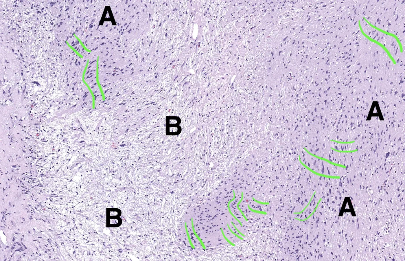
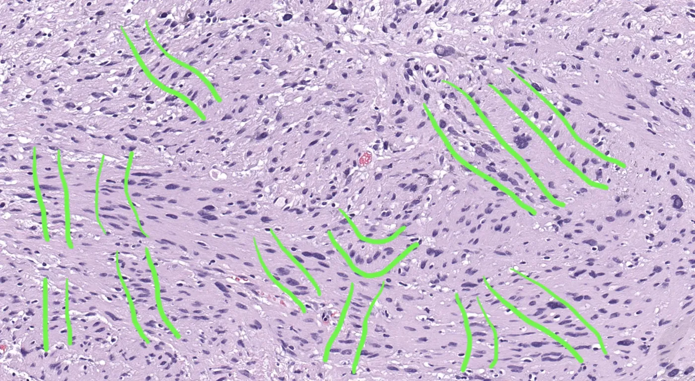
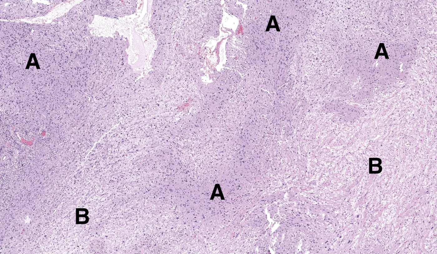
- 一部分神经鞘瘤没有 Verocay 小体,但多找找通常能看到隐约的核栅栏:细胞核大致聚成一条模糊的线,旁边是粉染、细胞较少的区带,可弯曲、成簇或不呈直线。
- 识别隐约栅栏也有助于发现梭形细胞/多形性脂肪瘤等其他病变。
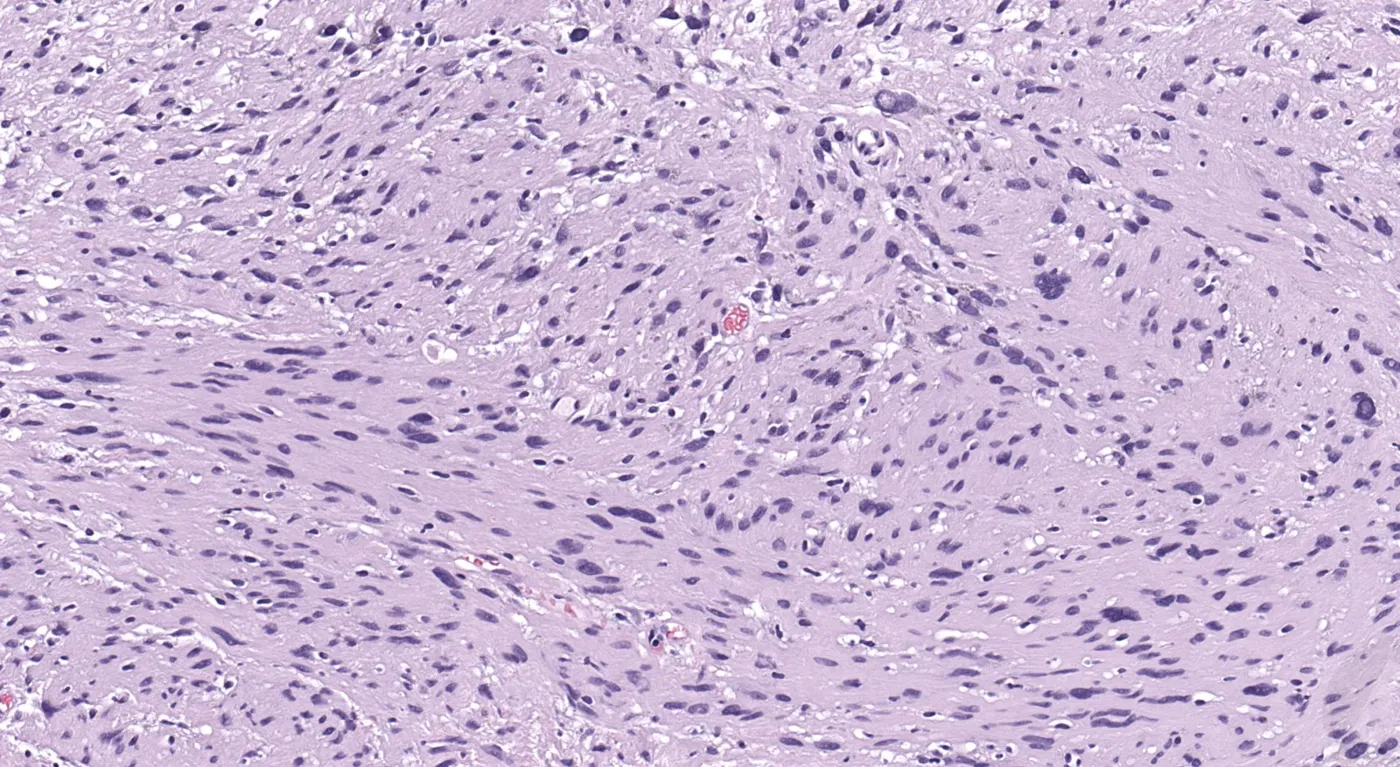
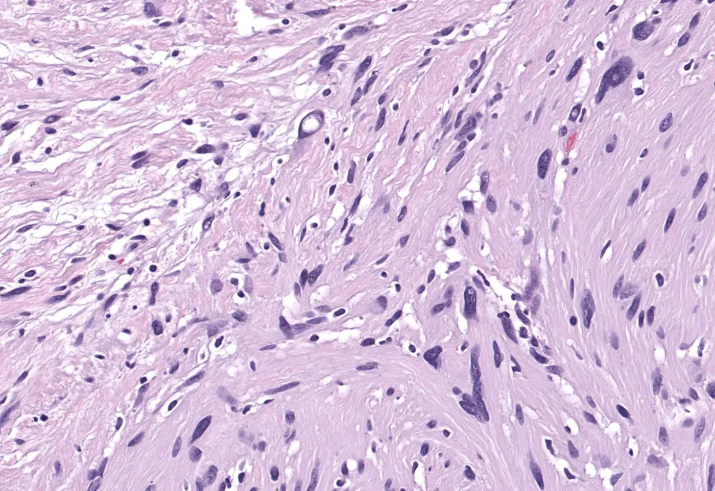
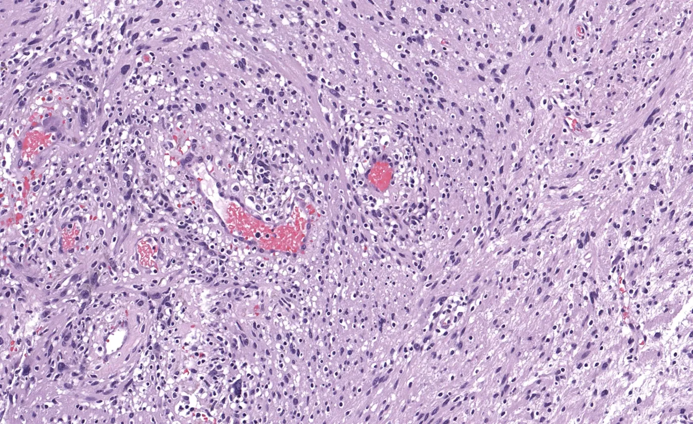
- 注意随机分布的核异型(散在深染、多形核):大多数神经鞘瘤都有一定程度,本例这样的陈旧性神经鞘瘤尤为显著。
收录范围:KiKo 帖子全文、帖内 8 幅图及资料链接,中英对照。原帖图片无图注,图注由本站据原帖说明添加。短视频与数字切片请经链接观看。
本帖短视频讲解:https://kikoxp.com/posts/25630
本例 WSI 数字切片:https://kikoxp.com/posts/11804
神经鞘瘤入门视频:https://kikoxp.com/posts/4093
识别隐约的栅栏状排列
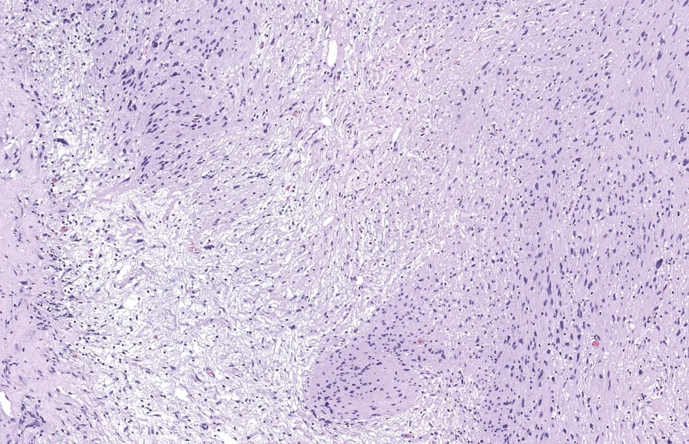
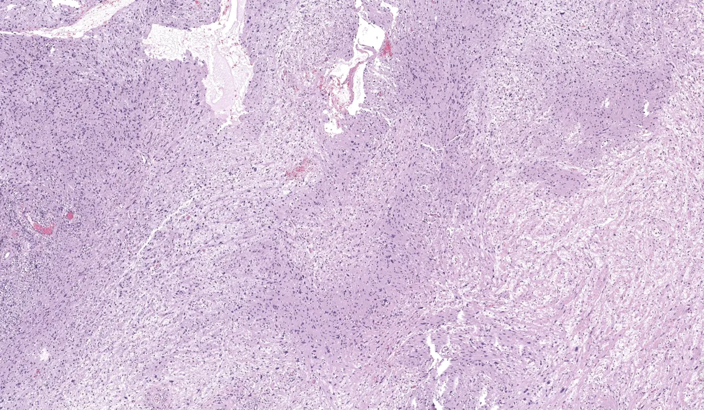
有 Verocay 小体时当然很好,但有一部分神经鞘瘤没有 Verocay 小体。不过只要多找一找,通常能发现一些隐约、模糊的核栅栏状排列区。这些不明显的栅栏灶也许不如经典 Verocay 小体那种完美平行的醒目栅栏好看,但识别它们能帮助你得出正确诊断。学会识别隐约栅栏需要时间和练习,但非常值得。要寻找的是:细胞核大致聚集成一条模糊的线,旁边是粉染、细胞较少的区带。有时这些平行排列的模糊栅栏会弯曲、成簇,或者不呈直线。刚开始你可能得眯起眼睛,甚至有点「脑补」,但慢慢地这些不明显的区域就会凸显出来(识别隐约栅栏对发现神经鞘瘤以外的其他病变也有用,例如梭形细胞/多形性脂肪瘤及相关病变)。
我把其中几张图复制了一份,并在(至少在我看来)呈模糊栅栏状的区域两侧加了绿线。试着在标注图和原图之间来回切换,看看栅栏是否开始显现出来。
这些图中还有其他栅栏状区域……我只标出了最典型的几处。我还在部分图上标了 A 和 B,用来指出 Antoni A 区和 Antoni B 区。
请注意随机分布的核异型(散在深染、多形性细胞核)。大多数神经鞘瘤都会有一定程度的核异型,但在像本例这样的陈旧性神经鞘瘤中尤为显著。
关于陈旧性神经鞘瘤的更多讲解:https://kikoxp.com/posts/12126
外周神经鞘肿瘤更多资料
神经鞘瘤(施万细胞瘤)
- 视频(入门):https://kikoxp.com/posts/4093
- 视频(短片 1):https://kikoxp.com/posts/4530
- 视频(短片 2):https://kikoxp.com/posts/4555
- 视频(在该综述视频中讨论):https://kikoxp.com/posts/4999
- 视频(在该综述视频中讨论):https://kikoxp.com/posts/4927
- 视频(伴陈旧性改变,在该综述视频中讨论):https://kikoxp.com/posts/6553
- 图片(皮肤神经鞘瘤——少见部位):https://kikoxp.com/posts/8892
神经纤维瘤(NF)
- 视频(NF 入门、神经纤维瘤病/NF1 及低级别 MPNST):https://kikoxp.com/posts/4091
- 视频(短片——NF1 中发生的丛状神经纤维瘤):https://kikoxp.com/posts/4584
- 视频(色素性、弥漫性及丛状神经纤维瘤):https://kikoxp.com/posts/4999
- 视频(伴圆形细胞、虎纹样图案和 Wagner-Meissner 小体的弥漫性神经纤维瘤,在该综述视频中讨论):https://kikoxp.com/posts/7318
- 图片(弥漫性神经纤维瘤中的 Wagner-Meissner 小体):https://kikoxp.com/posts/8156
- 图片(史上最佳的弥漫性神经纤维瘤 Wagner-Meissner 小体):https://kikoxp.com/posts/8494
神经束膜瘤
- 视频(神经束膜瘤入门):https://kikoxp.com/posts/3694
- 视频(13:28 处):https://kikoxp.com/posts/3784
- 视频(硬化性神经束膜瘤,在该综述视频中讨论):https://kikoxp.com/posts/4999
- 视频(短片——神经束膜瘤及免疫组化):https://kikoxp.com/posts/10073
- WSI 数字切片(硬化性神经束膜瘤):https://kikoxp.com/posts/3676
- WSI 数字切片(神经束膜瘤及免疫组化):https://kikoxp.com/posts/8021
颗粒细胞瘤
- 图片(累及神经周围的颗粒细胞瘤——良性):https://kikoxp.com/posts/8186
- 视频(累及神经周围的颗粒细胞瘤——良性):https://kikoxp.com/posts/11641
- 视频(舌颗粒细胞瘤类似鳞癌,1:09:59 处):https://kikoxp.com/posts/4999
- 视频(短片 1):https://kikoxp.com/posts/4550
- 视频(短片 2):https://kikoxp.com/posts/4617
- 视频(短片 3——颗粒细胞瘤伴假上皮瘤样增生类似鳞癌):https://kikoxp.com/posts/8113
- 视频(在该综述视频中讨论):https://kikoxp.com/posts/4827
真皮神经鞘黏液瘤(旧称经典型/黏液型神经鞘黏液瘤 neurothekeoma)
- 视频(神经鞘黏液瘤与富于细胞性神经鞘黏液瘤 neurothekeoma 鉴别入门):https://kikoxp.com/posts/4103
- 视频(短片):https://kikoxp.com/posts/4616
- 视频(56:07 处):https://kikoxp.com/posts/3784
- 视频(在该综述视频中讨论):https://kikoxp.com/posts/4999
孤立性局限性神经瘤(栅栏状包膜性神经瘤)(SCN/PEN)
- 视频(短片):https://kikoxp.com/posts/3844
- 视频(在该综述视频中讨论):https://kikoxp.com/posts/4999
- 图片:https://kikoxp.com/posts/8893
创伤性神经瘤
- 视频(在该综述视频中讨论):https://kikoxp.com/posts/4999
- 视频(在该综述视频中讨论):https://kikoxp.com/posts/6553
- WSI 数字切片(发生于大神经):https://kikoxp.com/posts/8018
恶性外周神经鞘瘤(MPNST)
- 视频(经典型 MPNST 入门):https://kikoxp.com/posts/4092
- 视频(低级别 MPNST——38:00 处):https://kikoxp.com/posts/4091
- 视频(伴横纹肌母细胞分化的 Triton 瘤 MPNST——1:00:07 处):https://kikoxp.com/posts/3784
作者全部视频、数字切片、图片及病理报告范例的完整分类资料库:https://kikoxp.com/posts/5084(皮肤病理)与 https://kikoxp.com/posts/5083(骨/软组织肉瘤病理)。
- Jerad Gardner’s KiKo soft tissue teaching post (shared again on X on 30 October 2026) with 8 images, several duplicated with green lines and A/B labels for comparison with the originals.
- A subset of schwannomas lack Verocay bodies, but subtle palisading can usually be found: nuclei clustered in a vague line with adjacent pink, less cellular zones, sometimes curved, clustered or not straight.
- Recognizing subtle palisading also helps with other entities such as spindle cell/pleomorphic lipoma.
- Note the random nuclear atypia (scattered hyperchromatic pleomorphic nuclei): present to some extent in most schwannomas, especially dramatic in an ancient schwannoma like this one.
Scope: the full KiKo post with its 8 images and resource links. The original images have no captions; captions were added by this site based on the post text. Videos and the digital slide are available via the links.
Short video explanation of this post: https://kikoxp.com/posts/25630
WSI digital slide of this case here: https://kikoxp.com/posts/11804
Schwannoma 101 video: https://kikoxp.com/posts/4093
Recognizing subtle palisading
Verocay bodies are great when you have them, but a subset of schwannomas do not. However, if you look around a bit, you can usually find subtle areas of vague nuclear palisading. These foci of subtle palisading may not be as aesthetically pleasing as the dramatic perfectly parallel palisading seen in classic Verocay bodies, but recognizing these subtle areas can help you arrive at the correct diagnosis. Learning to recognize the subtle palisading takes time and practice, but it's well worth the effort. Look for nuclei that are clustered together in a vague line with adjacent zones of pink with lower cellularity. Sometimes these parallel arrays of vague palisading may be curved or clustered or otherwise not in a straight line. You may have to squint your eyes and even hallucinate a bit at first, but eventually, these subtle areas will start to stand out (recognizing subtle palisading is also useful for picking up on other entities aside from schwannoma, such as spindle cell/pleomorphic lipoma and related entities).
I've duplicated a few of the images and added green lines flanking the areas that are vaguely palisaded (to my eye, at least). Try to flip back and forth between the annotated image and the original image and see if the palisades begin to stand out.
There are other areas in these images that are also palisaded...I've just highlighted some of the best ones. I've also added A and B to some of the images to point out the Antoni A and Antoni B areas.
Notice the random nuclear atypia (scattered hyperchromatic pleomorphic nuclei). This is present in most schwannomas to some extent, but it's especially dramatic in a case of ancient schwannoma like this one.
More explanation of ancient schwannoma here: https://kikoxp.com/posts/12126
More info about Peripheral Nerve Sheath Tumors
Schwannoma (Neurilemmoma)
- Video (101): https://kikoxp.com/posts/4093
- Video (short 1): https://kikoxp.com/posts/4530
- Video (short 2): https://kikoxp.com/posts/4555
- Video (discussed in this review video): https://kikoxp.com/posts/4999
- Video (discussed in this review video): https://kikoxp.com/posts/4927
- Video (with ancient change - discussed in this review video): https://kikoxp.com/posts/6553
- Images (schwannoma in skin – uncommon location): https://kikoxp.com/posts/8892
Neurofibroma (NF)
- Video (NF 101, Neurofibromatosis/NF1, & Low Grade MPNST): https://kikoxp.com/posts/4091
- Video (short – plexiform NF arising in NF1): https://kikoxp.com/posts/4584
- Video (pigmented, diffuse, & plexiform neurofibroma): https://kikoxp.com/posts/4999
- Video (diffuse neurofibroma with round cells, tiger stripe pattern, and Wagner-Meissner bodies - discussed in this review video): https://kikoxp.com/posts/7318
- Images (Wagner-Meissner bodies in diffuse neurofibroma): https://kikoxp.com/posts/8156
- Images (BEST EVER Wagner-Meissner bodies in diffuse neurofibroma): https://kikoxp.com/posts/8494
Perineurioma
- Video (perineurioma 101): https://kikoxp.com/posts/3694
- Video (at 13:28): https://kikoxp.com/posts/3784
- Video (sclerosing perineurioma discussed in this review video): https://kikoxp.com/posts/4999
- Video (short - perineurioma with immunostains): https://kikoxp.com/posts/10073
- WSI digital slide (sclerosing perineurioma): https://kikoxp.com/posts/3676
- WSI digital slide (perineurioma with immunostains): https://kikoxp.com/posts/8021
Granular cell tumor
- Images (granular cell tumor with perineural involvement – benign): https://kikoxp.com/posts/8186
- Video (granular cell tumor with perineural involvement – benign): https://kikoxp.com/posts/11641
- Video (granular cell tumor of tongue mimicking SCC at 1:09:59): https://kikoxp.com/posts/4999
- Video (short 1): https://kikoxp.com/posts/4550
- Video (short 2): https://kikoxp.com/posts/4617
- Video (short 3 – granular cell tumor with PEH mimicking SCC): https://kikoxp.com/posts/8113
- Video (discussed in this review video): https://kikoxp.com/posts/4827
Dermal nerve sheath myxoma (formerly conventional/myxoid neurothekeoma)
- Video (nerve sheath myxoma vs cellular neurothekeoma 101): https://kikoxp.com/posts/4103
- Video (short): https://kikoxp.com/posts/4616
- Video (at 56:07): https://kikoxp.com/posts/3784
- Video (discussed in this review video): https://kikoxp.com/posts/4999
Solitary circumscribed neuroma (palisaded encapsulated neuroma) (SCN/PEN)
- Video (short): https://kikoxp.com/posts/3844
- Video (discussed in this review video): https://kikoxp.com/posts/4999
- Images: https://kikoxp.com/posts/8893
Traumatic neuroma
- Video (discussed in this review video): https://kikoxp.com/posts/4999
- Video (discussed in this review video): https://kikoxp.com/posts/6553
- WSI digital slide (arising from large nerve): https://kikoxp.com/posts/8018
Malignant peripheral nerve sheath tumor (MPNST)
- Video (Conventional MPNST 101): https://kikoxp.com/posts/4092
- Video (Low grade MPNST - 38:00): https://kikoxp.com/posts/4091
- Video (Triton MPNST w rhabdomyoblastic diff. – at 1:00:07): https://kikoxp.com/posts/3784
A complete organized library of all my videos, digital slides, pics, & sample pathology reports is available here: https://kikoxp.com/posts/5084 (dermpath) & https://kikoxp.com/posts/5083 (bone/soft tissue sarcoma pathology).