- 问题:特发性炎性肌病(IIM)的肌活检免疫组化通常依赖速冻标本,很多资源有限的科室拿不到冰冻肌肉。作者建立可重复的 FFPE 肌肉切片免疫组化流程,评估其诊断价值。
- 方法:20 例(皮肌炎 5、包涵体肌炎 2、免疫介导坏死性肌病 5、重叠性肌炎 1、未分类肌病 3,及线粒体肌病、LGMD R2 dysferlin 相关肌营养不良、面肩肱型肌营养不良、神经源性改变各 1);3 μm FFPE 切片,柠檬酸/EDTA 热修复,4°C 过夜孵育 MHC-I、MHC-II、MAC、MxA、p62,冰冻切片平行对照。
- 结果:MHC-II、MxA、p62 在 FFPE 上可获得与冰冻相当的强度与模式;8 例各有 1 个标志物不一致,但差异无统计学意义(P 值 0.48–0.62),一致率 MHC-II 与 MxA 90%、p62 80%,不一致可能源于 IIM 节段性、斑片状受累。
- 局限与实用点:MHC-I 与 MAC 在 FFPE 上染色失败,仍需冰冻;在无法速冻的条件下,MHC-II、MxA、p62 三项可作为 IIM 诊断的可行补充。图 1–5、表 1–4 中英对照。
收录范围:PMC 开放获取全文(CC BY 4.0)——摘要、引言、材料与方法、结果、讨论、结论,图 1–5 与表 1–4 中英对照;作者贡献、资助、利益冲突、补充材料与参考文献未收录。
摘要
本研究旨在为福尔马林固定石蜡包埋(FFPE)肌肉切片建立一种可重复的免疫组织化学方法,并评估其在无法获得速冻肌肉活检的条件下对特发性炎性肌病(IIM)的诊断价值。
共分析20例病例:皮肌炎(n = 5)、包涵体肌炎(n = 2)、免疫介导坏死性肌病(n = 5)、重叠性肌炎(n = 1)、未特指肌病(n = 3)、线粒体肌病(n = 1)、LGMD R2 dysferlin相关肌营养不良(n = 1)、面肩肱型肌营养不良(n = 1)以及肌肉神经源性改变(n = 1)。在3 μm厚的FFPE肌肉切片上进行免疫组织化学染色,采用柠檬酸盐/EDTA缓冲液进行热诱导抗原修复,并使用针对MHC‐I、MHC‐II、MAC、MxA和p62的抗体于4°C孵育过夜。同时对冰冻切片进行平行分析以作比较。
FFPE肌肉切片中MHC‐II、MxA和p62的染色强度及模式与冰冻切片相当。尽管8例病例的冰冻切片与FFPE切片之间出现差异,且每例仅涉及一种标志物,但MHC‐II(P = 0.4795)、MxA(P = 0.4795)和p62(P = 0.6171)的免疫组织化学结果均未见统计学显著差异。两种方法的一致率较高,MHC‐II和MxA均达90%,p62为80%。这些个别差异可能反映了与IIM特征性的节段性和斑片状受累相关的抗原表达变异。然而,FFPE切片中的MHC‐I和MAC染色未获成功。
在FFPE肌肉切片中成功实现了MHC‐II、MxA和p62的免疫标记。这些结果表明,FFPE肌肉组织能够可靠地评估多种对IIM诊断具有信息价值的标志物,为资源受限环境提供了一种实用的诊断选择。
引言
特发性炎性肌病(IIM)是一组具有多样性的免疫介导肌肉疾病,其特征为肌肉炎症和进行性肌无力。近期共识进一步完善了IIM分类,将其分为5种不同亚型,包括皮肌炎(DM)、包涵体肌炎(IBM)、免疫介导坏死性肌病(IMNM)、抗合成酶综合征和重叠性肌炎(OM)。 1 , 2 , 3 , 4 , 5 , 6 多发性肌炎曾被视为一种独立疾病实体,如今越来越多地被重新归类至其他肌病亚型,目前已被认为是一个过时的诊断。 1 , 2 , 3 , 4 临床信息、自身抗体谱和肌肉活检对IIM诊断至关重要。速冻肌肉活检被认为是金标准,因为它可保留对准确组织病理学评估至关重要的酶活性和组织抗原性。 7 然而,在资源受限环境中,其可及性有限仍是一项重大挑战。泰国虽有77个府,目前仅曼谷少数几家三级医院提供速冻肌肉活检。 8 , 9 这种有限的可及性限制了患者获得及时、准确诊断的机会,最终延误适当治疗。相比之下,采用福尔马林固定石蜡包埋(FFPE)肌肉组织的免疫组织化学(IHC)更容易获得。然而,探讨FFPE肌肉标本在IIM中诊断潜力的研究仍然有限。肌营养不良诊断也面临类似挑战,这促使我们建立了一套可靠且可重复的FFPE肌肉组织IHC方案,用于检测肌膜相关蛋白,以诊断常见的肌营养不良。 10 在此成功经验基础上,我们旨在建立一种灵敏且可重复的IHC方法,用于检测FFPE肌肉样本中的炎症标志物,使其与速冻技术相当,从而克服IIM诊断中的类似限制。本研究重点关注主要组织相容性复合体I类(MHC‐I)、主要组织相容性复合体II类(MHC‐II)、膜攻击复合物(MAC或C5b‐9)、黏液病毒抗性蛋白A(MxA)和p62,它们分别反映异常MHC表达、补体沉积、I型干扰素活性和自噬空泡。 11 第二个目标是评估这些标志物在FFPE肌肉活检标本中对IIM的诊断价值。
材料与方法
研究设计与病例选择
本研究获得玛希隆大学拉玛提博迪医院医学院人体研究伦理委员会的伦理批准(COA. MURA2022/301),其中包括允许使用生物样本库组织而无需知情同意。本研究纳入20例患者,诊断如下:DM(n = 5)、IBM(n = 2)、IMNM(n = 5)、OM(n = 1)、未特指肌病(n = 3)、线粒体肌病(n = 1)、LGMD R2 dysferlin相关肌营养不良(原称LGMD2B)(n = 1)、面肩肱型肌营养不良(n = 1)和肌肉神经源性改变(n = 1)。既往诊断根据临床表现、组织病理学评估和速冻肌肉组织免疫表型确定。详细病例信息见表 1。
| 病例 | 性别 | 年龄(岁) | 诊断 | 冰冻切片 | 福尔马林固定石蜡包埋切片 | ||||||||
|---|---|---|---|---|---|---|---|---|---|---|---|---|---|
| MHC‐I | MHC‐II | MAC | MxA | p62 | MHC‐I | MHC‐II | MAC | MxA | p62 | ||||
| 1 | 女 | 61 | 皮肌炎 | + | − | + | ++, PFA | +++ | 染色未成功 | − | 染色未成功 | ++, PFA | +++ |
| 2 | 男 | 55 | 皮肌炎(Tif 1 gamma) | − | − | − | − | − | − | − | + | ||
| 3 | 女 | 60 | 皮肌炎 | + | − | + | − | ++ | − | +++, PFA | ++ | ||
| 4 | 女 | 7 | 幼年型皮肌炎 | + | ++, PFA | + | − | ++ | ++, PFA | − | ++ | ||
| 5 | 女 | 14 | 幼年型皮肌炎 | + | ++ | + | +++, PFA | +++ | − | +++, PFA | +++ | ||
| 6 | 男 | 76 | 包涵体肌炎 | + | +++ | − | − | +++ | +++ | − | +++ | ||
| 7 | 男 | 67 | 包涵体肌炎 | − | +++ | − | − | ++ | +++ | − | ++ | ||
| 8 | 男 | 65 | 免疫介导坏死性肌病 | − | − | − | − | ++ | +++ | − | +++ | ||
| 9 | 女 | 10 | 免疫介导坏死性肌病 | + | − | + | − | ++ | − | − | − | ||
| 10 | 女 | 14 | 免疫介导坏死性肌病 | + | − | + | − | +++ | − | − | +++ | ||
| 11 | 女 | 79 | 免疫介导坏死性肌病 | + | +++ | + | ++ | +++ | ++ | ++ | +++ | ||
| 12 | 男 | 78 | 免疫介导坏死性肌病 | + | − | + | ++ | +++ | − | − | +++ | ||
| 13 | 女 | 56 | 重叠性肌炎 | + | − | + | − | ++ | − | − | ++ | ||
| 14 | 男 | 61 | 未特指肌病 | − | − | − | − | ++ | − | − | +++ | ||
| 15 | 女 | 61 | 未特指肌病 | − | − | − | − | − | − | − | +++ | ||
| 16 | 女 | 16 | 未特指肌病 | − | − | − | − | + | − | − | + | ||
| 17 | 男 | 31 | 线粒体肌病 | − | − | − | − | + | − | − | + | ||
| 18 | 女 | 42 | LGMD R2 dysferlin相关肌营养不良(原称LGMD2B) | − | − | − | − | ++ | − | − | ++ | ||
| 19 | 男 | 68 | 面肩肱型肌营养不良 | − | − | + | − | +++ | − | − | +++ | ||
| 20 | 男 | 67 | 肌肉神经源性改变 | − | − | − | − | +++ | − | − | − | ||
带有彩色背景的结果单元格表示结果不一致。
F:女性;M:男性;PFA:束周萎缩。
组织处理
根据临床指征,在局部麻醉下对股四头肌或肱二头肌进行肌肉活检。新鲜组织样本迅速送至病理实验室,以保持其完整性。每份标本分为两部分:一部分进行速冻,另一部分用10%中性缓冲福尔马林固定并制成石蜡包埋组织块。
免疫组织化学
从每个石蜡块切取3 μm厚的FFPE肌肉切片,贴附于载玻片上,于60°C孵育至少1 h。切片经脱蜡、复水后,先在柠檬酸盐缓冲液中于97°C进行热诱导抗原修复20 min,再在EDTA缓冲液中于97°C修复20 min。蒸馏水冲洗后,用5%过氧化氢处理30 min以阻断内源性过氧化物酶活性。室温下使用Bond一抗稀释液(Ventana)处理15 min,以尽量减少非特异性背景染色。使用EnVision FLEX洗涤缓冲液(1×)(Dako)清洗切片。针对MHC‐I、MHC‐II、MAC、MxA和p62的一抗(厂家、克隆号、目录号和稀释度详见表 2)于4°C孵育过夜。随后按照厂家方案,使用自动染色平台进行免疫染色(MHC‐II、MxA和p62采用Ventana;MHC‐I和MAC采用Dako的Autostainer Link 48)。之后对切片进行复染、脱水、透明和封片。
| 抗体 | 厂家 | 克隆号[目录号] | 冰冻切片用稀释度 | FFPE切片用稀释度 | 染色模式 |
|---|---|---|---|---|---|
| MHC‐I | NeoMarker | W6/32 [MS‐1218‐P] | 1:15,000 (Ventana) | 1:50 (Dako) | 肌膜和肌浆染色 |
| MHC‐II | Dako | CR3/43 [M0775] | 1:100 (Ventana) | 1:50 (Ventana) | 肌膜和肌浆染色;束周或非束周区域 |
| MAC (c5b‐9) | Abcam | aE11 [ab66768] | 1:300 (Ventana) | 1:50 (Dako) | 肌膜染色或肌内膜毛细血管血栓样模式 |
| MxA | EMD Millipore | M143 (CL143) [MABF938] | 1:300 (Ventana) | 1:200 (Ventana) | 肌浆染色;束周或非束周区域 |
| p62 (SQSTM1) | Santa cruz | D‐3 [sc‐28359] | 1:200 (Ventana) | 1:200 (Ventana) | 肌浆染色;镶边空泡内呈细颗粒状或聚团状染色 |
制备冰冻切片作为对照。新鲜肌肉标本在经液氮预冷的异戊烷(−150°C)中迅速冷冻,随后储存于−80°C。使用恒冷切片机切取10 μm厚切片,贴附于载玻片上,并于室温风干。IHC染色前不进行固定或抗原修复。使用Bond一抗稀释液(Leica)稀释的一抗孵育40 min。随后按照厂家方案,在Ventana自动平台上进行免疫染色。之后对切片进行苏木精复染、脱水、透明和封片。
选择这5种克隆抗体,是因为它们均常规用于我们的诊断工作。厂家仅验证了MHC‐I和MAC适用于冰冻组织切片,而MHC‐II、MxA和p62则已获验证,可用于冰冻和FFPE两种标本。
在载玻片上每张组织切片旁均设置相应的FFPE或冰冻切片阳性对照。对各病例编号进行设盲并随机重新排序后判读IHC。首先在不知晓临床信息的情况下独立评估IHC结果。阳性免疫反应的判定标准见表 2。免疫反应强度分级如下:–(阴性)、+(弱)、++(中等)和+++(强)。随后,将FFPE或冰冻肌肉切片的IHC结果与临床背景相结合以作出诊断。分别对FFPE切片和冰冻切片采用这一诊断方法。比较FFPE切片与冰冻切片的IHC结果。
统计学分析
采用针对配对分类数据、经Edwards连续性校正的McNemar检验,评估冰冻与FFPE肌肉切片之间免疫组织化学结果的差异(P值<0.05视为具有统计学意义)。
结果
表 1详细汇总了全部20例病例冰冻和FFPE切片中MHC‐I、MHC‐II、MAC、MxA及p62的全面IHC特征。对FFPE切片评估了多种抗体稀释度,以确定最佳染色条件。在最佳抗体稀释度分别为1:50、1:200和1:200时,FFPE切片中的MHC‐II、MxA和p62染色成功(表 2)。相比之下,即使稀释度为1:50,所有FFPE切片中仍均未能显示MHC‐I和MAC。
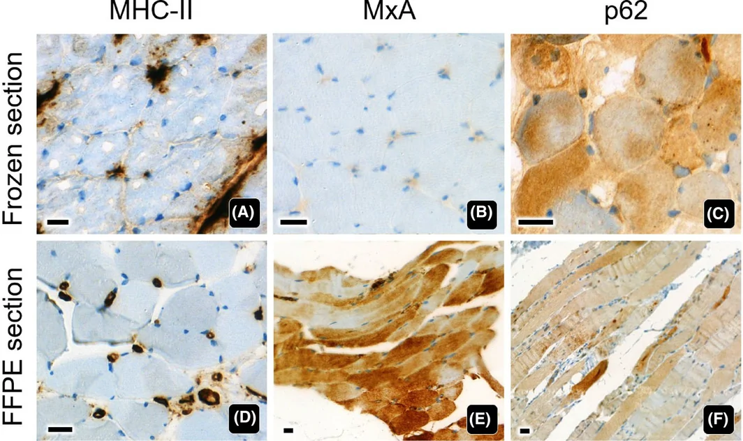
12例病例的MHC‐II、MxA和p62免疫反应染色相当(表 1)。病例6(IBM)、病例1(DM)和病例11(IMNM)的代表性显微照片分别见图 1、2、3,展示了这些一致的结果。MHC‐II在FFPE切片中显示清晰的肌膜和肌浆免疫反应(图 1D和3D),与冰冻切片中观察到的染色模式相符(图 1A和3A)。MxA在FFPE切片中显示明确的肌浆染色(图 2E和3E),与冰冻切片相当(图 1B和3B)。同样,p62在FFPE切片中表现为弥漫性细颗粒状肌浆染色(图 1F、2F和3F),与冰冻切片中观察到的染色一致(图 1C、2C和3C)。随后结合临床信息解释FFPE切片的免疫组织化学结果。病例6的FFPE切片(图 1D–F)显示MHC‐II强表达,p62在镶边空泡内呈聚团点状染色,同时MxA不表达,符合IBM的特征性免疫表型。病例1的FFPE切片(图 2D–F)在纵切方向显示束周萎缩,MxA和p62阳性表达,MHC‐II染色阴性,符合DM的典型免疫表型。病例11的FFPE切片(图 3D–F)显示MHC‐II、MxA和p62表达,符合IMNM的典型免疫表型。
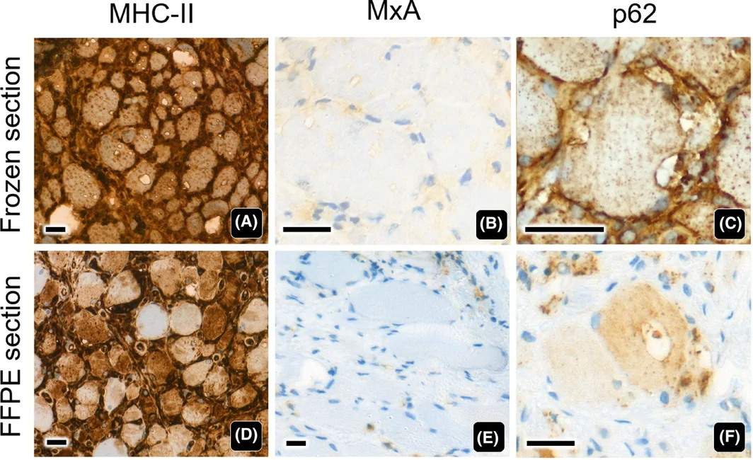
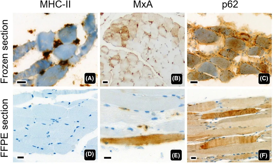
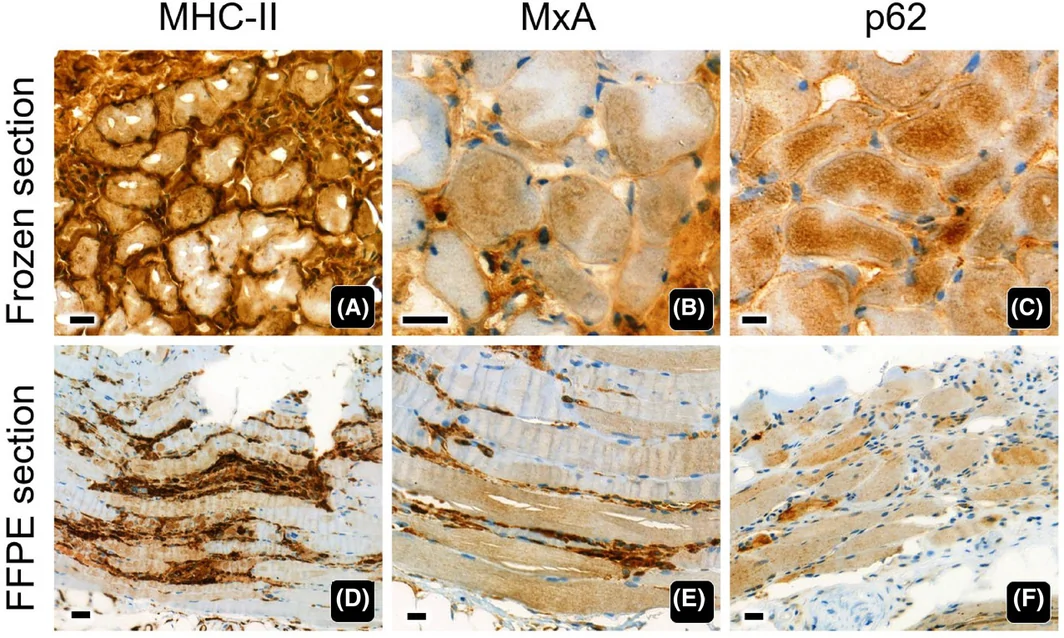
8例病例(病例2、3、5、8、9、12、15、20)的冰冻切片与FFPE切片之间存在免疫组织化学染色差异,每例均仅涉及一种标志物(表 1)。病例5(幼年型DM)仅冰冻切片显示MHC‐II阳性(图 4A),FFPE切片未见染色(图 4D)。相反,病例8(IMNM)仅FFPE切片显示MHC‐II强染色,冰冻切片未见染色。病例3(DM)仅FFPE切片显示MxA强阳性(图 5E),冰冻切片未见染色(图 5B),而病例12(IMNM)在冰冻切片中呈MxA中等强度染色,FFPE切片则完全不染色。对于p62,病例2(DM)和病例15(未特指肌病)仅FFPE切片呈阳性,而病例9(IMNM)和病例20(肌肉神经源性改变)在冰冻切片中呈强染色,对应FFPE切片则为阴性。为确认这些不一致的结果,对这8例不一致病例所有对应的存档冰冻切片进行了复查,其染色结果与本研究所评估冰冻切片中观察到的结果完全一致。这些不一致的IHC结果可能反映了IIM典型的节段性和斑片状抗原表达。
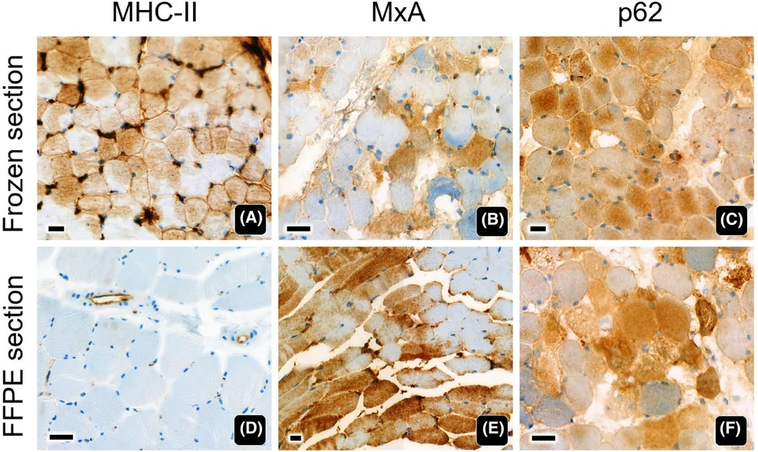
McNemar检验显示,冰冻和FFPE肌肉切片中这3种标志物的免疫组织化学阳性率均无统计学显著差异(表 3)。对于MHC‐II和MxA,每种标志物仅有2例结果不一致,冰冻与FFPE肌肉切片之间无显著差异(P = 0.4795)。对于p62,发现4例结果不一致。然而,这一差异同样无统计学意义(P = 0.6171),提示冰冻切片与FFPE切片之间不存在系统性偏倚。总体一致率较高,MHC‐II和MxA均达90%,p62为80%。综合来看,这些结果表明,FFPE切片的染色结果与冰冻切片相当,支持FFPE组织用于常规诊断工作中评估这些标志物的可靠性。
| 标志物 | 结果一致的病例(%) | 结果不一致的病例 a (%) | P值 | ||||
|---|---|---|---|---|---|---|---|
| 冰冻+/FFPE+ | 冰冻−/FFPE− | 合计 | 冰冻+/FFPE− | 冰冻−/FFPE+ | 合计 | ||
| MHC‐II | 4 (20%) | 14 (70%) | 18 (90%) | 1 (5%) | 1 (5%) | 2 (10%) | 0.4795 |
| MxA | 3 (15%) | 15 (75%) | 18 (90%) | 1 (5%) | 1 (5%) | 2 (10%) | 0.4795 |
| p62 | 16 (80%) | 0 (0%) | 16 (80%) | 2 (10%) | 2 (10%) | 4 (20%) | 0.6171 |
结果不一致的病例:MHC‐II(病例5、8);MxA(病例3、12);p62(病例2、9、15、20)。
比较冰冻和FFPE肌肉标本中的MHC‐II、MxA及p62染色,各IIM亚组的阳性结果总体一致,标志物阳性率存在轻微差异(表 4)。MHC‐II表达在IBM中完全一致(两种制备方式均为100%),在OM中均不表达,而DM中的检出率略低(冰冻为40%,FFPE为20%),使用FFPE组织的IMNM检出率则略有升高(20%对40%)。MxA表达在冰冻与FFPE切片中的总体分布相似。与其他亚组相比,DM的阳性率更高,IBM和OM在两种制备方式中均无表达。冰冻组织与FFPE组织的阳性率存在轻微差异(DM:40%对60%;IMNM:40%对20%)。p62染色在所有IIM亚组和组织类型中均持续呈高表达,尤其是IBM和OM(两种制备方式均为100%),仅DM和IMNM中存在轻微变化。总体而言,这些结果提示,FFPE肌肉切片对MHC‐II、MxA和p62的免疫组织化学检出能力与冰冻标本相当。这支持了在IIM诊断流程中采用FFPE评估的可行性。
| 标志物 | 冰冻肌肉标本 | FFPE肌肉标本 | ||||||
|---|---|---|---|---|---|---|---|---|
| DM (n = 5) | IBM (n = 2) | IMNM (n = 5) | OM (n = 1) | DM (n = 5) | IBM (n = 2) | IMNM (n = 5) | OM (n = 1) | |
| MHC‐II | 40% (2) | 100% (2) | 20% (1) | 0% (0) | 20% (1) | 100% (2) | 40% (2) | 0% (0) |
| MxA | 40% (2) | 0% (0) | 40% (2) | 0% (0) | 60% (3) | 0% (0) | 20% (1) | 0% (0) |
| p62 | 80% (4) | 100% (2) | 100% (5) | 100% (1) | 100% (5) | 100% (2) | 80% (4) | 100% (1) |
讨论
本研究比较了20例患者速冻和FFPE肌肉活检标本中5种诊断标志物MHC‐I、MHC‐II、MAC、MxA及p62的IHC染色结果。主要目的是评估在无法常规获得冰冻IHC切片的资源受限环境中使用FFPE切片的可行性。基于MHC‐II和MxA均为90%、p62为80%的高一致率,我们的结果表明,在保留这3种免疫组织化学标志物的抗原性方面,FFPE肌肉活检与冰冻切片表现相当。本研究采用3 μm厚的FFPE切片,因为其能够更好地显示肌纤维形态,而我们既往研究中较厚的8 μm FFPE切片出现了肌膜染色重叠。 10 尽管数据仍然有限,现有文献提示,在无法进行冰冻切片时,可以成功优化适用于FFPE肌肉组织的免疫组织化学方案。 10 , 12 , 13 , 14 Suriyonplengsaeng等证明了基于FFPE的IHC在肌营养不良中的成功应用,但对某些表位仍存在局限。 10 本研究通过证明FFPE样本中的MHC‐II、MxA和p62仍可被可靠检出,在IIM领域扩展了这一证据。在10–20%的病例中观察到的差异(染色不一致)未达到统计学显著性(P > 0.05),很可能反映了IIM特征性的节段性和斑片状受累。虽然冰冻和FFPE切片来自同一肌肉活检标本,但采用不同切面也可能促成观察到的差异。这些结果支持,不一致归因于生物学异质性和取样差异,而非技术变异。
第二个目的是评估使用FFPE肌肉标本诊断IIM的效能。在5例DM病例中,FFPE切片中MHC‐II、MxA和p62的免疫组织化学评估结果与冰冻组织相当(表 1)。值得注意的是,1例DM的FFPE切片显示束周MxA强阳性,而对应冰冻标本未见染色(图 5)。这一观察结果凸显了基于FFPE的MxA IHC的稳健性,并表明其能够检出对DM诊断至关重要的I型干扰素相关改变。这一发现与分子证据一致:活动期DM病例肌肉中的MX1基因表达显著升高,约为正常肌肉的280倍,反映了显著的I型干扰素特征。 15 在两种制备方式中,MHC‐II表达均为1至2例病例提供了支持性证据,与既往报告32%–55%的DM病例表达MHC‐II一致。 6 , 16 p62染色持续为阳性,但不具有疾病特异性。Milisenda等报告,DM、IMNM和IBM病例中p62的表达率分别为57%、87%和92%,其他肌病中为26%。 17 FFPE肌肉标本的免疫组织化学分析通过束周MxA表达使3例DM得以明确诊断,并通过束周MHC‐II表达使1例得以明确诊断(表 1)。不同研究报告的DM肌浆MxA表达频率存在差异,在Waisayarat等、 18 Uruha等 11 和Xing等 19 的队列中,分别有46%、71%和94%的病例检出表达。Soponkanaporn等报告,61%的幼年型DM病例有肌浆MxA表达。 20 尽管阳性率存在这种差异,MxA仍被广泛认为是DM相对特异的标志物,在Waisayarat等 18 和Uruha等 11 的研究中,特异性分别达到94%和98%。然而,其敏感性仍属中等,这两项研究分别报告为46%和71%。这些结果表明,MxA不表达并不能排除DM诊断。事实上,可能遇到MxA阴性的DM病例,例如本研究中的病例17和5。
在2例IBM病例中,弥漫性MHC‐II表达和肌膜下p62聚集在FFPE切片中同样得以保留,与冰冻组织结果一致,并保持了诊断判读价值(表 1)。结合相符的临床信息解读时,FFPE切片中的这些表达特征足以使这2例病例获得明确诊断。弥漫性MHC‐II表达是公认的IBM特征,Lessard等 6 和Rodríguez Cruz等 16 分别报告96%和100%的病例具有这一特征。尽管肌膜下p62聚集被认为是IBM的特征,但据报道,DM、IMNM和IBM病例中分别有28%、55%和76%出现此模式。 17 Milisenda等阐明了IBM中p62 mRNA表达下降,提示p62蛋白积聚更可能归因于降解受损,而非合成增强。 17 2例IBM病例在两种制备方式中均无MxA表达,与Uruha等的观察一致,后者在50例IBM病例组成的队列中证实MxA染色完全缺失。 11
在5例IMNM病例中,FFPE切片中MHC‐II、MxA和p62的免疫组织化学评估显示,其模式与冰冻组织中观察到的模式基本相当(表 1)。使用FFPE标本评估时,仅2例IMNM病例存在MHC‐II表达,与既往报告17%–44%的IMNM病例表达MHC‐II一致。 6 , 16 IMNM中MxA表达在两种组织类型中均不常见,与该亚型缺乏显著I型干扰素激活相符,并进一步支持其作用在于排除DM而非确认IMNM。在Uruha等 11 和Waisayarat等 18 的队列中,仅分别有2%和8%的IMNM病例检出肌浆MxA表达。FFPE标本中p62染色持续可检出,与冰冻切片结果一致,凸显了其显示肌纤维应激和退行性改变的可靠性。
在该例OM中,两种制备方式均无MHC‐II和MxA表达,与OM预期的免疫病理学特征相符(表 1)。尽管据报道,OM病例中MHC‐II表达率可高达56%–85%, 6 , 16 但MxA表达似乎并不常见,仅在14%的病例中检出。 18 在冰冻和FFPE标本中均持续可检出p62染色,凸显了其在显示肌纤维应激和退行性改变方面的稳健性,而非疾病特异性。
MHC‐I IHC被认为缺乏特异性,在IIM组和非IIM组中的检出率分别为98%和93%。 16 相比之下,同一研究显示,62%的IIM病例表达MHC‐II,而非IIM病例仅为10%,强调了其较MHC‐I具有更高的特异性。 16 尽管本研究中FFPE标本的MHC‐I染色未获成功,但MHC‐II IHC可在FFPE组织上可靠进行(表 1)。FFPE肌肉标本能够可靠进行MHC‐II IHC,加上其较MHC‐I具有更高特异性,支持在无法获得冰冻材料时,将其作为IIM常规诊断评估中的替代标志物。
以毛细血管为主的MAC沉积被广泛认为是DM的典型组织病理学特征。在Uruha等的研究中,肌浆MxA比毛细血管MAC沉积具有更高的敏感性(71%对35%),同时保持相当的特异性(98%对93%),提示MxA免疫染色在改善DM病理诊断方面具有潜在价值。 11 尽管MAC IHC未能在FFPE肌肉标本中显示染色,但MxA IHC已成功应用于FFPE组织(表 1)。鉴于其更高的敏感性、特异性及在FFPE标本中的技术可行性,当无法获得冰冻材料时,MxA可作为常规评估IIM的一种实用、可靠的辅助标志物。
本研究中MHC‐I和MAC免疫反应缺失,与其仅获验证适用于冰冻组织切片相符,可能反映了FFPE样本中的表位遮蔽或福尔马林诱导的抗原改变。使用其他经过FFPE验证的抗体可能改善染色表现,并进一步拓展FFPE肌肉病理学的适用范围。然而,由于资源和时间限制,进一步的抗体优化与评估超出了本研究范围。尽管MHC‐I和MAC的FFPE染色结果为阴性,我们的结果表明,在无法进行冰冻组织处理的资源受限环境中,FFPE肌肉切片中成功的MHC‐II、MxA和p62免疫染色足以支持IIM诊断。
与肌肉病理相关的多种炎症标志物,包括CD3、CD20、CD4、CD8、CD56和CD68,已被广泛确立可用于FFPE组织。我们的结果进一步证明,MHC‐II、MxA和p62可以在FFPE标本中被可靠检出。综合来看,这些结果表明,FFPE肌肉组织可用于评估一组数量可观、对IIM诊断具有信息价值的标志物。FFPE组织允许制备更薄且更均匀的切片(FFPE切片为3 μm,冰冻切片为10 μm),从而提高形态学分辨率,更清晰地显示肌纤维内、肌内膜及肌束膜区域的炎症细胞分布。这一进展与诊断病理学的历史转变相呼应,尤其是在血液病理学的淋巴瘤分类中,免疫表型鉴定曾经依赖冰冻组织,而随着抗原修复技术的出现和免疫组织化学方法的进步,如今已可常规利用FFPE标本完成。 21 , 22 , 23
综合来看,这些结果表明,在IIM诊断中,FFPE肌肉组织对MHC‐II、MxA和p62的免疫组织化学表现与冰冻切片相当。考虑到FFPE处理具有常规可获得、处理流程标准化及长期存档稳定等实用优势,在冰冻肌肉标本有限或无法获得时,基于FFPE的IHC是一种可靠且具有临床实用价值的替代方法。目前尚缺乏广泛确立的肌肉活检FFPE IHC方案,这突显了亟须制定并标准化专门针对肌肉病理的操作流程,尤其是在资源受限环境中。
结论
FFPE肌肉活检切片可在大多数IIM病例中可靠地显示MHC‐II、MxA和p62的免疫组织化学染色,且与冰冻切片具有较高一致率。鉴于两种方法之间无统计学显著差异,基于FFPE的IHC为资源受限环境提供了一种实用的替代方法,有助于在不影响诊断完整性的情况下实现准确诊断。今后建议对用于IIM的FFPE方案开展更广泛的验证,包括标准化抗原修复方法和扩展标志物组合,以提高诊断精确性和可及性。此外,需要更大规模的研究,以更精确地估计FFPE相对于冰冻切片在各标志物上的敏感性和特异性。
- Problem: immunohistochemistry for idiopathic inflammatory myopathies (IIM) usually needs snap-frozen muscle, which many resource-constrained laboratories lack. The authors developed a reproducible IHC protocol for FFPE muscle sections and assessed its diagnostic utility.
- Methods: 20 cases (dermatomyositis 5, inclusion body myositis 2, IMNM 5, overlap myositis 1, unspecified myopathy 3, and one each of mitochondrial myopathy, LGMD R2 dysferlin-related dystrophy, FSHD and neurogenic change); 3-μm FFPE sections, citrate/EDTA heat-induced retrieval, overnight 4°C incubation with MHC-I, MHC-II, MAC, MxA and p62, with frozen sections in parallel.
- Results: MHC-II, MxA and p62 gave staining intensity and patterns comparable to frozen sections; eight cases had a single-marker discrepancy, without significant differences (P 0.48–0.62); concordance 90% for MHC-II and MxA and 80% for p62, discrepancies possibly reflecting patchy, segmental involvement.
- Limits and practice point: MHC-I and MAC failed on FFPE and still require frozen tissue; where snap-freezing is unavailable, MHC-II, MxA and p62 offer a practical diagnostic option. Figures 1–5 and Tables 1–4 included.
Scope: PMC open-access full text (CC BY 4.0) — abstract, introduction, materials and methods, results, discussion and conclusions, Figures 1–5 and Tables 1–4. Author contributions, funding, conflicts, supporting information and references are not included.
Abstract
This study aimed to develop a reproducible immunohistochemistry method for formalin‐fixed, paraffin‐embedded (FFPE) muscle sections and to assess its diagnostic utility for idiopathic inflammatory myopathies (IIM) in settings where snap‐frozen muscle biopsy is unavailable.
Twenty cases were analysed: dermatomyositis (n = 5), inclusion body myositis (n = 2), immune‐mediated necrotizing myopathy (n = 5), overlap myositis (n = 1), unspecified myopathy (n = 3), mitochondrial myopathy (n = 1), LGMD R2 dysferlin‐related muscular dystrophy (n = 1), facioscapulohumeral muscular dystrophy (n = 1), and neurogenic muscle change (n = 1). Immunohistochemistry was performed on 3‐μm FFPE muscle sections using heat‐induced antigen retrieval with citrate/EDTA buffer and overnight incubation at 4°C with antibodies against MHC‐I, MHC‐II, MAC, MxA, and p62. Frozen sections were analysed in parallel for comparison.
Comparable staining intensities and patterns for MHC‐II, MxA, and p62 were achieved in FFPE muscle sections. Although discrepancies between frozen and FFPE sections occurred in eight cases, each involving a single marker, no statistically significant differences in immunohistochemical results were observed for MHC‐II (P = 0.4795), MxA (P = 0.4795), and p62 (P = 0.6171). Concordance between methods was high, reaching 90% for MHC‐II and MxA and 80% for p62. These isolated discrepancies may reflect variability in antigen expression associated with the segmental and patchy involvement characteristic of IIM. However, MHC‐I and MAC staining were unsuccessful in FFPE sections.
Successful immunolabelling of MHC‐II, MxA, and p62 was achieved in FFPE muscle sections. These findings indicate that several diagnostically informative markers for IIM can be reliably assessed in FFPE muscle, providing a practical diagnostic option in resource‐constrained settings.
Introduction
Idiopathic inflammatory myopathies (IIM) are a diverse group of immune‐mediated muscle diseases characterized by muscle inflammation and progressive muscle weakness. Recent consensus has refined IIM classification into five distinct subtypes, including dermatomyositis (DM), inclusion body myositis (IBM), immune‐mediated necrotizing myopathy (IMNM), anti‐synthetase syndrome, and overlap myositis (OM). 1 , 2 , 3 , 4 , 5 , 6 Polymyositis, once regarded as a distinct entity, is increasingly being reclassified under other myopathy subtypes and is now considered an obsolete diagnosis. 1 , 2 , 3 , 4 Clinical information, autoantibody profile, and muscle biopsy are essential in the diagnosis of IIM. Snap‐frozen muscle biopsy is considered the gold standard, as it preserves enzyme activity and tissue antigenicity critical for accurate histopathological assessment. 7 However, its limited availability in resource‐constrained settings remains a significant challenge. In Thailand, snap‐frozen muscle biopsy is currently offered at only a few tertiary care hospitals in Bangkok, 8 , 9 despite the country comprising 77 provinces. This limited accessibility restricts patients' access to timely and accurate diagnosis, ultimately delaying appropriate treatment. In contrast, immunohistochemistry (IHC) using formalin‐fixed, paraffin‐embedded (FFPE) muscle tissue is far more accessible. However, studies investigating the diagnostic potential of FFPE muscle specimens in IIM remain limited. A similar challenge exists in muscular dystrophy diagnosis, which prompted us to develop a reliable and reproducible IHC protocol using FFPE muscle tissue to detect sarcolemmal membrane‐associated proteins for diagnosing common muscular dystrophies. 10 Building on this success, we aim to overcome similar limitations in IIM diagnosis by developing a sensitive and reproducible IHC method for detecting inflammatory markers in FFPE muscle samples that is comparable to the snap‐frozen technique. This study focused on major histocompatibility complex class I (MHC‐I), major histocompatibility complex class II (MHC‐II), membrane attack complex (MAC or C5b‐9), myxovirus resistance protein A (MxA), and p62, which reveal aberrant MHC expression, complement deposition, type I interferon activity, and autophagic vacuoles, respectively. 11 The second objective was to evaluate the diagnostic utility of these markers for IIM using FFPE muscle biopsy specimens.
Materials and Methods
Study Design and Case Selection
Ethical approval for this study was granted by the Human Research Ethics Committee, Faculty of Medicine, Ramathibodi Hospital, Mahidol University (COA. MURA2022/301), including permission to use biobank tissue in lieu of informed consent. The study included 20 patients diagnosed as follows: DM (n = 5), IBM (n = 2), IMNM (n = 5), OM (n = 1), unspecified myopathy (n = 3), mitochondrial myopathy (n = 1), LGMD R2 dysferlin‐related muscular dystrophy (formerly LGMD2B) (n = 1), facioscapulohumeral dystrophy (n = 1), and neurogenic change of muscle (n = 1). Diagnoses were previously determined based on clinical presentation, histopathological evaluation, and immunophenotyping of snap‐frozen muscle tissue. Detailed case information was presented in Table 1.
| Case | Sex | Age (years) | Diagnosis | Frozen section | Formalin‐fixed, paraffin‐embedded section | ||||||||
|---|---|---|---|---|---|---|---|---|---|---|---|---|---|
| MHC‐I | MHC‐II | MAC | MxA | p62 | MHC‐I | MHC‐II | MAC | MxA | p62 | ||||
| 1 | F | 61 | Dermatomyositis | + | − | + | ++, PFA | +++ | Unsuccessful Staining | − | Unsuccessful Staining | ++, PFA | +++ |
| 2 | M | 55 | Dermatomyositis (Tif 1 gamma) | − | − | − | − | − | − | − | + | ||
| 3 | F | 60 | Dermatomyositis | + | − | + | − | ++ | − | +++, PFA | ++ | ||
| 4 | F | 7 | Dermatomyositis, juvenile | + | ++, PFA | + | − | ++ | ++, PFA | − | ++ | ||
| 5 | F | 14 | Dermatomyositis, juvenile | + | ++ | + | +++, PFA | +++ | − | +++, PFA | +++ | ||
| 6 | M | 76 | Inclusion body myositis | + | +++ | − | − | +++ | +++ | − | +++ | ||
| 7 | M | 67 | Inclusion body myositis | − | +++ | − | − | ++ | +++ | − | ++ | ||
| 8 | M | 65 | Immune‐mediated necrotizing myopathy | − | − | − | − | ++ | +++ | − | +++ | ||
| 9 | F | 10 | Immune‐mediated necrotizing myopathy | + | − | + | − | ++ | − | − | − | ||
| 10 | F | 14 | Immune‐mediated necrotizing myopathy | + | − | + | − | +++ | − | − | +++ | ||
| 11 | F | 79 | Immune‐mediated necrotizing myopathy | + | +++ | + | ++ | +++ | ++ | ++ | +++ | ||
| 12 | M | 78 | Immune‐mediated necrotizing myopathy | + | − | + | ++ | +++ | − | − | +++ | ||
| 13 | F | 56 | Overlap myositis | + | − | + | − | ++ | − | − | ++ | ||
| 14 | M | 61 | Unspecified myopathy | − | − | − | − | ++ | − | − | +++ | ||
| 15 | F | 61 | Unspecified myopathy | − | − | − | − | − | − | − | +++ | ||
| 16 | F | 16 | Unspecified myopathy | − | − | − | − | + | − | − | + | ||
| 17 | M | 31 | Mitochondrial myopathy | − | − | − | − | + | − | − | + | ||
| 18 | F | 42 | LGMD R2 dysferlin‐related muscular dystrophy (formerly LGMD2B) | − | − | − | − | ++ | − | − | ++ | ||
| 19 | M | 68 | Facioscapulohumeral dystrophy | − | − | + | − | +++ | − | − | +++ | ||
| 20 | M | 67 | Neurogenic change of muscle | − | − | − | − | +++ | − | − | − | ||
Result cells with a coloured background indicate discordant findings.
F: Female; M: Male; PFA: Perifascicular atrophy.
Tissue Processing
Muscle biopsies were performed under local anaesthesia, targeting either the quadriceps femoris or the biceps brachii, depending on clinical indication. Fresh tissue samples were promptly transported to the pathology laboratory to preserve integrity. Each specimen was bisected: one portion underwent snap freezing, while the other was fixed in 10% neutral‐buffered formalin and processed into paraffin‐embedded blocks.
Immunohistochemistry
FFPE muscle sections were cut at 3 μm from each paraffin block, mounted on glass slides, and incubated at 60°C for at least 1 h. Slides were deparaffinized, rehydrated, and subjected to heat‐induced antigen retrieval at 97°C for 20 min in citrate buffer followed by 97°C for 20 min in EDTA buffer. After rinsing in distilled water, endogenous peroxidase activity was blocked with 5% hydrogen peroxide for 30 min. Non‐specific background staining was minimized using Bond Primary Antibody Diluent (Ventana) for 15 min at room temperature. Slides were washed with EnVision FLEX Wash Buffer (1×) (Dako). Primary antibodies against MHC‐I, MHC‐II, MAC, MxA, and p62 (manufacturer, clone, catalogue number, and dilution detailed in Table 2) were incubated overnight at 4°C. Immunostaining was subsequently processed using automated staining platforms (Ventana for MHC‐II, MxA, and p62; Autostainer Link 48, Dako, for MHC‐I and MAC) according to the manufacturers' protocols. Slides were then counterstained, dehydrated, cleared, and coverslipped.
| Antibody | Manufacturer | Clone [catalogue number] | Dilution for frozen section | Dilution for FFPE section | Staining pattern |
|---|---|---|---|---|---|
| MHC‐I | NeoMarker | W6/32 [MS‐1218‐P] | 1:15,000 (Ventana) | 1:50 (Dako) | Sarcolemmal and sarcoplasmic staining |
| MHC‐II | Dako | CR3/43 [M0775] | 1:100 (Ventana) | 1:50 (Ventana) | Sarcolemmal and sarcoplasmic staining; Perifascicular or non‐perifascicular area |
| MAC (c5b‐9) | Abcam | aE11 [ab66768] | 1:300 (Ventana) | 1:50 (Dako) | Sarcolemmal staining or endomysial capillary thrombotic pattern |
| MxA | EMD Millipore | M143 (CL143) [MABF938] | 1:300 (Ventana) | 1:200 (Ventana) | Sarcoplasmic staining; Perifascicular or non‐perifascicular area |
| p62 (SQSTM1) | Santa cruz | D‐3 [sc‐28359] | 1:200 (Ventana) | 1:200 (Ventana) | Sarcoplasmic staining; Fine granular or clumping staining in rimmed vacuoles |
Frozen sections were prepared as controls. Fresh muscle specimens were rapidly frozen in isopentane precooled in liquid nitrogen (−150°C) and subsequently stored at −80°C. Cryostat sections (10 μm) were cut, mounted on glass slides, and air‐dried at room temperature. IHC was performed without prior fixation or antigen retrieval. Primary antibodies diluted in Bond Primary Antibody Diluent (Leica) were applied for 40 min. Subsequent immunostaining was performed on an automated Ventana platform according to the manufacturer's protocols. Slides were then counterstained with haematoxylin, dehydrated, cleared, and coverslipped.
All five clones were selected because they are routinely employed in our diagnostic practice. MHC‐I and MAC have been manufacturer‐validated exclusively for frozen tissue sections, whereas MHC‐II, MxA, and p62 are validated for use in both frozen and FFPE specimens.
Positive controls for FFPE and frozen sections were included alongside each tissue section on the glass slide. Each case number was blinded and randomly reordered for IHC interpretation. IHC results were initially evaluated independently, without clinical information. Positive immunoreactivity criteria were defined as shown in Table 2. The intensity of immunoreactivity was graded as follows: – (negative), + (weak), ++ (moderate), and +++ (strong). Subsequently, the IHC findings from either FFPE or frozen muscle sections were correlated with the clinical context to render a diagnosis. This diagnostic approach was applied separately to FFPE and frozen sections. A comparison of IHC results between FFPE and frozen sections was performed.
Statistical Analysis
Differences in immunohistochemical results between frozen and FFPE muscle sections were evaluated using McNemar's test with Edwards continuity correction for paired categorical data (P‐value <0.05 considered statistically significant).
Results
A comprehensive IHC characterization of MHC‐I, MHC‐II, MAC, MxA, and p62 using frozen and FFPE sections in all 20 cases was meticulously summarized in Table 1. Multiple antibody dilutions were evaluated for FFPE sections to determine the optimal staining conditions. Successful staining of MHC‐II, MxA, and p62 in FFPE sections was achieved at the optimal antibody dilution of 1:50, 1:200, and 1:200, respectively (Table 2). In contrast, MHC‐I and MAC were not demonstrable in any FFPE sections, even at a dilution of 1:50.
Equivalent immunoreactivity staining for MHC‐II, MxA, and p62 was observed in 12 cases (Table 1). Representative photomicrographs from Case 6 (IBM), Case 1 (DM), and Case 11 (IMNM), illustrating these consistent findings, were presented in Figures 1, 2, 3, respectively. MHC‐II demonstrated clear sarcolemmal and sarcoplasmic immunoreactivity in FFPE sections (Figures 1D and 3D), matching the staining patterns observed in frozen sections (Figures 1A and 3A). MxA showed distinct sarcoplasmic staining in FFPE sections (Figures 2E and 3E) comparable to the frozen sections (Figures 1B and 3B). Similarly, p62 exhibited diffuse fine granular sarcoplasmic staining in FFPE sections (Figures 1F, 2F and 3F), consistent with the staining observed in frozen sections (Figures 1C, 2C and 3C). The immunohistochemical results in the FFPE sections were subsequently interpreted in the context of clinical information. In Case 6, the FFPE sections (Figure 1D–F) showed strong MHC‐II expression and clumped dot‐like staining within rimmed vacuoles for p62, along with absent MxA expression, which is consistent with the characteristic immunophenotype of IBM. In Case 1, the FFPE sections (Figure 2D–F) demonstrated perifascicular atrophy in longitudinal orientation, with positive MxA and p62 expression and absent MHC‐II staining, consistent with the typical immunophenotype of DM. In Case 11, the FFPE sections (Figure 3D–F) displayed MHC‐II, MxA, and p62 expression, consistent with the typical immunophenotype of IMNM.
Discrepancies in immunohistochemical staining between frozen and FFPE sections were observed in eight cases (Cases 2, 3, 5, 8, 9, 12, 15, 20), each involving a single marker (Table 1). Case 5 (juvenile DM) demonstrated MHC‐II positivity only in the frozen section (Figure 4A), with no staining in the FFPE section (Figure 4D). In contrast, Case 8 (IMNM) exhibited strong MHC‐II staining exclusively in the FFPE section but not in the frozen section. Case 3 (DM) showed strong MxA positivity exclusively in the FFPE section (Figure 5E) without staining in the frozen section (Figure 5B), whereas Case 12 (IMNM) exhibited moderate MxA staining in the frozen section with complete absence in the FFPE section. For p62, Case 2 (DM) and Case 15 (unspecified myopathy) displayed positivity only in the FFPE section, while Case 9 (IMNM) and Case 20 (neurogenic change of muscle) showed strong staining in the frozen section with negativity in the FFPE counterpart. To confirm these discrepant results, all corresponding archived frozen sections from the eight discordant cases were re‐examined and demonstrated identical staining findings to those observed in the frozen sections evaluated in the present study. These discordant IHC results may reflect the typical segmental and patchy antigen expression in IIM.
McNemar's test demonstrated no statistically significant differences in immunohistochemical positivity between frozen and FFPE muscle sections for all three markers (Table 3). For MHC‐II and MxA, only two discordant cases were observed for each marker, without significant difference between frozen and FFPE muscle sections (P = 0.4795). For p62, four discordant cases were identified. However, this difference also remained statistically non‐significant (P = 0.6171), indicating no systematic bias between frozen and FFPE sections. Overall concordance was high, reaching 90% for MHC‐II and MxA and 80% for p62. Collectively, these findings demonstrate that FFPE sections provide staining results comparable to frozen sections, supporting the reliability of FFPE tissue for assessing these markers in routine diagnostic practice.
| Markers | Concordant cases (%) | Discordant cases a (%) | P‐value | ||||
|---|---|---|---|---|---|---|---|
| Frozen+/FFPE+ | Frozen−/FFPE− | Total | Frozen+/FFPE− | Frozen−/FFPE+ | Total | ||
| MHC‐II | 4 (20%) | 14 (70%) | 18 (90%) | 1 (5%) | 1 (5%) | 2 (10%) | 0.4795 |
| MxA | 3 (15%) | 15 (75%) | 18 (90%) | 1 (5%) | 1 (5%) | 2 (10%) | 0.4795 |
| p62 | 16 (80%) | 0 (0%) | 16 (80%) | 2 (10%) | 2 (10%) | 4 (20%) | 0.6171 |
Discordant cases: MHC‐II (Cases 5, 8); MxA (Cases 3, 12); p62 (Cases 2, 9, 15, 20).
Comparison of MHC‐II, MxA, and p62 staining between frozen and FFPE muscle specimens yielded largely consistent positivity results across IIM subgroups, with minor variability in marker prevalence (Table 4). MHC‐II expression showed complete concordance in IBM (100% in both preparations) and absence in OM, while slightly lower detection rates were observed in DM (40% frozen vs. 20% FFPE) and a modest increase in IMNM using FFPE tissue (20% vs. 40%). MxA expression showed a similar overall distribution between frozen and FFPE sections. Positivity was higher in DM compared with other subgroups and was absent in IBM and OM in both preparations. Minor differences in prevalence were observed between frozen and FFPE tissues (DM: 40% vs. 60%; IMNM: 40% vs. 20%). p62 staining showed consistently high expression across all IIM subgroups and tissue types, particularly in IBM and OM (100% in both preparations), with only slight variation in DM and IMNM. Overall, these findings suggest that FFPE muscle sections provide comparable immunohistochemical detection of MHC‐II, MxA, and p62 relative to frozen specimens. This supports the feasibility of FFPE‐based evaluation in the diagnostic workup of IIM.
| Markers | Frozen muscle specimen | FFPE muscle specimen | ||||||
|---|---|---|---|---|---|---|---|---|
| DM (n = 5) | IBM (n = 2) | IMNM (n = 5) | OM (n = 1) | DM (n = 5) | IBM (n = 2) | IMNM (n = 5) | OM (n = 1) | |
| MHC‐II | 40% (2) | 100% (2) | 20% (1) | 0% (0) | 20% (1) | 100% (2) | 40% (2) | 0% (0) |
| MxA | 40% (2) | 0% (0) | 40% (2) | 0% (0) | 60% (3) | 0% (0) | 20% (1) | 0% (0) |
| p62 | 80% (4) | 100% (2) | 100% (5) | 100% (1) | 100% (5) | 100% (2) | 80% (4) | 100% (1) |
Discussion
In this study, we compared IHC staining outcomes for five diagnostic markers, MHC‐I, MHC‐II, MAC, MxA, and p62, between snap‐frozen and FFPE muscle biopsy specimens of 20 patients. The primary aim was to evaluate the feasibility of using FFPE sections in resource‐constrained settings lacking routine access to frozen IHC cuttings. Based on high concordance rates of 90% for both MHC‐II and MxA and 80% for p62, our findings indicate that FFPE muscle biopsies perform comparably to frozen sections in preserving antigenicity for these three immunohistochemical markers. FFPE sections of 3 μm were utilized in the current study because they enable superior morphological visualization of myofibers, whereas thicker 8‐μm FFPE sections in our previous study demonstrated overlapping sarcolemmal staining. 10 Although data remain limited, available literature suggests that immunohistochemical protocols can be successfully optimized for FFPE muscle tissue when frozen sectioning is not feasible. 10 , 12 , 13 , 14 Suriyonplengsaeng et al. demonstrated successful FFPE‐based IHC utilization in muscular dystrophy, though limitations existed for certain epitopes. 10 The findings in the present study build on this evidence in the context of IIM by demonstrating that MHC‐II, MxA, and p62 remain reliably detectable in FFPE samples. The observed discrepancy in 10–20% of cases (discordant staining), which did not reach statistical significance (P > 0.05), likely reflects the characteristic segmental and patchy involvement of IIM. The use of different cutting planes for the frozen and FFPE sections, although derived from the same muscle biopsy specimen, may also have contributed to the observed differences. These findings support that the discordance is attributable to biological heterogeneity and sampling variation rather than technical variability.
The second aim was to evaluate the diagnostic efficacy of using FFPE muscle specimens for IIM. In five DM cases, immunohistochemical assessment of MHC‐II, MxA, and p62 in FFPE sections demonstrated findings comparable to frozen tissue (Table 1). Notably, one DM case demonstrated strong perifascicular MxA positivity on FFPE sections despite absent staining in the corresponding frozen specimen (Figure 5). This observation underscores the robustness of FFPE‐based MxA IHC and highlights its ability to detect type I interferon‐associated changes critical for DM diagnosis. This finding is consistent with molecular evidence showing that MX1 gene expression in muscle from active DM cases is profoundly elevated with an approximately 280‐fold increase relative to normal muscle reflecting the prominent type I interferon signature. 15 MHC‐II expression provided supportive evidence in one to two cases of both preparations, consistent with previous reports demonstrating MHC‐II expression in 32%–55% of DM cases. 6 , 16 p62 staining was consistently positive but not disease‐specific. Milisenda et al. reported p62 expression in 57%, 87%, and 92% of DM, IMNM, and IBM cases, respectively, as well as in 26% of other myopathies. 17 Immunohistochemical analysis of FFPE muscle specimens enabled definitive diagnosis of DM in three cases through perifascicular MxA expression and in one case through perifascicular MHC‐II expression (Table 1). The reported frequency of sarcoplasmic MxA expression in DM varies across studies, being identified in 46%, 71%, and 94% of cases in the cohorts of Waisayarat et al., 18 Uruha et al., 11 and Xing et al., 19 respectively. Soponkanaporn et al. reported sarcoplasmic MxA expression in 61% of juvenile DM cases. 20 Despite this variability in prevalence, MxA is widely recognized as a relatively specific marker for DM, with specificity reaching 94% and 98% in the studies by Waisayarat et al. 18 and Uruha et al., 11 respectively. Nevertheless, its sensitivity remains moderate, reported at 46% and 71% in these two studies. These findings indicate that absence of MxA expression does not exclude the diagnosis of DM. Indeed, MxA‐negative DM cases may be encountered, as exemplified by Case 17 and 5 in the present study.
In two IBM cases, diffuse MHC‐II expression and subsarcolemmal p62 aggregates were equally preserved in FFPE sections, mirroring frozen tissue findings and maintaining diagnostic interpretability (Table 1). These expression profiles in FFPE sections were sufficient to establish a definitive diagnosis in both cases when interpreted in conjunction with compatible clinical information. Diffuse MHC‐II expression is a well‐recognized feature of IBM, reported in 96% and 100% of cases by Lessard et al. 6 and Rodríguez Cruz et al., 16 respectively. Although subsarcolemmal p62 aggregation is considered characteristic of IBM, this pattern has been reported in 28%, 55%, and 76% of DM, IMNM, and IBM cases, respectively. 17 Milisenda et al. elucidated decreased p62 mRNA expression in IBM, suggesting that p62 protein accumulation is more likely attributable to impaired degradation than to enhanced synthesis. 17 MxA expression was uniformly absent in both IBM cases across both preparations, consistent with the observations of Uruha et al., who demonstrated a complete lack of MxA staining in a cohort of 50 IBM cases. 11
In five IMNM cases, immunohistochemical assessment of MHC‐II, MxA, and p62 in FFPE sections demonstrated patterns largely comparable to those observed in frozen tissue (Table 1). MHC‐II expression was present in only two IMNM cases evaluated using FFPE specimens, consistent with previous reports demonstrating MHC‐II expression in 17%–44% of IMNM cases. 6 , 16 MxA expression remained infrequent in IMNM across both tissue types, consistent with the absence of prominent type I interferon activation in this subtype and reinforcing its role in excluding DM rather than confirming IMNM. The frequency of sarcoplasmic MxA expression in IMNM was identified in only 2% and 8% of cases in the cohorts of Uruha et al., 11 and Waisayarat et al., 18 respectively. p62 staining was consistently detectable in FFPE specimens and paralleled frozen section findings, highlighting its reliability in demonstrating myofiber stress and degenerative changes.
In the OM case, MHC‐II and MxA expression were absent in both preparations, aligning with the expected immunopathologic profile of OM (Table 1). Although MHC‐II expression has been reported in up to 56%–85% of OM cases, 6 , 16 MxA expression appears to be uncommon, identified in only 14% of cases. 18 p62 staining was consistently detectable across frozen and FFPE specimens, highlighting its robustness in demonstrating myofiber stress and degenerative changes rather than disease specificity.
MHC‐I IHC is considered non‐specific, being detected in 98% and 93% of IIM and non‐IIM groups, respectively. 16 In contrast, the same study demonstrated MHC‐II expression in 62% of IIM cases compared with only 10% of non‐IIM cases, emphasizing its greater specificity relative to MHC‐I. 16 Although MHC‐I staining is unsuccessful in FFPE specimens in this study, MHC‐II IHC can be reliably performed on FFPE tissue (Table 1). The ability to reliably perform MHC‐II IHC on FFPE muscle specimens, coupled with its higher specificity relative to MHC‐I, supports its use as a surrogate marker in routine diagnostic evaluation of IIM when frozen material is unavailable.
Capillary‐predominant MAC deposition is widely recognized as a typical histopathological feature of DM. In the study by Uruha et al., sarcoplasmic MxA demonstrated a higher sensitivity than capillary MAC deposition (71% vs. 35%), while maintaining comparable specificity (98% vs. 93%), indicating the potential value of MxA immunostaining in improving the pathological diagnosis of DM. 11 Although MAC IHC failed to demonstrate staining in FFPE muscle specimens, MxA IHC was successfully applied to FFPE tissue (Table 1). Given its higher sensitivity, specificity, and technical feasibility in FFPE specimens, MxA serves as a practical and reliable adjunct marker for the routine evaluation of IIM when frozen material is unavailable.
The absence of MHC‐I and MAC immunoreactivity in this study is consistent with their validation restricted to frozen tissue sections and may reflect epitope masking or formalin‐induced antigen alteration in FFPE samples. The use of alternative FFPE‐validated antibodies may improve staining performance and further expand the applicability of FFPE muscle pathology. Nevertheless, further antibody optimization and evaluation were beyond the scope of the current study due to resource and time constraints. Despite the negative FFPE staining results for MHC‐I and MAC, our findings demonstrated that successful MHC‐II, MxA, and p62 immunostaining in FFPE muscle sections is sufficient to support the diagnosis of IIM in resource‐limited settings where frozen tissue processing is unavailable.
Several inflammatory markers relevant to muscle pathology, including CD3, CD20, CD4, CD8, CD56, and CD68, are well established for use in FFPE tissue. Our findings further demonstrate that MHC‐II, MxA, and p62 can be reliably detected in FFPE specimens. Taken together, these findings indicate that a substantial panel of diagnostically informative markers for IIM can be evaluated in FFPE muscle. FFPE tissue permits the preparation of thinner and more uniform sections (3 μm in FFPE sections compared with 10 μm in frozen sections), providing improved morphological resolution and clearer visualization of inflammatory cell distribution within muscle fibres, the endomysial, and perimysial compartments. This progression mirrors the historical shift in diagnostic pathology, particularly for lymphoma classification in hematopathology, where immunophenotypic characterization once dependent on frozen tissue is now routinely achieved using FFPE specimens following the advent of antigen retrieval and advances in immunohistochemical methodology. 21 , 22 , 23
Collectively, these findings indicate that FFPE muscle tissue maintains comparable immunohistochemical performance to frozen sections for MHC‐II, MxA, and p62 in IIM diagnosis. Given the practical advantages of FFPE processing, including routine availability, standardized handling, and long‐term archival stability, FFPE‐based IHC represents a reliable and clinically useful alternative when frozen muscle specimens are limited or unavailable. The current absence of widely established FFPE IHC protocols for muscle biopsy highlights the urgent need to develop and standardize procedures specifically tailored to muscle pathology, particularly in resource‐constrained settings.
Conclusions
FFPE muscle biopsy sections can reliably demonstrate immunohistochemical staining for MHC‐II, MxA, and p62 in most cases of IIM, with high concordance compared with frozen sections. Given the lack of statistically significant differences between modalities, FFPE‐based IHC offers a practical alternative in resource‐limited settings, facilitating accurate diagnosis without compromising diagnostic integrity. Moving forward, broader validation of FFPE protocols for IIM including standardized antigen retrieval methods and expanded marker panels is recommended to enhance diagnostic precision and accessibility. Additionally, larger studies are warranted to estimate more precisely the marker‐specific sensitivity and specificity of FFPE compared with frozen sections.