- 背景:神经元中央染色质溶解(CC)是糙皮病脑病(烟酸等维生素缺乏所致)的组织学标志,形态上与逆行性变性及神经变性病中的气球样无色神经元相似,机制差别不明。
- 材料:10 例糙皮病脑病尸检(酒精使用障碍 5 例、FTLD-TDP 2 例、癫痫 3 例,有重叠),与轴索损伤、皮质基底节变性(CBD)、Pick 病等疾病对照比较免疫组化,5 例做透射电镜,并用 DAB 光密度定量。
- 鉴别要点:糙皮病 CC 细胞 SMI-31、αB-crystallin 及 AT8/RD3/RD4 均阴性、SMI-32 弱或阴性;轴索损伤与神经变性病的气球样神经元则细胞骨架标志物阳性。CC 细胞多见于中央前回、动眼神经核、中缝核、脑桥核及延髓网状结构等,海马未见。
- 机制线索:CC 细胞 COX-IV 与线粒体分裂因子(Mff)免疫反应更强,电镜见线粒体增多、线粒体内无定形致密物和碎裂成小球形、嵴减少,提示 NAD 缺乏致能量不足引发线粒体碎裂;4/10 例有区别于酒精戒断谵妄的长期意识障碍。图 1–7、表 1–3 中英对照。
收录范围:PMC 开放获取全文(CC BY 4.0)——摘要、引言、方法、结果、讨论、结论,图 1–7 与表 1–3 中英对照;致谢、资助、利益冲突、补充材料与参考文献未收录。
摘要
神经元中央染色质溶解(CC)是糙皮病脑病的组织病理学标志,后者是维生素缺乏所致的神经功能障碍。糙皮病 CC 神经元在形态上与其他疾病中的气球样无色神经元相似,但其各自的发病机制仍不清楚。我们对 10 例糙皮病脑病尸检病例进行了临床神经病理学分析。通过免疫组织化学,将糙皮病脑病病例与疾病对照病例进行比较,后者包括轴索损伤和神经退行性疾病病例。对一个代表性病例进行了电镜评估以及针对线粒体碎片化的免疫组织化学检查。10 例糙皮病脑病患者中有 4 例出现了可与酒精戒断谵妄区分的持续性意识障碍。糙皮病 CC 神经元的细胞骨架标志物呈阴性,而疾病对照病例中的气球样无色神经元呈阳性。线粒体标志物的免疫组织化学分析显示,与疾病对照相比,CC 神经元的 COX-IV 和线粒体分裂因子免疫反应性更强。对这些 CC 神经元进行透射电镜检查发现,含无定形致密物的线粒体显著增多。这些发现表明,糙皮病 CC 的发病机制与其他病因所致气球样无色神经元的发病机制不同。糙皮病 CC 神经元的线粒体改变提示,烟酰胺腺嘌呤二核苷酸缺乏导致的神经元能量不足可诱发线粒体碎片化。
引言
糙皮病由维生素 B3(烟酸)、B6(吡哆醛)及色氨酸缺乏引起。其临床特征为“4Ds”:腹泻等胃肠道症状、皮炎、痴呆以及(未经治疗时的)死亡。1 糙皮病脑病是一种神经系统表现,除痴呆外,还包括意识障碍、幻觉、妄想及惊厥。2,3 尽管糙皮病在发达国家相对少见,但营养状况不良可导致该病,尤其见于慢性酒精滥用、2–4神经性厌食症5,6以及以玉米为主的饮食。1 某些药物也可引起糙皮病,例如硫唑嘌呤,7该药抑制由色氨酸合成烟酰胺腺嘌呤二核苷酸(NAD+)的途径;以及异烟肼,8,9该药是一种维生素 B3 类似物,可减少 NAD+ 合成所需的维生素 B6。
酒精依赖综合征是发达国家糙皮病常见的背景因素,与酒精戒断症状有关。10 停止饮酒后的酒精戒断谵妄在适当治疗下通常可于数日内恢复。11 相比之下,糙皮病脑病未经治疗可能致命,而且由于它可能参与酒精使用障碍患者的持续性意识障碍,即使在今天仍具有重要的临床意义。为更有效地区分酒精戒断谵妄和以意识障碍为表现的糙皮病脑病,必须深入了解糙皮病脑病的发病机制。这些知识将阐明为何特定病变会出现在中枢神经系统的特征性部位。
据推测,糙皮病脑病的病理生理机制涉及多条通路的异常,包括 NAD+ 合成通路、色氨酸–犬尿喹啉酸通路、线粒体 ATP 生成相关通路、聚(ADP–核糖)聚合酶(PARP)通路、BDNF–TrkB 轴,以及易受烟酸缺乏影响的遗传背景。12 然而,仍有许多未知之处。糙皮病脑病发病机制尚未完全阐明的原因之一,是难以建立糙皮病脑病动物模型。这种困难主要源于实验小鼠和大鼠由色氨酸合成 NAD+ 的效率比人类高数十倍。13 尽管已有少数糙皮病动物模型的报道,14,15但据我们所知,尚无模型专门复现糙皮病脑病。因此,要阐明糙皮病脑病的发病机制,对人类患者进行组织病理学研究至关重要。
推测糙皮病脑病中存在 NAD+ 缺乏。NAD+ 缺乏也见于其他脑损伤,例如脑梗死,16其组织切片中可观察到线粒体碎片化。17 线粒体碎片化是一种细胞应激反应,通过线粒体分裂因子(Mff)介导的线粒体分裂发生。18,19 在培养细胞中,使用引起线粒体去极化的药物也可观察到线粒体碎片化;已知这一过程伴随嵴结构以及维持该结构所必需的动力蛋白样 120k-Da 蛋白(OPA1)动态的改变。20–23 然而,线粒体碎片化是否参与糙皮病脑病仍不清楚。
糙皮病脑病在组织病理学上以中央染色质溶解(CC)著称,通常见于脑桥核及其他脑干核团。糙皮病 CC 是一种神经元组织病理学表现,以细胞核边缘化、染色质移位及胞体肿胀为特征。24 形态学上相似的表现常被称为无色神经元或气球样神经元(BN),见于轴索损伤后的逆行性改变,25以及皮质基底节变性(CBD)26和 Pick 病(PiD)27等神经退行性疾病。目前,尚未建立明确的标准来区分这些形态相似的表现。这些具有相似组织病理学特征的不同疾病,其病变形成机制的差异也仍不清楚。尤其是 NAD+ 缺乏和线粒体碎片化是否参与糙皮病 CC 的形成,目前尚不明确。
本研究对糙皮病脑病患者尸检脑组织中的糙皮病 CC 细胞进行了详细的临床神经病理学研究。本研究旨在阐明支持 NAD+ 缺乏与糙皮病 CC 之间存在关联的神经病理学证据,并将糙皮病 CC 与其他病因所致的气球样无色神经元加以区分。
方法
研究对象
东京都医学综合研究所和东京都立松泽医院的尸检档案中登记了 10 例糙皮病脑病病例(表 1)。糙皮病脑病病例的定义为:具有营养障碍临床病史,且神经病理学检查发现中枢神经系统内存在 CC 细胞。通过患者临床病历,对每例糙皮病脑病病例进行了回顾性复核。为进行比较,分析了 1 例轴索损伤、3 例 CBD 和 3 例 PiD 病例的免疫组织化学表现,这些病例均存在 BN,并将其与糙皮病 CC 的表现进行比较。10 例糙皮病脑病病例中有 3 例曾在一项临床病理学研究中报道。28
| 病例编号 | 1 | 2 | 3 | 4 | 5 | 6 | 7 | 8 | 9 | 10 |
|---|---|---|---|---|---|---|---|---|---|---|
| 性别 | 男 | 男 | 男 | 男 | 男 | 女 | 男 | 男 | 女 | 男 |
| 年龄(岁) | 64 | 59 | 55 | 71 | 54 | 72 | 63 | 57 | 不详 | 72 |
| 临床诊断 | 酒精使用障碍、癫痫 | 酒精使用障碍 | 酒精使用障碍 | 酒精使用障碍 | 酒精使用障碍 | bvFTD | bvFTD | 癫痫 | 癫痫、智力障碍 | 精神分裂症 |
| 持续性意识障碍 | + | + | + | + | − | 难以评估 | 难以评估 | − | − | − |
| 糙皮病相关临床症状 | 腹泻、皮肤色素沉着 | − | − | 进食减少、腹泻 | 腹泻、恶心 | 进食减少、恶心、皮炎 | 进食减少 | 进食减少、恶心、腹泻 | − | 进食减少 |
| 营养供给状况 | 鼻胃管营养 3 个月 | 因酒精使用障碍而进食量减少 | 因酒精使用障碍而进食量减少 | 因酒精使用障碍而进食量减少 | 因酒精使用障碍而进食量减少 | 鼻胃管营养数年 | 鼻胃管营养一年 | 严重便秘及进食不足 | 中心静脉营养一个月 | 中心静脉营养 6 个月 |
| 合并的神经病理学诊断 | − | − | 硬膜下血肿 | 陈旧性梗死 | 陈旧性梗死 | FTLD-TDP | FTLD-TDP | 陈旧性梗死 | 小脑萎缩 | 陈旧性梗死 |
10 例糙皮病脑病患者的临床和人口统计学资料。7 例患者出现了糙皮病经典“4Ds”中的部分症状:胃肠道症状、皮炎、痴呆,以及严重病例中的死亡。4 例患者有持续性意识障碍的临床病史。
缩写:bvFTD,行为变异型额颞叶痴呆。
组织学与免疫组织化学
脑组织经 10% 甲醛固定并进行石蜡包埋。从海马、中央前回、额上皮质、眶额皮质、中脑、脑桥和延髓制备切片(厚度 8 μm)(表 2)。切片采用苏木精–伊红(H&E)染色和 Klüver-Barrera(KB)染色。免疫组织化学检查使用以下一抗:抗磷酸化神经丝(SMI-31;单克隆,BioLegend)、抗非磷酸化神经丝(SMI-32;单克隆,BioLegend)、抗 α-晶状体蛋白 Β 链(αΒ-crystallin;单克隆,Stress Marq)、抗磷酸化 tau(AT8 [ps202, pt205];单克隆,Invitrogen)、抗 3 重复 tau(RD3;单克隆,Sigma–Aldrich)、抗 4 重复 tau(RD4;单克隆,Sigma–Aldrich)、抗 α-突触核蛋白(pSyn#64 [ps129];单克隆,Abcam)、抗磷酸化 TDP-43(ps409-410;单克隆,Cosmo Bio)、抗胶质纤维酸性蛋白(GFAP;单克隆,PROGEN)、抗细胞色素 c 氧化酶(COX-IV;单克隆,Cell Signaling)、抗 Mff(多克隆,Proteintech)以及抗动力蛋白样 120k-Da 蛋白(OPA1;多克隆,Proteintech)。采用 DAB 辣根过氧化物酶聚合物检测试剂盒(Nichirei Biosciences)显示一抗标记。各病例接受免疫组织化学检查的具体脑区详见表 3。
| 病例编号 | 1 | 2 | 3 | 4 | 5 | 6 | 7 | 8 | 9 | 10 |
|---|---|---|---|---|---|---|---|---|---|---|
| 中央前回 | 1 | 3 | 3 | 3 | 2 | 1 | 2 | 2 | 0 | 2 |
| 动眼神经核 | 3 | 2 | – | 1 | 2 | 2 | 3 | 1 | – | – |
| 黑质 | 0 | 0 | 0 | 0 | 0 | 0 | 0 | 0 | 0 | 0 |
| 蓝斑 | 0 | 0 | 0 | 0 | 0 | 0 | 0 | 0 | 0 | 0 |
| 中缝核 | 1 | 1 | 3 | 0 | 1 | 2 | 2 | 2 | 3 | 1 |
| 脑桥核 | 0 | 3 | 3 | 3 | 3 | 3 | 3 | 3 | 3 | 2 |
| 脑桥脚核 | 0 | 3 | 3 | 3 | 2 | 0 | 3 | 0 | 2 | 0 |
| 舌下神经核 | 0 | 0 | 0 | 0 | 0 | – | 0 | 0 | 0 | 0 |
| 迷走神经背核 | 0 | 1 | 2 | 1 | 2 | 1 | 0 | 2 | 2 | 2 |
| 楔束核 | 2 | 2 | 3 | 3 | 3 | 2 | – | 2 | 1 | 2 |
| 薄束核 | 2 | 3 | 3 | – | 2 | 2 | – | – | 0 | – |
| 三叉神经脊束核 | 2 | 2 | 3 | 2 | 2 | 3 | 2 | 2 | 1 | 2 |
| 下橄榄核 | 0 | 0 | 0 | 0 | 0 | 0 | 0 | 0 | 0 | 0 |
| 脑桥和延髓网状结构 | 1 | 3 | 3 | 3 | 2 | 2 | 2 | 1 | – | 1 |
| 弓状核 | 0 | 2 | 2 | 3 | 2 | 2 | – | 3 | 0 | 2 |
10 例糙皮病脑病病例中中央染色质溶解(CC)细胞的分布。
缩写:3,重度(几乎所有细胞均发生染色质溶解);2,中度(存在明显的染色质溶解细胞);1,轻度(少数细胞疑似发生染色质溶解);0,无;(–),未评估。
| 糙皮病 | 轴索损伤 | CBD | PiD | ||||||||||||||
|---|---|---|---|---|---|---|---|---|---|---|---|---|---|---|---|---|---|
| (伴酒精使用障碍) | (不伴酒精使用障碍) | ||||||||||||||||
| 病例编号 | 1 | 2 | 3 | 4 | 5 | 6 | 7 | 8 | 9 | 10 | 11 | 12 | 13 | 14 | 15 | 16 | 17 |
| 评估区域 | ON | PN | PN | PN | PN | ON | PN | PN | PN | PN | OFG | SFG | SFG | SFG | CA | CA | CA |
| 抗体 | |||||||||||||||||
| SMI-31 | 0 | 0 | 0 | 0 | 0 | 0 | 0 | 0 | 0 | 0 | 2 | 2 | 2 | 2 | 0 | 1 | 2 |
| SMI-32 | 1 | 1 | 1 | 1 | 1 | 1 | 1 | 1 | 1 | 1 | 2 | 2 | 2 | 2 | – | 2 | 2 |
| αΒ-crystallin | 0 | 0 | 0 | 0 | 0 | 0 | 0 | 0 | 0 | 0 | 2 | 2 | 2 | 2 | – | 2 | 2 |
| AT8 | 0 | 0 | 0 | 0 | 0 | 0 | 0 | 0 | 0 | 0 | 0 | 1 | 1 | 1 | 2 | 2 | 2 |
| RD3 | 0 | 0 | 0 | 0 | 0 | 0 | 0 | 0 | 0 | 0 | 0 | 0 | 0 | 0 | 2 | 2 | 2 |
| RD4 | 0 | 0 | 0 | 0 | 0 | 0 | 0 | 0 | 0 | 0 | 0 | 1 | 1 | 1 | 0 | 0 | 0 |
| COX-IV | 2 | 2 | 2 | 2 | 2 | 2 | 2 | 2 | 2 | 2 | 1 | 1 | 1 | 1 | – | 1 | 1 |
| Mff | 2 | 2 | 2 | 2 | 2 | 2 | 2 | 2 | 2 | 2 | 1 | 2 | 1 | 1 | – | 0 | 1 |
| OPA1 | 0 | 1 | 0 | 1 | 1 | 1 | 1 | 1 | 1 | 1 | 1 | 1 | 1 | 1 | – | 0 | 0 |
糙皮病脑病、轴索损伤、CBD 和 PiD 病例中受累神经元的免疫组织化学表现。受累神经元的染色强度按以下标准分级:2,强阳性。1,弱阳性。0,阴性。(–),未进行。
缩写:CA,海马;CBD,皮质基底节变性;Mff,线粒体分裂因子;OFG,眶额回;ON,动眼神经核;PiD,Pick 病;PN,脑桥核;SFG,额上回。
DAB 染色光密度分析与统计学方法
对 5 例酒精使用障碍相关糙皮病脑病病例(病例 1 至 5)的 CC 与 4 例逆行性变性病例(病例 11 至 14)的 BN 进行 DAB 染色光密度的统计学比较。使用 Fiji(ImageJ 版本 1.54f)29勾画用于光密度测量的感兴趣区域,重点为肿胀细胞的细胞质。光密度测量所针对细胞的解剖部位,与接受线粒体标志物评估的区域相对应(表 3)。统计学分析采用 Mann–Whitney U 检验,在 R(版本 4.4.3;https://www.R-project.org/)中进行。
透射电镜检查
将经 10% 甲醛固定的脑组织(病例 1:中脑;病例 2 至 5:脑桥)用 1% 四氧化锇再次固定,并进行树脂包埋。从动眼神经核病变部位制备超薄切片(厚度 70 nm)。制备超薄切片前,先在连续切片中进行光镜观察并识别 CC 细胞。使用透射电镜(JEM-1400;JEOL)分析所制备的切片。
结果
糙皮病脑病的临床表现
10 例糙皮病脑病患者包括 5 例酒精使用障碍患者(病例 1 至 5)、2 例神经退行性疾病患者(伴 TAR DNA 结合蛋白 43 的额颞叶变性;FTLD-TDP)(病例 6 和 7),以及 3 例癫痫患者(病例 1、8 和 9)(表 1)。1 例患者(病例 1)正在接受包括异烟肼在内的抗结核药物治疗。7 例患者出现胃肠道症状(病例 1、4、5、6、7、8 和 10);2 例出现皮肤病变(病例 1、5)。4 例患者出现持续性意识障碍(病例 1、2、3、4)。无一例患者同时出现全部经典“4Ds”表现。在营养状况方面,5 例患者(病例 1、5、6、8 和 9)接受过数月或更长时间的鼻胃管营养或中心静脉营养。4 例患者(病例 2 至 5)因严重酒精使用障碍而进食量显著减少,1 例患者(病例 9)存在严重便秘和长期进食量减少。
糙皮病脑病中 CC 细胞的分布
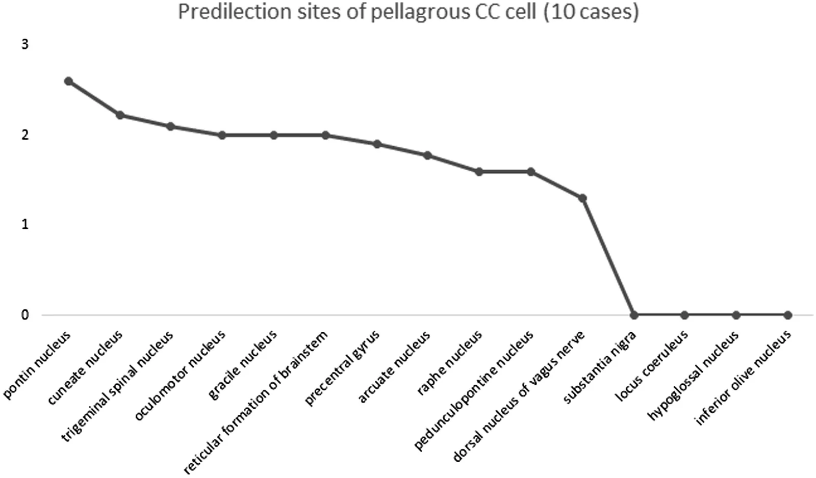
在 10 例糙皮病脑病病例中,CC 细胞常见于中央前回、动眼神经核、中缝核、脑桥核、脑桥脚核、三叉神经脊束核、楔束核、薄束核、弓状核以及脑桥和延髓网状结构(表 2,图 1)。海马、前扣带皮质及额上皮质中未发现 CC 细胞。
CC 细胞的免疫组织化学表现
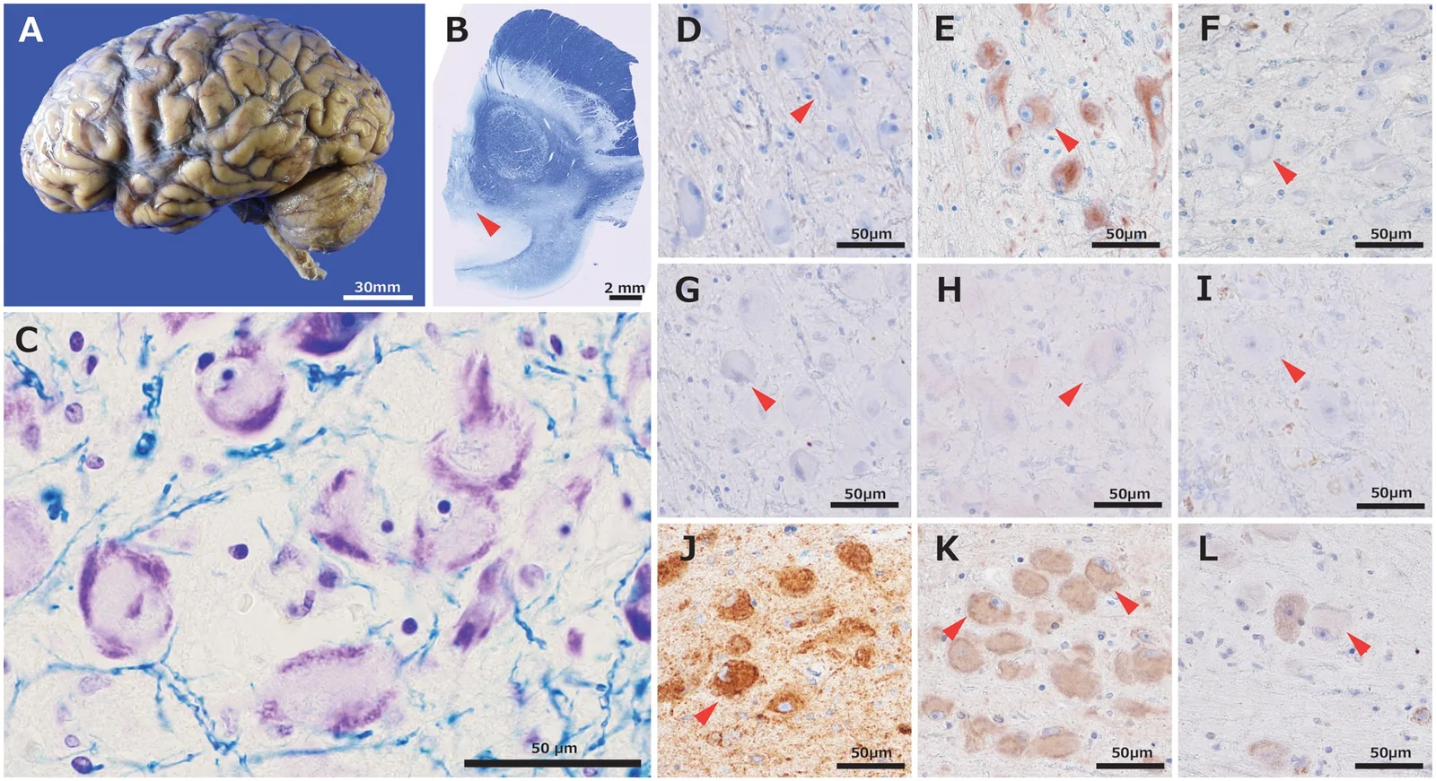
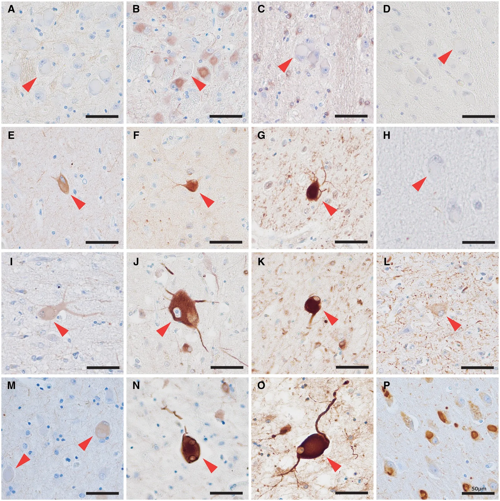
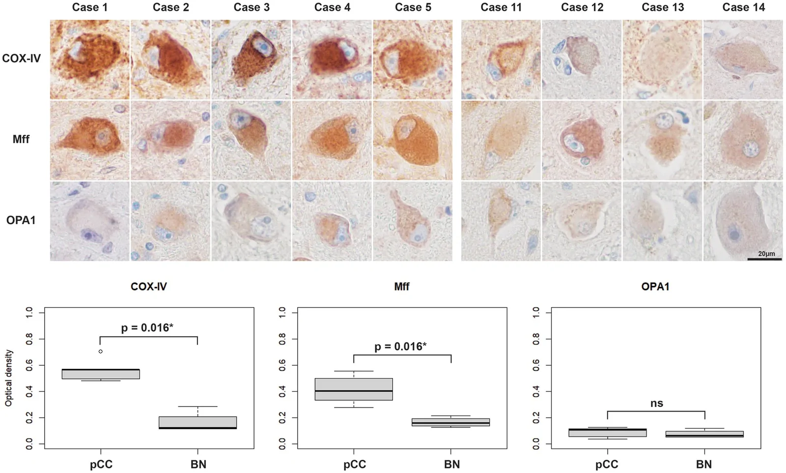
在全部 10 例糙皮病脑病病例中,CC 细胞的 SMI-31、αΒ-crystallin 以及所有接受检查的抗 tau 抗体(AT8、RD3 和 RD4)均呈阴性(表 3,图 2D、F-I,图 3A、C、D)。糙皮病 CC 的 SMI-32 免疫反应性较弱或缺失(表 3,图 2E 和 3B)。轴索损伤病例中的气球样无色神经元对 SMI-31、SMI-32 和 αΒ-crystallin 呈阳性,但抗 tau 抗体呈阴性(表 3,图 3E-H)。CBD 病例中的气球样无色神经元对 SMI-31、SMI-32 和 αΒ-crystallin 呈阳性,AT8 和 RD4 呈中度阳性,但 RD3 呈阴性(表 3,图 3I-L)。CBD 病例的 AT8 免疫染色显示,BN 的染色强度低于神经原纤维缠结。在 CBD 病例中可观察到 SMI-31、32 和 αΒ-crystallin 免疫阳性细胞,尤其见于额上回。PiD 病例中的气球样无色神经元对 SMI-31、SMI-32 和 αΒ-crystallin 呈中度阳性,AT8 和 RD3 呈强阳性,但 RD4 呈阴性(表 3,图 3M-P)。PiD 病例中 SMI-31、32 和 αΒ-crystallin 阳性细胞的数量少于 AT8 和 RD3 阳性细胞。PiD 病例中的 SMI-31、32 和 αΒ-crystallin 阳性细胞往往见于海马周围严重变性的病变部位。对 COX-IV、Mff 和 OPA1 的 DAB 染色强度进行形态计量学定量分析发现,与 BN 相比,发生糙皮病 CC 的胞体具有显著更高的 COX-IV 和 Mff 染色强度(COX-IV:P = .016;Mff:P = .016;Mann–Whitney U 检验;图 4)。
CC 细胞的电镜表现
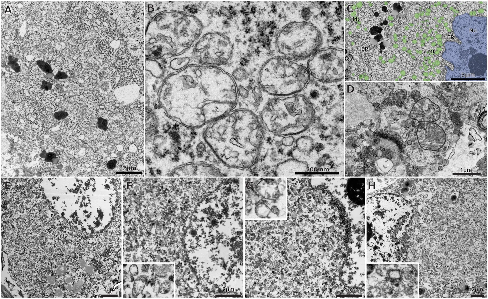
对 5 例糙皮病脑病病例进行超微结构检查发现,出现 CC 的神经元内存在线粒体增生,并伴有线粒体内无定形致密物。部分线粒体可见碎片化,形成小球形结构,同时伴有嵴减少。这些形态改变的严重程度在病例间存在显著差异;其表现从几乎所有线粒体均出现广泛的嵴丢失和碎片化(病例 1、5;图 5A-C 和 H),到虽有线粒体密度增加及无定形致密物,但嵴保存良好且大量桶状线粒体显著的病例(病例 2、3、4;图 5E-G)不等。
代表性病例的临床和神经病理学表现
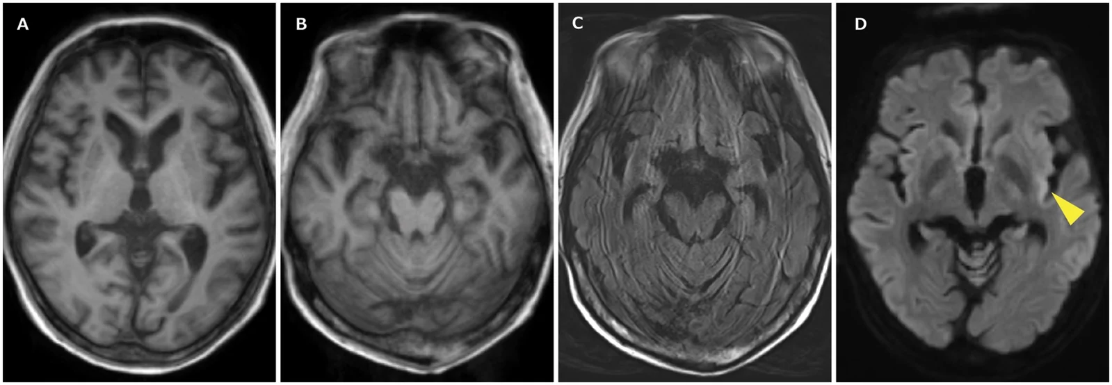
病例 1:患者为男性,死亡时 64 岁。既往有肺结核治疗史。约 43 岁退出工作岗位后,其饮酒量增加,并在 50 多岁时被诊断为酒精使用障碍。他还于 52 岁时被诊断为癫痫。63 岁时肺结核复发,入我院接受治疗。入院时血液检查显示维生素 B1 水平为 4.6 μg/dL,维生素 B3 水平为 5.1 μg/mL。入院当天,患者意识完全清楚。然而,开始使用包括异烟肼在内的抗结核药物后,其意识水平逐渐下降。开始抗结核治疗时即给予了维生素 B 补充剂。住院期间,他出现了持续性意识水平下降、顽固性腹泻及四肢皮肤色素沉着。入院后发生了 3 次全身性惊厥发作。发作后即刻的脑电图显示,以左侧顶叶区域为主的 δ 活动。同时进行的脑 MRI 显示,弥散加权成像中左侧岛叶尾侧信号强度轻度增高(图 6A-D)。尽管给予抗癫痫药物后意识略有改善,但在长达数月的发作间期内,他始终未恢复完全清醒。终末期可见肝酶显著升高。入院 4 个月后,他因脓毒症和多器官衰竭死亡。
尸检显示脑重 1294 g。大体检查未见明显的大脑和小脑萎缩(图 2A)。镜下,在动眼神经核、三叉神经脊束核、薄束核和延髓网状结构中发现 CC 细胞(表 2,图 2C)。CC 细胞的 SMI-31、αΒ-crystallin、AT8、RD3 和 RD4 呈阴性(图 2D、F-I)。CC 细胞的 COX-IV 免疫染色呈强阳性(图 2J)。本例免疫组织化学检查中,CC 细胞的 Mff 染色强度与未发生染色质溶解的细胞相似(图 2K)。相比之下,CC 细胞的 OPA1 染色弱于周围未发生染色质溶解的细胞(图 2L)。左侧岛叶可见 GFAP 阳性的反应性星形胶质细胞(图 S1),与生前癫痫发作后 T2 加权成像高信号区域相对应。年龄相关的神经退行性改变极轻微。透射电镜显示,CC 细胞的神经元胞体内,小圆形线粒体数量显著增多,其嵴减少且形态不成熟(图 5A-C)。此外,游离核糖体数量增多,而粗面内质网减少并移位至胞体周边。胞体内神经丝未见明显增多。在用于分析 CC 细胞的同一切片中,突触前终末和轴索内可识别出嵴保存完好的线粒体(图 5D)。基于这些发现,该患者的病理诊断为糙皮病脑病。
讨论
本研究揭示了以下主要发现。首先,本研究中的糙皮病 CC 神经元常见于特定的脑区,其分布与既往研究结果一致。24 这一发现提示,我们的糙皮病脑病病例可能与既往研究所描述的人群相似。24 其次,本研究中的糙皮病 CC 细胞对 SMI-31、αΒ-crystallin 和 tau 相关抗体(AT8、RD3 和 RD4)呈免疫阴性,而逆行性变性的神经元对 SMI-31、SMI-32、αΒ-crystallin 呈免疫阳性,CBD 或 PiD 中的气球样无色神经元则对 SMI-31、SMI-32、αΒ-crystallin 以及数种 tau 相关抗体呈免疫阳性。这一发现提示,糙皮病 CC 神经元的形成机制与细胞骨架改变无关,且不同于其他病因所致的气球样无色神经元。第三,我们对线粒体标志物的免疫组织化学分析显示,与发生逆行性变性的 BN 相比,出现糙皮病 CC 的神经元具有显著更高的 COX-IV 和 Mff 免疫反应性。尤其是对代表性病例的详细神经病理学分析发现,该例糙皮病 CC 细胞中,与线粒体嵴维持有关的 OPA1 免疫反应强度较低,而 COX-IV 和 Mff 的免疫反应性得以保留。此外,超微结构检查显示,糙皮病 CC 神经元的细胞质内含有大量线粒体及线粒体内无定形致密物。另外,部分线粒体出现碎片化和嵴排列紊乱,尤其见于代表性病例 1。在糙皮病 CC 中,电镜下所见嵴紊乱程度的差异与 OPA1 染色强度的差异一致,后者相较于 BN 未显示出统计学显著的降低。这些发现提示,糙皮病脑病中推测存在的 NAD+ 缺乏所导致的神经元能量不足,可能诱发线粒体碎片化。第四,我们的糙皮病脑病患者不仅包括酒精使用障碍患者,也包括因严重躯体疾病或进展性神经退行性疾病而经口摄入受损的患者。此外,无一病例出现全部经典“4Ds”表现。这些发现揭示了糙皮病脑病患者背景及临床表现的多样性。此外,我们的发现提示,糙皮病脑病可发生于身体储备能力低下的患者,例如严重全身性疾病或终末期神经退行性疾病患者,而它可通过引起意识障碍使这些患者的预后恶化。
糙皮病脑病相关 CC 中的线粒体改变
神经元所需的大部分能量由线粒体提供。30 为维持细胞稳态,线粒体通过融合、分裂、线粒体自噬及转运主动改变其形态。31 线粒体碎片化是细胞周围环境应激因素诱导的一种形态改变。这一过程以线粒体分裂19和嵴结构改变为特征。20,21,23 线粒体碎片化由线粒体去极化20,21及 NAD+ 合成受损所致。16 此外,已知细胞内 ADP/ATP 比值升高会下调轴索内的线粒体转运。32
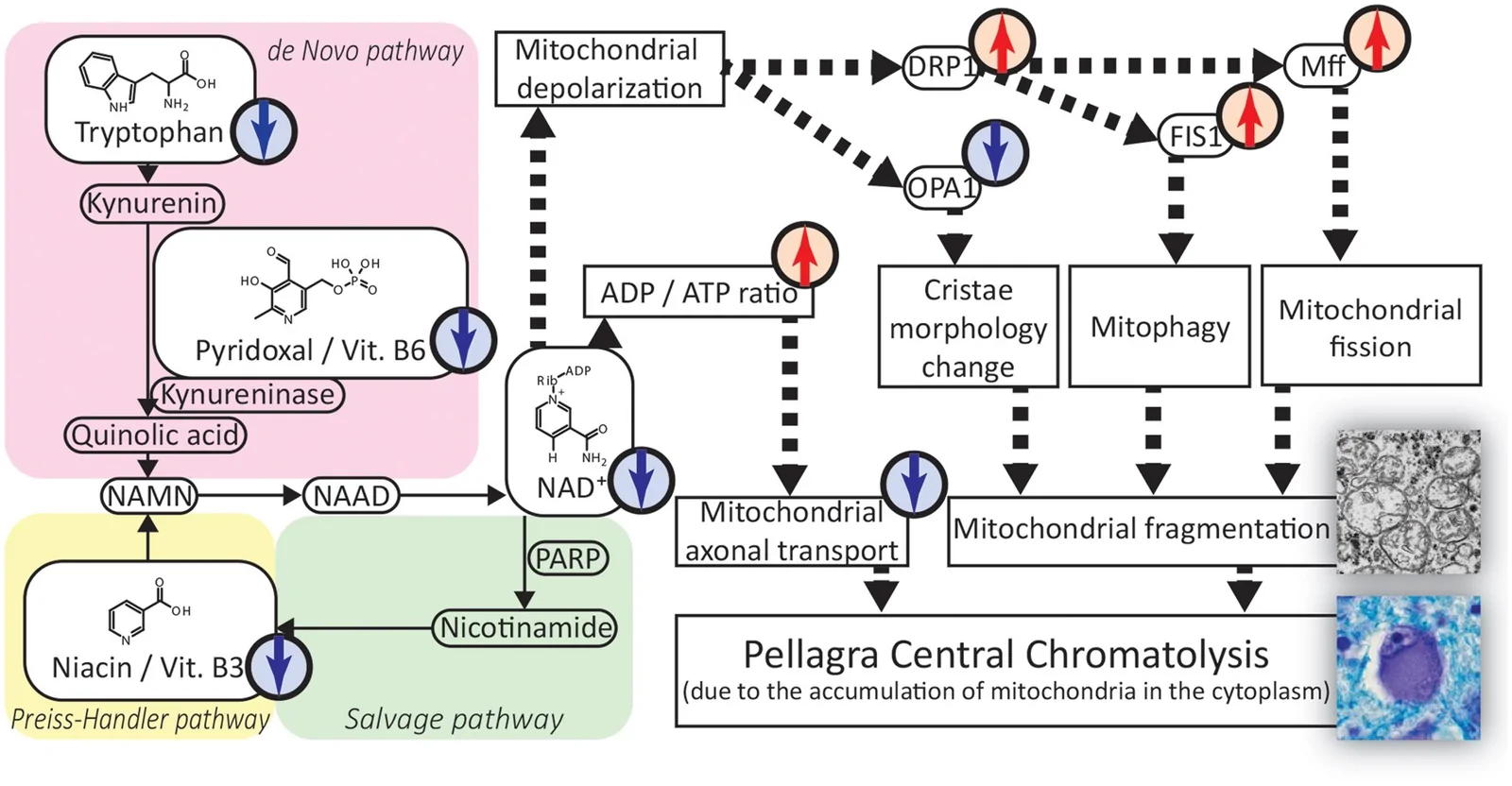
NAD+ 合成受损还可由维生素 B3 和维生素 B6 缺乏引起,前者是 Preiss–Handler 通路的底物,后者是色氨酸–犬尿喹啉酸通路的辅因子。此外,推测糙皮病脑病中的线粒体形态改变源于 NAD+ 缺乏。COX-IV 和 Mff 的免疫组织化学分析显示,出现糙皮病 CC 的神经元内存在线粒体增生(图 4)。在代表性病例中,与未发生染色质溶解的细胞相比,CC 细胞对 COX-IV(图 2J)和 Mff(图 2K)呈强免疫阳性,但 OPA1 免疫反应性降低(图 2L)。超微结构检查显示,细胞质内积聚了小圆形线粒体,其内部可见无定形致密物及紊乱的嵴。既往报道指出,线粒体内无定形致密物是受损细胞的特征性表现,包括遭受缺血性损伤的细胞。33 这些发现支持我们基于既往文献提出的假说(图 7):糙皮病脑病中的 NAD+ 缺乏及细胞能量状态不足,可导致线粒体碎片化以及线粒体在神经元胞体内积聚。线粒体碎片化被认为代表细胞死亡过程中的一个过渡阶段。给予维生素 B3 有望恢复细胞内 NAD+ 水平,可能阻止糙皮病脑病中的细胞死亡进程。这与临床实践中观察到的糙皮病脑病症状经维生素 B3 治疗后消退的现象一致。
本研究中,糙皮病 CC 细胞主要分布于中央前回、动眼神经核、中缝核、脑桥核、脑桥脚核、三叉神经脊束核、楔束核、薄束核、弓状核以及脑桥和延髓网状结构。糙皮病 CC 细胞的分布模式与既往一项糙皮病脑病神经病理学研究的发现一致。24 这些特定解剖区域的选择性受累在各病例中保持一致,不仅提示我们的患者与既往报道的糙皮病脑病人群具有可比性,也提示不同区域对 NAD+ 缺乏的易感性可能存在差异。
糙皮病 CC 细胞与逆行性变性或神经退行性疾病中气球样无色神经元的差异
轴索损伤神经元中逆行性变性的特征已得到充分确立。既往脊髓前根横断动物研究中对运动神经元的观察显示,轴索被切断的运动神经元胞体内总蛋白和 RNA 增多,且增加的蛋白几乎全部位于细胞骨架中。34 一项轴索切断实验研究采用多种神经丝抗体进行免疫组织化学评估,发现横断后胞体内神经丝免疫标记显著增强,同时轴索转运减少,横断部位近端的轴索直径减小。35 这些发现被解释为胞体内神经丝增多,以及其向轴索转位受阻。CBD 和 PiD 等神经退行性疾病中观察到的 BN 也可被神经丝抗体标记,提示其机制与轴索损伤中的逆行性变性相似。26
相比之下,既往一项针对糙皮病脑病 CC 细胞的免疫组织化学研究报道,抗磷酸化神经丝抗体仅呈弱免疫标记。36 本研究中,糙皮病 CC 细胞未被抗磷酸化神经丝抗体或任何抗 tau 抗体免疫组织化学标记。这一结果不同于上述轴索损伤神经元中的逆行性改变,也不同于 CBD 和 PiD 中的 BN。本研究所观察到的糙皮病脑病 CC 细胞的免疫组织化学特征与既往报道一致。36
既往关于轴索损伤中逆行性变性的超微结构研究报道,胞体内神经丝、游离核糖体和线粒体增多。37,38 另一方面,在少数病例报告中,糙皮病 CC 细胞的电镜检查显示,小型线粒体显著增多,游离核糖体轻度增多,但神经丝并未增多。39,40 本研究的电镜表现也以大量含无定形致密物的线粒体、游离核糖体增多及神经丝不明显为特征。这些发现与既往报道的糙皮病 CC 细胞电镜图像一致。因此,可依据电镜特征,尤其是神经丝及线粒体形态,将糙皮病脑病中的 CC 细胞与因轴索损伤而发生逆行性变性的神经元区分开来。
综上,这些免疫组织化学和电镜发现提示,糙皮病脑病 CC 细胞的病变形成机制,与轴索损伤以及 CBD 和 PiD 等神经退行性疾病中的病理机制不同。
糙皮病脑病患者的临床背景
本研究检查的 10 例糙皮病脑病病例中,5 例有酒精使用障碍病史。这一发现与发达国家糙皮病脑病的已知危险因素一致。此外,本研究还包括因进展性神经退行性疾病或严重躯体疾病而经口摄入受损的病例。另外,病例 1 使用了异烟肼,推测这可能是该患者发生糙皮病的促成因素。既往研究提示,长期住院照护的老年人可能存在维生素缺乏风险,41也指出老年人维生素 B3 摄入量低与认知能力下降有关。42,43 因此,老年人及长期照护患者可能存在发生糙皮病脑病的风险。糙皮病脑病常表现为意识障碍。虽然它常与酒精使用障碍及神经性厌食症病例相关,但也可发生于其他脆弱人群,例如老年人或严重躯体疾病患者。此外,糙皮病脑病还可能进一步恶化这些患者的预后。因此,密切的临床观察及及时给予维生素,对于预防糙皮病脑病的发生十分重要。
局限性
本研究存在以下局限性。首先,我们的详细临床病理学分析集中于单个病例(病例 1),其意识障碍可能受到多种混杂因素的影响。例如,其在长达数月的发作间期内意识状态未能完全恢复,可能归因于非惊厥性癫痫持续状态。此外,糙皮病脑病本身也可能加重癫痫。3
另一项局限性涉及分子和遗传学分析。由于无法获得新鲜冷冻脑组织样本,未能进行遗传学或生化分析。因此,无法分析受累区域与线粒体改变相关的基因表达。由此,我们对糙皮病脑病中线粒体反应的讨论基于对既往文献的解读,而非直接来自糙皮病脑病病例样本的基因表达数据。
本次免疫组织化学研究中的轴索损伤病例数量有限。这是由于脑外伤继发轴索损伤后出现无色神经元的时间窗有限,且本机构档案中仅有 1 例包含可观察到、适于研究的无色神经元。我们在该轴索损伤病例中的发现,与既往关于轴索损伤后逆行性变性的研究一致。35,36
用于电镜观察的样本先经甲醛固定,随后再经锇固定。这一固定过程,加上样本可能随时间推移而发生的劣化,可能引入了伪影。因此,不能排除观察到的线粒体膜结构发生了非预期的人为改变。此外,受样本可获得性限制,接受电镜评估的病例数量有限。
结论
糙皮病脑病 CC 细胞的发病机制可能涉及线粒体改变,以及糙皮病脑病中推测存在的 NAD+ 缺乏所致的神经元能量不足。该机制与轴索损伤中观察到的逆行性反应或神经退行性疾病中观察到的气球样细胞有根本区别。我们的发现提示,神经功能障碍可源于原发性细胞代谢改变,而不只是机械性创伤或神经退行性疾病。
在当今发达国家,糙皮病脑病被认为是一种罕见疾病,仅在有限的条件下发生。然而,如果缺乏适当的评估、诊断和治疗,该病可能致命。糙皮病脑病不仅需要在有慢性酒精使用障碍或神经性厌食症病史的患者中受到重视,也需要在因严重躯体疾病而经口摄入困难的患者以及接受抗结核药等药物治疗的患者中受到重视。糙皮病脑病仍是持续性意识障碍患者的一项重要鉴别诊断。
通过研究较既往报道更大规模的人类糙皮病脑病病例群体,36我们为糙皮病 CC 神经元与其他病因所致的气球样无色神经元提供了有力的免疫组织化学比较。此外,据我们所知,本研究首次通过免疫组织化学和电镜证据表明,依据临床病史、病变分布及细胞骨架标志物缺失而诊断的多个糙皮病脑病病例,均一致表现出线粒体增生标志物升高及线粒体碎片化。
需要进一步积累病例并开展研究,以阐明有无意识障碍等临床表现与糙皮病 CC 病变组织病理学分布之间的关系。
- Background: neuronal central chromatolysis (CC) is the histological hallmark of pellagra encephalopathy, a vitamin-deficiency disorder; pellagrous CC neurons resemble ballooned achromatic neurons of retrograde degeneration and neurodegenerative disease, but the mechanisms differ in ways that were unclear.
- Material: 10 autopsy cases of pellagra encephalopathy (5 alcohol use disorder, 2 FTLD-TDP, 3 epilepsy, overlapping), compared immunohistochemically with axonal injury, CBD and Pick disease controls; electron microscopy in 5 cases and DAB optical-density quantification.
- Diagnostic point: pellagrous CC cells were negative for SMI-31, αB-crystallin and AT8/RD3/RD4, with weak or absent SMI-32, whereas ballooned neurons in axonal injury and neurodegeneration were positive for cytoskeletal markers. CC cells clustered in precentral gyrus, oculomotor, raphe and pontine nuclei and medullary reticular formation, not the hippocampus.
- Mechanistic clue: CC cells showed stronger COX-IV and mitochondrial fission factor (Mff) immunoreactivity; EM showed mitochondrial proliferation, intramitochondrial amorphous densities, fragmentation into small spheres and loss of cristae — consistent with NAD-deficiency energy failure driving mitochondrial fragmentation. Four of 10 patients had prolonged impaired consciousness distinct from alcohol withdrawal delirium. Figures 1–7 and Tables 1–3 included.
Scope: PMC open-access full text (CC BY 4.0) — abstract, introduction, methods, results, discussion and conclusion, Figures 1–7 and Tables 1–3. Acknowledgments, funding, conflicts, supplementary material and references are not included.
Abstract
Neuronal central chromatolysis (CC) is the histopathological hallmark of pellagra encephalopathy, a neurological deficit resulting from vitamin deficiencies. Pellagrous CC neurons are morphologically similar to ballooned achromatic neurons in other conditions but the distinct pathomechanisms remain unclear. We performed a clinico-neuropathological analysis of 10 autopsy cases of pellagra encephalopathy. The pellagra encephalopathy cases were immunohistochemically compared with disease controls, including cases of axonal injury and neurodegenerative diseases. Electron microscopic evaluation and immunohistochemical examinations targeting mitochondrial fragmentation were performed for a representative case. Four of 10 pellagra encephalopathy patients exhibited prolonged impairment of consciousness distinguishable from alcohol withdrawal delirium. Pellagrous CC neurons were negative for cytoskeletal markers whereas ballooned achromatic neurons in the disease control cases were positive. Immunohistochemical analysis of mitochondrial markers revealed that CC neurons exhibited more intense immunoreactivity for COX-IV and mitochondrial fissure factor compared to the disease controls. Transmission electron microscopy of these CC neurons revealed a marked increase in the mitochondria with amorphous densities. These findings indicate that the pathomechanism of pellagrous CC is distinct from that of the ballooned achromatic neurons of other etiologies. Mitochondrial alterations in pellagrous CC neurons suggest that neuronal energy deficits resulting from nicotinamide adenine dinucleotide deficiency induce mitochondrial fragmentation.
Introduction
Pellagra is caused by deficiencies in vitamin B3 (niacin), B6 (pyridoxal), and tryptophan. Clinically, it is characterized by the “4Ds”: gastrointestinal symptoms such as diarrhea, dermatitis, dementia, and (if untreated) death.1 Pellagra encephalopathy is a neurological manifestation consisting of disturbances of consciousness, hallucinations, delusions, and convulsions in addition to dementia.2,3 Although pellagra is relatively infrequent in developed countries, it can be caused by low nutritional status, particularly chronic alcohol abuse,2–4 anorexia nervosa,5,6 and corn diet.1 Pellagra can also be caused by certain medications, such as azathioprine,7 which inhibits the synthetic pathway of nicotinamide adenine dinucleotide (NAD+) from tryptophan, and isoniazid,8,9 which is a vitamin B3 analogue that reduces the vitamin B6 required for NAD+ synthesis.
Alcohol dependence syndrome, a common background factor for pellagra in developed countries, is associated with alcohol withdrawal symptoms.10 Alcohol withdrawal delirium after drinking cessation usually recovers within a few days with appropriate treatment.11 In contrast, pellagra encephalopathy is a potentially fatal disease if left untreated and has important clinical significance even today in terms of its possible involvement in prolonged consciousness disturbances in cases of alcohol use disorder. To more effectively differentiate alcohol withdrawal delirium from pellagra encephalopathy presenting with impaired consciousness, a thorough understanding of the pathomechanisms of pellagra encephalopathy is essential. Such knowledge would elucidate why specific lesions develop in characteristic locations within the central nervous system.
The pathophysiological mechanisms of pellagra encephalopathy are postulated to involve abnormalities in several pathways, including the NAD+ synthesis pathway, the tryptophan-kynurenic acid pathway, the mitochondrial ATP generation related pathway, the Poly (ADP–ribose) polymerase (PARP) pathway, the BDNF–TrkB axis, and a genetic background with vulnerability to niacin deficiency.12 However, much remains unknown. One of the reasons why the pathogenesis of pellagra encephalopathy is not fully understood is the difficulty in establishing animal models of pellagra encephalopathy. This difficulty arises mainly because laboratory mice and rats can synthesize NAD+ from tryptophan dozens of times more efficiently than humans.13 Although a few animal models of pellagra have been reported,14,15 to the best of our knowledge, none has specifically replicated pellagra encephalopathy. Consequently, to elucidate the pathomechanism of pellagra encephalopathy, histopathological studies of human patients are essential.
In pellagra encephalopathy, NAD+ deficiency is presumed to occur. NAD+ deficiency also occurs in other brain injuries, such as cerebral infarction,16 where mitochondrial fragmentation is observed in histological sections.17 Mitochondrial fragmentation is a cellular stress response through mitochondrial fissure factor (Mff)-mediated mitochondrial fission.18,19 Mitochondrial fragmentation has also been observed with agents that cause mitochondrial depolarization in cultured cells, a process that is known to be accompanied by changes in cristae structure and the dynamics of dynamin-like 120k-Da protein (OPA1), which is required for its maintenance.20–23 However, the involvement of mitochondrial fragmentation in pellagra encephalopathy remains unclear.
Pellagra encephalopathy is known histopathologically for central chromatolysis (CC), which is typically observed in pontine nuclei and other brainstem nuclei. Pellagrous CC is a neuronal histopathological finding characterized by nuclear marginalization, chromatin displacement, and cell body swelling.24 Morphologically similar findings, often referred to as achromatic or ballooned neurons (BN), are observed in retrograde changes following axonal injury,25 and in neurodegenerative diseases such as corticobasal degeneration (CBD)26 and Pick disease (PiD).27 Currently, clear criteria to differentiate these morphologically similar findings are not well established. The differences in the mechanisms of lesion formation under each of these conditions with similar histopathological features also remain unclear. In particular, the potential involvement of NAD+ deficiency and mitochondrial fragmentation in the development of pellagrous CC remains unknown.
In this study, we present a detailed clinico-neuropathological study of pellagra CC cells, from the autopsy brains of patients with pellagra encephalopathy. The aims of this study were to elucidate the neuropathological evidence supporting an association between NAD+ deficiency and pellagrous CC, and to differentiate pellagrous CC from ballooned achromatic neurons of other etiologies.
Methods
Participants
Ten cases of pellagra encephalopathy were registered in the autopsy archives of Tokyo Metropolitan Institute of Medical Science and Tokyo Metropolitan Matsuzawa Hospital (Table 1). Cases of pellagra encephalopathy were defined as those with a clinical history of a nutritional disorders and neuropathological findings of CC cells in the central nervous system. Each case of pellagra encephalopathy was retrospectively reviewed via patient clinical records. For comparison, immunohistochemical findings of 1 axonal injury case, 3 CBD cases, and 3 PiD cases, all of which exhibited BN, were analyzed and compared with those of pellagrous CC. Three out of 10 pellagra encephalopathy cases have been previously reported in a clinico-pathological study.28
| Case no. | 1 | 2 | 3 | 4 | 5 | 6 | 7 | 8 | 9 | 10 |
|---|---|---|---|---|---|---|---|---|---|---|
| Sex | M | M | M | M | M | F | M | M | F | M |
| Age (years) | 64 | 59 | 55 | 71 | 54 | 72 | 63 | 57 | Unknown | 72 |
| Clinical diagnosis | Alcohol use disorder, epilepsy | Alcohol use disorder | Alcohol use disorder | Alcohol use disorder | Alcohol use disorder | bvFTD | bvFTD | Epilepsy | Epilepsy, intellectual disability | Schizophrenia |
| Prolonged disturbance of consciousness | + | + | + | + | − | Difficult to evaluate | Difficult to evaluate | − | − | − |
| Pellagra related clinical symptoms | Diarrhea, skin pigmentation | − | − | Hypophagia, diarrhea | Diarrhea, nausea | Hypophagia, nausea, dermatitis | Hypophagia | Hypophagia, nausea, diarrhea | − | Hypophagia |
| Nutritional administration status | 3 months of nasogastric tube nutrition | Reduced food intake due to alcohol disorder | Reduced food intake due to alcohol disorder | Reduced food intake due to alcohol disorder | Reduced food intake due to alcohol disorder | Several years of nasogastric tube nutrition | A year of nasogastric tube nutrition | Severe constipation and insufficient food intake | A month of central venous nutrition | 6 months of central venous nutrition |
| Comorbid neuropathological diagnosis | − | − | Subdural hematoma | Obsolete infarction | Obsolete infarction | FTLD-TDP | FTLD-TDP | Obsolete infarction | Cerebellar atrophy | Obsolete infarction |
Clinical and demographic data of 10 patients with pellagra encephalopathy. Seven patients presented with some of the classic “4Ds” symptoms of pellagra: gastrointestinal symptoms, dermatitis, dementia, and, in severe cases, death. Four patients had a clinical history of prolonged disturbance of consciousness.
Abbreviations: bvFTD, behavioral variant frontotemporal dementia.
Histology and immunohistochemistry
Brain tissue was fixed in 10% formaldehyde and embedded in paraffin. Sections (8-μm thick) were cut from the hippocampus, precentral gyrus, superior frontal cortex, orbitofrontal cortex, midbrain, pons, and medulla oblongata (Table 2). The sections were stained with hematoxylin and eosin (H&E) and Klüver-Barrera (KB) stains. Immunohistochemical examination was performed using the following primary antibodies: anti-phosphorylated neurofilament (SMI-31; monoclonal, BioLegend), anti-non-phosphorylated neurofilament (SMI-32; monoclonal, BioLegend), anti-α-crystallin Β chain (αΒ-crystallin; monoclonal, Stress Marq), anti-phosphorylated tau (AT8 [ps202, pt205]; monoclonal, Invitrogen), anti-3-repeat tau (RD3; monoclonal, Sigma–Aldrich), anti-4-repeat tau (RD4; monoclonal, Sigma–Aldrich), anti-α-synuclein (pSyn#64 [ps129]; monoclonal, Abcam), anti-phosphorylated TDP-43 (ps409-410; monoclonal, Cosmo Bio), anti-glial fibrillary acidic protein (GFAP; monoclonal, PROGEN), anti-cytochrome c oxidase (COX-IV; monoclonal, Cell Signaling), anti-Mff ( polyclonal, Proteintech), and anti-dynamin-like 120k-Da protein (OPA1; polyclonal, Proteintech). Primary antibody labeling was visualized using DAB horseradish peroxidase polymer detection kits (Nichirei Biosciences). The specific brain regions examined by immunohistochemistry for each case are detailed in Table 3.
| Case no. | 1 | 2 | 3 | 4 | 5 | 6 | 7 | 8 | 9 | 10 |
|---|---|---|---|---|---|---|---|---|---|---|
| Precentral gyrus | 1 | 3 | 3 | 3 | 2 | 1 | 2 | 2 | 0 | 2 |
| Oculomotor nucleus | 3 | 2 | – | 1 | 2 | 2 | 3 | 1 | – | – |
| Substantia nigra | 0 | 0 | 0 | 0 | 0 | 0 | 0 | 0 | 0 | 0 |
| Locus coeruleus | 0 | 0 | 0 | 0 | 0 | 0 | 0 | 0 | 0 | 0 |
| Raphe nucleus | 1 | 1 | 3 | 0 | 1 | 2 | 2 | 2 | 3 | 1 |
| Pontin nucleus | 0 | 3 | 3 | 3 | 3 | 3 | 3 | 3 | 3 | 2 |
| Pedunculopontine nucleus | 0 | 3 | 3 | 3 | 2 | 0 | 3 | 0 | 2 | 0 |
| Hypoglossal nucleus | 0 | 0 | 0 | 0 | 0 | – | 0 | 0 | 0 | 0 |
| Dorsal nucleus of vagus nerve | 0 | 1 | 2 | 1 | 2 | 1 | 0 | 2 | 2 | 2 |
| Cuneate nucleus | 2 | 2 | 3 | 3 | 3 | 2 | – | 2 | 1 | 2 |
| Gracile nucleus | 2 | 3 | 3 | – | 2 | 2 | – | – | 0 | – |
| Trigeminal spinal nucleus | 2 | 2 | 3 | 2 | 2 | 3 | 2 | 2 | 1 | 2 |
| Inferior olive nucleus | 0 | 0 | 0 | 0 | 0 | 0 | 0 | 0 | 0 | 0 |
| Reticular formation of pons and medulla oblongata | 1 | 3 | 3 | 3 | 2 | 2 | 2 | 1 | – | 1 |
| Arcuate nucleus | 0 | 2 | 2 | 3 | 2 | 2 | – | 3 | 0 | 2 |
Distribution of central chromatolysis (CC) cells in 10 cases of pellagra encephalopathy.
Abbreviations: 3, severe (almost all cells are chromatolytic); 2, moderate (obvious chromatolytic cells are present); 1, slightly (a few cells are suspected to be chromatolytic); 0, absent; (–), not assessed.
| Pellagra | Axonal injury | CBD | PiD | ||||||||||||||
|---|---|---|---|---|---|---|---|---|---|---|---|---|---|---|---|---|---|
| (with alcohol disorder) | (without alcohol disorder) | ||||||||||||||||
| Case no. | 1 | 2 | 3 | 4 | 5 | 6 | 7 | 8 | 9 | 10 | 11 | 12 | 13 | 14 | 15 | 16 | 17 |
| Evaluated region | ON | PN | PN | PN | PN | ON | PN | PN | PN | PN | OFG | SFG | SFG | SFG | CA | CA | CA |
| Antibodies | |||||||||||||||||
| SMI-31 | 0 | 0 | 0 | 0 | 0 | 0 | 0 | 0 | 0 | 0 | 2 | 2 | 2 | 2 | 0 | 1 | 2 |
| SMI-32 | 1 | 1 | 1 | 1 | 1 | 1 | 1 | 1 | 1 | 1 | 2 | 2 | 2 | 2 | – | 2 | 2 |
| αΒ-crystallin | 0 | 0 | 0 | 0 | 0 | 0 | 0 | 0 | 0 | 0 | 2 | 2 | 2 | 2 | – | 2 | 2 |
| AT8 | 0 | 0 | 0 | 0 | 0 | 0 | 0 | 0 | 0 | 0 | 0 | 1 | 1 | 1 | 2 | 2 | 2 |
| RD3 | 0 | 0 | 0 | 0 | 0 | 0 | 0 | 0 | 0 | 0 | 0 | 0 | 0 | 0 | 2 | 2 | 2 |
| RD4 | 0 | 0 | 0 | 0 | 0 | 0 | 0 | 0 | 0 | 0 | 0 | 1 | 1 | 1 | 0 | 0 | 0 |
| COX-IV | 2 | 2 | 2 | 2 | 2 | 2 | 2 | 2 | 2 | 2 | 1 | 1 | 1 | 1 | – | 1 | 1 |
| Mff | 2 | 2 | 2 | 2 | 2 | 2 | 2 | 2 | 2 | 2 | 1 | 2 | 1 | 1 | – | 0 | 1 |
| OPA1 | 0 | 1 | 0 | 1 | 1 | 1 | 1 | 1 | 1 | 1 | 1 | 1 | 1 | 1 | – | 0 | 0 |
Immunohistochemical findings in affected neurons from cases of pellagra encephalopathy, axonal injury, CBD, and PiD. Staining intensity in the affected neurons was graded on the following scale: 2; strongly positive. 1; weakly positive. 0; negative. (–); not done.
Abbreviations: CA, hippocampus; CBD, corticobasal degeneration; Mff, mitochondrial fissure factor; OFG, orbitofrontal gyrus; ON, oculomotor nucleus; PiD, Pick disease; PN; pontine nucleus; SFG, superior frontal gyrus.
DAB staining optical density analysis and statistical methods
Statistical comparisons of the optical density of DAB staining were performed between the CC from 5 cases of alcohol use disorder-associated pellagra encephalopathy (Cases 1 to 5) and the BN from 4 cases of retrograde degeneration (Cases 11 to 14). The regions of interest for optical density measurement were delineated using Fiji (ImageJ version 1.54f),29 focusing on the cytoplasm of the swollen cells. The anatomical location of the cells targeted for optical density measurement corresponds to the regions evaluated for mitochondrial markers (Table 3). Statistical analysis was conducted using the Mann–Whitney U test in R (version 4.4.3; https://www.R-project.org/).
Transmission electron microscopic examination
Brain tissue, which was fixed in 10% formaldehyde (Case 1; midbrain, Cases 2 to 5; pons), was refixed with 1% osmium tetroxide and embedded in resin. Ultrathin sections (70-nm thick) were cut from the lesions in the oculomotor nucleus. Light microscopic observation and identification of CC cells in sequential sections were performed prior to the preparation of ultrathin sections. The prepared sections were analyzed by transmission electron microscopy (JEM-1400; JEOL).
Results
Clinical findings in pellagra encephalopathy
The 10 pellagra encephalopathy patients included 5 with alcohol use disorders (Cases 1 to 5), 2 with neurodegenerative diseases (frontotemporal lobar degeneration with TAR DNA-binding protein 43; FTLD-TDP) (Cases 6 and 7), and 3 with epilepsy (Cases 1, 8, and 9) (Table 1). One patient (Case 1) was receiving treatment with antitubercular medications, including isoniazid. Seven patients exhibited gastrointestinal symptoms (Cases 1, 4, 5, 6, 7, 8, and 10); 2 exhibited skin disorders (Cases 1, 5). Prolonged disturbance of consciousness was observed in 4 patients (Cases 1, 2, 3, 4). None of the patients presented with all of the classic “4Ds.” Regarding nutritional status, 5 patients (Cases 1, 5, 6, 8, and 9) had received nasogastric or central venous nutrition for several months or longer. Four patients (Cases 2 to 5) had a markedly reduced food intake due to severe alcohol use disorder, and one patient (Case 9) had severe constipation and chronically reduced food intake.
Distribution of CC cells in pellagra encephalopathy
CC cells were frequently observed in the precentral gyrus, oculomotor nucleus, raphe nucleus, pontine nucleus, pedunculopontine nucleus, trigeminal spinal nucleus, cuneate nucleus, gracile nucleus, arcuate nucleus, and reticular formation of the pons and medulla oblongata in 10 cases of pellagra encephalopathy (Table 2, Figure 1). No CC cells were found in the hippocampus, anterior cingulate cortex, or superior frontal cortex.
Immunohistochemical findings in CC cells
In all 10 pellagra encephalopathy cases, the CC cells were negative for SMI-31, αΒ-crystallin, and all anti-tau antibodies examined (AT8, RD3, and RD4) (Table 3, Figure 2D, F-I, Figure 3A, C, D). SMI-32 immunoreactivity was weak or absent in the pellagrous CC (Table 3, Figures 2E and 3B). Ballooned achromatic neurons in the axonal injury case were positive for SMI-31, SMI-32, and αΒ-crystallin but negative for anti-tau antibodies (Table 3, Figure 3E-H). Ballooned achromatic neurons in the CBD cases were positive for SMI-31, SMI-32, and αΒ-crystallin, and moderately positive for AT8 and RD4, but negative for RD3 (Table 3, Figure 3I-L). AT8 immunostaining in CBD cases revealed that BN were less intensely stained than neurofibrillary tangles. In CBD cases, SMI-31, 32, and αΒ-crystallin-immunopositive cells were observed, particularly in the superior frontal gyrus. Ballooned achromatic neurons in the PiD cases were moderately positive for SMI-31, SMI-32, and αΒ-crystallin, and strongly positive for AT8 and RD3, but negative for RD4 (Table 3, Figure 3M-P). The number of SMI-31, 32, and αΒ-crystallin-positive cells in the PiD cases was lower than that of AT8- and RD3-positive cells. SMI-31, 32, and αΒ-crystallin-positive cells in the PiD cases tended to be observed in severely degenerated lesions around the hippocampus. Morphometrical quantitative analysis of DAB staining intensity for COX-IV, Mff, and OPA1 revealed that cell bodies with pellagrous CC exhibited significantly higher staining intensity for COX-IV and Mff compared to BNs (COX-IV: P = .016; Mff: P = .016; Mann–Whitney U test; Figure 4).
Electron microscopy findings in CC cells
Ultrastructural examination of the 5 pellagra encephalopathy cases revealed mitochondrial proliferation within neurons exhibiting CC, along with the presence of intramitochondrial amorphous densities. In some mitochondria, fragmentation into small spherical structures was observed, accompanied by a reduction in cristae. The severity of these morphological alterations varied significantly between cases; findings ranged from widespread cristae loss and fragmentation in nearly all mitochondria (Cases 1, 5; Figure 5A-C, and H) to cases where, despite increased mitochondrial density and amorphous densities, cristae were well-preserved and numerous barrel-shaped mitochondria were prominent (Cases 2, 3, 4; Figure 5E-G).
Clinical and neuropathological findings in a representative case
Case 1: The patient was a male who died at 64 years of age. He had a medical history of treatment for pulmonary tuberculosis. His alcohol consumption increased after he left the workforce at approximately 43 years of age, and he was diagnosed with alcohol use disorder in his 50s. He was also diagnosed with epilepsy at the age of 52 years. At 63 years of age, his pulmonary tuberculosis recurred, and he was admitted to our institution for treatment. Blood tests on admission revealed a vitamin B1 level of 4.6 μg/dL and a vitamin B3 level of 5.1 μg/mL. On the day of admission, the patient was fully conscious. However, his level of consciousness gradually deteriorated after initiation of anti-tuberculosis medication, including isoniazid. Vitamin B supplements were administered at the start of his anti-tuberculosis treatment. During his admission, he experienced a prolonged decline in consciousness, intractable diarrhea, and skin pigmentation of his extremities. He experienced generalized convulsive seizures 3 times post-admission. An electroencephalogram immediately after the seizure revealed delta activity predominantly in the left parietal region. A brain MRI performed at the same time revealed a mildly increased signal intensity in the diffusion-weighted image of the left caudal insula (Figure 6A-D). Although his consciousness improved slightly after administration of anti-epileptic medication, he did not regain full consciousness during the interictal phase over several months. Markedly elevated liver enzymes were observed in the terminal phase. He died 4 months after admission from sepsis and multiple organ failure.
Post-mortem examination revealed that the brain weighed 1294 g. Macroscopically, atrophy of the cerebrum and cerebellum was not evident (Figure 2A). Microscopically, CC cells were found in the oculomotor nucleus, trigeminal spinal nucleus, gracile nucleus, and reticular formation of the medulla oblongata (Table 2, Figure 2C). The CC cells were negative for SMI-31, αΒ-crystallin, AT8, RD3, and RD4 (Figure 2D, F-I). CC cells were intensely immunopositive for COX-IV (Figure 2J). In this case, immunohistochemically, CC cells exhibited Mff staining intensity similar to that of non-chromatolytic cells (Figure 2K). In contrast, CC cells demonstrated weaker staining for OPA1 compared to the surrounding non-chromatolytic cells (Figure 2L). GFAP-positive reactive astrocytes were observed in the left insula (Figure S1), corresponding to the region of premortem postictal T2-weighted hyperintensity. Age-related neurodegenerative changes were minimal. Transmission electron microscopy revealed a marked increase in the number of small round mitochondria with reduced and immature cristae morphology in the neuronal cell bodies of CC cells (Figure 5A-C). Additionally, an increase in the number of free ribosomes was observed, while the rough endoplasmic reticulum was reduced and displaced to the periphery of the cell body. An increase in neurofilaments in the cell body was not evident. In the same sections used for the analysis of CC cells, mitochondria with preserved cristae were identified within presynaptic terminals and axons (Figure 5D). Based on these findings, this patient was pathologically diagnosed with pellagra encephalopathy.
Discussion
This study revealed the following key findings. First, the pellagrous CC neurons in our study were frequently observed in the specific brain distribution consistent with previous study findings.24 This finding suggests that our pellagra encephalopathy cases were likely a population similar to those described in the previous study.24 Second, the pellagrous CC cells in our study were immunonegative for SMI-31, αΒ-crystallin, and the tau-related antibodies (AT8, RD3, and RD4), while retrograde degenerated neurons were immunopositive for SMI-31, SMI-32, αΒ-crystallin and ballooned achromatic neurons in CBD or PiD were immunopositive for SMI-31, SMI-32, αΒ-crystallin, and several tau-related antibodies. This finding suggests that the formation mechanism of pellagrous CC neurons is unrelated to cytoskeletal alterations and is distinct from that of ballooned achromatic neurons with other etiologies. Third, our immunohistochemical analysis of mitochondrial markers revealed that neurons exhibiting pellagrous CC possessed significantly higher immunoreactivity for COX-IV and Mff compared to BNs undergoing retrograde degeneration. Especially, our detailed neuropathological analysis for the representative case revealed that the pellagrous CC cells in this case had lower intensity of immunoreactivity for OPA1 which is associated with mitochondrial cristae maintenance, while COX-IV and Mff immunoreactivity was preserved. Furthermore, ultrastructural examination revealed that the cytoplasm of pellagrous CC neurons contained numerous mitochondria and intramitochondrial amorphous densities. Additionally, some of these mitochondria exhibited fragmentation and disorganized cristae, particularly in Case 1, a representative case. In pellagrous CC, the variability in cristae disorganization observed under electron microscopy is consistent with the variability in OPA1 staining intensity, which showed no statistically significant reduction relative to BNs. These findings suggest that neuronal energy deficits resulting from NAD+ deficiency postulated in pellagra encephalopathy may induce mitochondrial fragmentation. Fourth, our pellagra encephalopathy patients included not only alcohol use disorders but also those with impaired oral intake due to severe physical illness or progressive neurodegenerative disease. In addition, none of our cases presented with all classic “4Ds.” These findings reveal the diversity of patient backgrounds and clinical manifestations in pellagra encephalopathy. Furthermore, our findings suggest that pellagra encephalopathy can occur in patients with low physical reserve, such as those with severe systemic diseases or end-stage neurodegenerative diseases, although it can worsen their prognosis by causing impaired consciousness.
Mitochondrial alterations in pellagra encephalopathy-associated CC
The majority of the energy required by neurons is supplied by mitochondria.30 To maintain cellular homeostasis, mitochondria actively change their shape via fusion, fission, mitophagy, and transport.31 Mitochondrial fragmentation is a morphological change induced by environmental stressors surrounding the cell. This process is characterized by mitochondrial fission19 and alteration of the cristae structure.20,21,23 Mitochondrial fragmentation results from mitochondrial depolarization20,21 and impairment of NAD+ synthesis.16 Furthermore, an increased intracellular ADP/ATP ratio is known to downregulate mitochondrial transport in axons.32
Impairment of NAD+ synthesis can also result from a deficiency of vitamin B3, a substrate of the Preiss–Handler pathway, and vitamin B6, a cofactor in the tryptophan–kynurenic acid pathway. Furthermore, mitochondrial morphological alterations are hypothesized to result from NAD+ deficiency in pellagra encephalopathy. Immunohistochemical analysis of COX-IV and Mff demonstrated mitochondrial proliferation in neurons exhibiting pellagrous CC (Figure 4). In the representative case, CC cells were intensely immunopositive for COX-IV (Figure 2J) and Mff (Figure 2K), but reduced OPA1 immunoreactivity (Figure 2L), compared to non-chromatolytic cells. Ultrastructural findings revealed an accumulation of small, round mitochondria with intramitochondrial amorphous densities and disorganized cristae in the cytoplasm. Intramitochondrial amorphous densities have been reported as a characteristic finding in injured cells, including those affected by ischemic insult.33 These findings support our hypothesis, based on the previous literature (Figure 7), that NAD+ deficiency and insufficient cellular energy status in pellagra encephalopathy can cause mitochondrial fragmentation and the accumulation of mitochondria in the neuronal cell body. Mitochondrial fragmentation is thought to represent a transitional period in the process of cell death. The administration of vitamin B3, which is expected to restore cellular NAD+ levels, may halt the process of cell death in pellagra encephalopathy. This is consistent with the clinical observation that symptoms of pellagra encephalopathy resolve with vitamin B3 treatment in clinical settings.
In the present study, pellagrous CC cells were predominantly found in the precentral gyrus, oculomotor nucleus, raphe nucleus, pontine nucleus, pedunculopontine nucleus, trigeminal spinal nucleus, cuneate nucleus, gracile nucleus, arcuate nucleus, and reticular formation of the pons and medulla oblongata. The distribution pattern of pellagra CC cells is consistent with findings from a previous neuropathological study of pellagra encephalopathy.24 The preferential involvement of these specific anatomical regions, which remains consistent across cases, not only suggests that our patients are comparable to previously reported populations of pellagra encephalopathy but also implies the possibility of regional differences in vulnerability to NAD+ deficiency.
Differences between pellagrous CC cells and ballooned achromatic neurons in retrograde degeneration or neurodegenerative diseases
The characteristics of retrograde degeneration in neurons with axonal injury have been well-established. Previously reported observations of motor neurons in animal studies of spinal cord anterior root transection revealed an increase in total protein and RNA in the cell bodies of axotomized motor neurons, with almost all of the increased protein being in the cytoskeleton.34 A study of axotomy experiments in which immunohistochemical evaluation was performed using various neurofilament antibodies revealed that after transection, the cell body showed a marked enhancement of neurofilament immunolabeling, while axonal transport was reduced and the axonal diameter proximal to the transection site was decreased.35 These findings are interpreted as an increase in neurofilaments in the cell body and a block in their translocation to the axon. BN observed in neurodegenerative diseases such as CBD and PiD were also labelled with neurofilament antibodies, suggesting a mechanism similar to retrograde degeneration in axonal injury.26
In contrast, a previous immunohistochemical study in CC cells with pellagra encephalopathy reported weak immunolabeling with anti-phosphorylated neurofilament antibodies.36 In the present study, pellagrous CC cells were not immunohistochemically labelled with anti-phosphorylated neurofilament antibodies or with any of the anti-tau antibodies. This result differs from the aforementioned retrograde changes observed in neurons with axonal injury, as well as BN in the CBD and PiD. The immunohistochemical features of CC cells in pellagra encephalopathy observed in the present study were consistent with previous reports.36
Previous ultrastructural studies on retrograde degeneration in axonal injury reported increases in neurofilaments, free ribosomes, and mitochondria in the cell bodies.37,38 On the other hand, in a few case reports, electron microscopic examination of pellagrous CC cells revealed a marked increase in small mitochondria and a mild increase in free ribosomes but no increase in neurofilaments.39,40 The electron microscopic findings in our study were also characterized by numerous mitochondria with amorphous densities, an increase in free ribosomes, and inconspicuous neurofilaments. These findings were consistent with previously reported electron microscopy images of pellagrous CC cells. Therefore, CC cells in pellagra encephalopathy can be distinguished from neurons undergoing retrograde degeneration due to axonal injury on the basis of electron microscopic features, particularly neurofilaments and mitochondrial morphology.
Taken together, these immunohistochemical and electron microscopic findings suggest that CC cells in pellagra encephalopathy have a different pathological mechanism of lesion formation from those involved in axonal injury and neurodegenerative diseases such as CBD and PiD.
Clinical background of the pellagra encephalopathy patients
The 10 pellagra encephalopathy cases examined in this study included 5 with a history of alcohol use disorder. This finding is consistent with the known risk factors for pellagra encephalopathy in developed countries. Additionally, the study included cases in which oral intake was impaired due to progressive neurodegenerative disease or severe physical illness. Moreover, isoniazid was administered in Case 1, which was presumed to be a contributing factor to pellagra in this patient. Previous studies have suggested that elderly individuals in long-term hospital care may be at risk of vitamin deficiencies41 and have also indicated a relationship between low vitamin B3 intake and cognitive decline in elderly individuals.42,43 Therefore, elderly individuals and long-term care patients may be at risk of developing pellagra encephalopathy. Pellagra encephalopathy often presents with disturbance of consciousness. Although it is frequently associated with alcohol use disorder cases and anorexia nervosa cases, it can also occur in other vulnerable populations, such as the elderly or patients with severe physical illness. Moreover, pellagra encephalopathy may further worsen the prognosis of these patients. Therefore, close clinical observation and timely vitamin administration are important for preventing the development of pellagra encephalopathy.
Limitations
This study has the following limitations. First, our detailed clinicopathological analysis is focused on a single case (Case 1), whose impaired consciousness could have been influenced by several confounding factors. For instance, the lack of full recovery in his conscious state over several months of the interictal phase might be attributed to non-convulsive status epilepticus. Moreover, pellagra encephalopathy itself may also exacerbate epilepsy.3
A further limitation relates to molecular and genetic analysis. Fresh frozen brain samples were unavailable for genetic or biochemical analysis. Therefore, it was not possible to analyze gene expression related to mitochondrial alterations in affected regions. Consequently, our discussion of the mitochondrial response in pellagra encephalopathy is based on an interpretation of previous literature, rather than on gene expression data directly derived from the sample of the pellagra encephalopathy cases.
The number of axonal injury cases in the present immunohistochemical study was limited. This is due to the limited time window in which achromatic neurons appear following axonal injury secondary to cerebral trauma, and only one case in our institution’s archive contained observable achromatic neurons suitable for investigation. Our findings in this case of axonal injury are consistent with those of previous studies on retrograde degeneration following axonal injury.35,36
For electron microscopic observations, the samples were fixed with formaldehyde and later refixed with osmium. This fixation process, combined with potential sample deterioration over time, may have introduced artifacts. Therefore, it cannot be ruled out that the observed mitochondrial membrane structures were unintentionally and artificially altered. Furthermore, the number of cases evaluated by electron microscopy is limited due to sample availability.
Conclusion
The pathomechanism of CC cells in pellagra encephalopathy likely involves mitochondrial alterations and neuronal energy deficit resulting from NAD+ deficiency postulated in pellagra encephalopathy. This mechanism is fundamentally distinct from the retrograde reactions observed in axonal injury or ballooned cells observed in neurodegenerative diseases. Our findings suggest that neurological deficits can arise from primary cellular metabolic alterations, not solely from mechanical trauma or neurodegenerative diseases.
In developed countries today, pellagra encephalopathy is considered a rare disease and occurs only under limited conditions. However, without proper assessment, diagnosis, and treatment, it can be fatal. Pellagra encephalopathy is not only a concern for patients with a history of chronic alcohol use disorder or anorexia nervosa, but also for patients with oral intake difficulties due to severe physical illness and those receiving medications such as antituberculosis drugs. Pellagra encephalopathy remains an important differential diagnosis in patients with prolonged impairment of consciousness.
By investigating a larger population of human pellagra encephalopathy cases than previously reported,36 we provide a robust immunohistochemical comparison between pellagrous CC neurons and ballooned achromatic neurons of other etiologies. Furthermore, to the best of our knowledge, this study is the first to show that multiple cases of pellagra encephalopathy—diagnosed based on clinical history, lesion distribution, and the absence of cytoskeletal markers—consistently exhibit elevated mitochondrial proliferation markers and fragmentation, as evidenced by immunohistochemistry and electron microscopy.
Further case accumulation and studies are required to elucidate the relationship between clinical manifestations, such as the presence or absence of impaired consciousness, and the histopathological distribution of pellagrous CC lesions.