- 两种少见的嗜酸性肾细胞肿瘤常难单凭形态与免疫组化区分:作者比较本院 9 例嗜酸性实性囊性肾细胞癌(ESC-RCC,8 例患者)与 6 例 TFEB 扩增型肾细胞癌。
- ESC-RCC:患者较年轻(平均 50 岁)、女性为主、多为 pT1;实性与囊性结构、紧密排列小管、絮状至粗颗粒状胞质伴嗜碱性点彩(9/9),混有明显泡沫样巨噬细胞,囊腔衬覆鞋钉样细胞。
- TFEB 扩增型 RCC:患者较年长(平均 66 岁)、分期更高、更易复发/转移;实性生长伴假乳头与疏松小管,致密嗜酸颗粒状胞质而无嗜碱性点彩,假乳头区核偏向腔面,可见细胞外品红色小球。
- 免疫组化重叠大:二者 vimentin、P504S 弥漫阳性,SDHB 保留,CK7 多阴性;ESC-RCC 更常 CK20 弥漫、Melan-A 阴性/斑片,TFEB 扩增型多相反,但差异无统计学意义;PD-L1 表达不一。分子检测是确诊关键:ESC-RCC 为 TSC1/TSC2 致病变异,TFEB 扩增型为 6p 含 TFEB 位点的局灶扩增。
收录范围:PMC 开放获取全文(CC BY 4.0)——摘要、引言、方法、结果、讨论、作者贡献与知情同意声明,图 1–5、表 1–5 中英对照;补充材料、利益冲突声明、数据可用性声明与参考文献未收录(正文保留文献序号)。
摘要
嗜酸性实性囊性肾细胞癌(ESC‐RCC)和 TFEB 扩增型肾细胞癌(TFEB 扩增型 RCC,属于 TFEB 改变型 RCC 的一个亚型)是罕见的嗜酸性肾细胞肿瘤,仅凭形态学和免疫组织化学可能难以区分。本研究比较了这两类肿瘤的临床病理学和分子特征,以确定可用于鉴别的特点。
我们对本机构档案中的 9 例 ESC‐RCC 和 6 例 TFEB 扩增型肾细胞癌的临床、形态学、免疫组织化学及分子特征进行了评估。尽管形态学存在重叠,两类肿瘤各自仍呈现一定的独特特征。ESC‐RCC 常呈实性和囊性结构,伴紧密排列的小管、絮状至粗颗粒状胞质及嗜碱性点彩,并混有显著的巨噬细胞。相比之下,TFEB 扩增型肾细胞癌更常呈假乳头和疏松小管构成的实性生长,胞质呈致密嗜酸性颗粒状,无嗜碱性点彩,局部细胞核位于顶端,并可见细胞外洋红色小球。然而,形态学及免疫组织化学的特异性均不足以完全区分这两类肿瘤。辅助分子检测在 ESC‐RCC 中检出致病性 TSC1 或 TSC2 基因变异,在 TFEB 扩增型肾细胞癌中检出 TFEB 基因座扩增。
尽管 ESC‐RCC 和 TFEB‐RCC 的形态学及免疫组织化学特征存在重叠,但各自均表现出一定的特征性组织学所见。明确区分二者需要通过分子检测确定其潜在分子改变。随着分子检测在 RCC 诊断与分类中的作用日益重要,认识这些罕见肿瘤及其重叠和鉴别特征,对于合理选择检测和准确诊断至关重要。
引言
过去半个世纪中,肾细胞癌(RCC)的分类体系经历了多次变更,反映出我们对肾细胞肿瘤生物学认识的不断深化。1,2 最新的世界卫生组织(WHO)分类将肾细胞肿瘤分为六组:透明细胞、乳头状、嗜酸细胞和嫌色细胞、集合管、分子定义的肾细胞癌以及其他肾细胞癌。透明细胞肾细胞癌、乳头状肾细胞癌和嫌色细胞肾细胞癌约占肾细胞肿瘤诊断的 95%。3,4 这些较常见的 RCC 亚型具有特征性的形态学及免疫组织化学(IHC)染色模式,大多数情况下无需进一步辅助检测即可诊断。
相比之下,较罕见的 RCC 亚型,尤其是嗜酸性或嗜酸细胞性肾肿瘤,可能存在显著的形态学和免疫表型重叠,常需采用高级检测方法才能明确诊断。5,6 尽管此类病例往往不可避免地需要辅助检测,但识别罕见 RCC 亚型的特定形态学和 IHC 特征,可能有助于更有效地开展罕见肾细胞肿瘤的检查与诊断,尤其是在诊断材料有限时。因此,尽管分子检测在肾细胞肿瘤诊断和分类中的作用不断扩大,充分描述并识别罕见 RCC 亚型的特定特征,对改善这些肿瘤患者的诊断、治疗及结局仍至关重要。7,8
嗜酸性实性囊性肾细胞癌(ESC‐RCC)和 TFEB 扩增型肾细胞癌(TFEB 扩增型 RCC,属于 TFEB 改变型 RCC 的一个亚型)是罕见的嗜酸性 RCC 亚型,均与 TSC/mTOR 通路调控失常有关,仅凭形态学和免疫组织化学可能难以区分。9,10,11,12,13 大体上,两类肿瘤均边界清楚,切面呈黄色,可伴或不伴出血及坏死区域;组织学上,这些肿瘤由大细胞构成,胞质丰富、呈颗粒状嗜酸性,细胞核大且核仁显著。14 此外,尽管有人提出可依据 Melan‐A 和 CK20 的 IHC 染色差异区分这两类肿瘤,但不同报道对该策略实用性的评价并不一致。15,16,17,18 因此,即使经过形态学和 IHC 检查,ESC‐RCC 和 TFEB 扩增型肾细胞癌仍可能同时列入嗜酸性肿瘤的鉴别诊断。区分这两类肿瘤至关重要,因为据报道,ESC‐RCC 的预后优于 TFEB 扩增型肾细胞癌,这可能影响临床病程及治疗策略。19,20,21
由于 ESC‐RCC 和 TFEB 扩增型肾细胞癌均较罕见,且采用相同标准直接比较这两类肿瘤的论文较少,现有比较研究仍然有限。本研究比较了单一机构的 9 例 ESC‐RCC 和 6 例 TFEB 扩增型肾细胞癌的临床、形态学、IHC 及分子特征。除拓展有关这些少见肾细胞肿瘤的已发表知识外,我们还展示了应用杂交捕获二代测序(NGS)识别各类肿瘤特征性分子改变的方法,包括通过拷贝数分析检测 TFEB 基因座扩增。通过这种整合临床病理学与分子分析的方法,我们旨在确定有助于识别这些肿瘤的特征,同时强调当形态学和 IHC 所见不明确时,分子检测在确立明确诊断中的作用。
方法
研究人群
本研究经芝加哥大学机构审查委员会批准,方案编号为 IRB‐24‐1223。我们回顾性检索了芝加哥大学病理科 2005 至 2025 年的数据库,筛选出疑似可重新分类为 ESC‐RCC 或 TFEB 扩增型肾细胞癌的未分类或不典型嗜酸性肾肿瘤,以及已确诊为 ESC‐RCC 或 TFEB 扩增型肾细胞癌的肿瘤。本研究纳入的 7 例病例(ESC‐RCC 病例 I、IIA、IIB、III、IV、V,以及 TFEB 扩增型肾细胞癌病例 II)此前已有报道。13,14 ESC‐RCC 病例的最初诊断为 ESC‐RCC(n = 1)、乳头状肾细胞癌(n = 1)和未分类肾细胞癌(n = 7)。TFEB 扩增型肾细胞癌病例的最初诊断为肉瘤样肾细胞癌(n = 1)、肾嗜酸细胞腺瘤(n = 1)和未分类肾细胞癌(n = 4)。临床资料通过查阅电子病历获得。
病理学分析与免疫组织化学染色
每例病例的原始病理诊断、苏木精-伊红(H&E)染色切片及 IHC 染色均由芝加哥大学的一名泌尿生殖病理医师复核。IHC 染色在芝加哥大学临床实验室采用自动免疫染色仪(Leica BOND‐III;Leica Biosystems,美国伊利诺伊州布法罗格罗夫;以及 BenchMark XT Ventana;Roche,美国亚利桑那州图森)完成,并设置适当对照。采用以下 IHC 染色组合:CK7(COV‐TL 12/30;Dako,美国加利福尼亚州卡平特里亚)、CK20(KS20.8;Dako,美国加利福尼亚州卡平特里亚)、Vimentin(V9;Dako,美国加利福尼亚州卡平特里亚)、消旋酶(P504S,CPK 200 AK、BK、CK;Biocare Medical,美国加利福尼亚州康科德)、CD10(56C6;Leica NCL,美国伊利诺伊州布法罗格罗夫)、SDHB(21A11AE7;Abcam,美国马萨诸塞州剑桥)、Melan‐A(A103;Dako,美国加利福尼亚州卡平特里亚)及 PD‐L1(22C3;Agilent,美国加利福尼亚州圣克拉拉)。
二代测序
9 例 ESC‐RCC 中的 5 例(I、IIA、III、V 和 VIII)及全部 6 例 TFEB 扩增型肾细胞癌(I–VI)有足够材料可用于 NGS。ESC‐RCC 病例 IIB 多次测序尝试均失败。ESC‐RCC 病例 IV 未进行测序,因为该病例发生于一名结节性硬化症患者,且呈现 ESC‐RCC 的典型形态学特征及 IHC 表型。对于接受测序的病例,选取具有代表性的福尔马林固定、石蜡包埋组织块,采用芝加哥大学医学中心 OncoPlus(UCM‐OncoPlus)检测面板进行 NGS;该面板为靶向 1005 个癌症相关基因的杂交捕获面板,其中 168 个基因用于临床报告。DNA 提取、DNA 定量、文库制备及 NGS 按照既往所述方法进行。22
数据分析在符合 HIPAA(《健康保险可携带性与责任法案》)要求的高性能计算系统(芝加哥大学研究信息学中心)上,采用自主开发的生物信息学流程完成。拷贝数结果通过 CNVkit 14 软件23与额外的内部批次内归一化相结合进行计算,通过与汇总的临床对照队列比较,消除特定运行批次的伪影。局灶性基因水平及染色体水平改变采用阈值与人工判读相结合的方法识别,具体方法如前所述。22,23,24 本研究采用的具体阈值为:log2(变化倍数)≥ 1.5 判定为扩增,log2(变化倍数)介于 1.0 与 1.5 之间判定为可疑扩增,与既往采用的阈值一致。25 使用整合基因组浏览器(IGV;麻省理工学院与哈佛大学博德研究所,美国马萨诸塞州剑桥)检查检出的体细胞变异。变异复核由分子病理医师完成,筛选依据包括人群频率(千人基因组计划,https://www.internationalgenome.org/)、癌症变异数据库中的频率(COSMIC:癌症体细胞突变目录,https://cancer.sanger.ac.uk/cosmic,以及 cBioPortal,https://www.cbioportal.org/)和编码效应。
结果
临床人口学特征
通过对既往未分类或不典型嗜酸性肾细胞肿瘤进行回顾性复核和分子分析,我们从本机构档案中确定了 9 例 ESC‐RCC(n = 9)和 6 例 TFEB 扩增型肾细胞癌(n = 6)(表 1)。两例 ESC‐RCC(病例 IIA 和 IIB)发生于同一患者,该患者因右肾肿块接受右侧根治性肾切除术(ESC‐RCC 病例 IIA),术后 2 个月发现对侧肾脏肿块(ESC‐RCC 病例 IIB)。一例 TFEB 扩增型肾细胞癌来自一名以脑转移就诊的患者;约 15 年前,该患者接受了原发肾肿块切除术,当时诊断为“肉瘤样肾细胞癌”。转移灶(TFEB 扩增型肾细胞癌病例 I)的组织学表现与原发肾肿瘤相似,并为明确诊断进行了测序。ESC‐RCC 队列中的 9 例肿瘤发生于 8 名患者(6 名女性、2 名男性),确诊年龄为 33 至 68 岁(平均 50 岁)。6 例 TFEB 扩增型肾细胞癌发生于 4 名女性和 2 名男性,确诊年龄为 53 至 76 岁(平均 66 岁)。ESC‐RCC 组有 1 名患者有结节性硬化症病史(ESC‐RCC 病例 IV)。在 TFEB 扩增型组中,1 名患者有 1 型戈谢病病史(TFEB 扩增型肾细胞癌病例 IV),另 1 名患者疑有 1 型多发性内分泌腺瘤病,但未进行确证检测(TFEB 扩增型肾细胞癌病例 II)。其余患者均无已知或疑似的潜在遗传性综合征。
| 病例 | 年龄(岁) | 性别 | 侧别 | 病灶数 | 最大径(cm) | 分期 | 原始分级 | 结构 | 遗传性疾病 | 原始诊断 |
|---|---|---|---|---|---|---|---|---|---|---|
| ESC‐RCC | ||||||||||
| I* | 52 | 女 | 右 | 单灶 | 5.5 | T1bNx | 3 | 实性和囊性 | 未检测 | 未分类肾细胞癌 |
| IIA* | 48 | 女 | 右 | 多灶 | 6 | T3bN2 | 高级别 | 实性和乳头状 | 未检测 | 2 型乳头状肾细胞癌 |
| IIB* | 49 | 女 | 左 | 多灶 | 2.5 | T1aN2 | 高级别(3–4) | 实性和乳头状 | 未检测 | 未分类肾细胞癌 |
| III* | 33 | 女 | 左 | 单灶 | 1.7 | T1aNx | 低级别 | 实性和囊性 | 未检测 | 未分类肾细胞癌 |
| IV* | 47 | 女 | 右 | 单灶 | 3 | T1aNx | 3 | 实性和囊性 | TSC | 未分类肾细胞癌 |
| V* | 65 | 女 | 左 | 单灶 | 11.5 | T3aNx | 4 | 实性和囊性 | 未检测 | 未分类嗜酸细胞性肾细胞癌 |
| VI | 37 | 女 | 左 | 单灶 | 2.2 | T1aNx | NA | 实性和囊性 | 未检测 | 嗜酸性实性囊性肾细胞癌 |
| VII | 68 | 男 | 右 | 单灶 | 2 | T1aNx | 2,局灶 3 | 实性和囊性 | 未检测 | 未分类肾细胞癌 |
| VIII | 51 | 男 | 左 | 单灶 | 4.1 | T3aNx | NA | 实性和囊性 | 未检测 | 未分类肾细胞癌 |
| TFEB‐amplified RCC | ||||||||||
| I | 53 | 男 | 左 | 单灶 | 11 | T3aNx | 4 | 实性 | 未检测 | 肉瘤样肾细胞癌 |
| II* | 67 | 女 | 右 | 单灶 | 2.9 | T3aNx | NA | 实性和乳头状 | MEN1(疑似) | 肾嗜酸细胞腺瘤 |
| III | 64 | 女 | 左 | 单灶 | 7.6 | T3aNx | 3,局灶 4 | 实性和乳头状 | 未检测 | 未分类肾细胞癌 |
| IV | 65 | 女 | 左 | 单灶 | 9.1 | T3aNx | 3 | 实性 | 戈谢病 | 未分类肾细胞癌 |
| V | 71 | 男 | 右 | 单灶 | 3.7 | T1aNx | 3 | 小管乳头状 | 未检测 | 未分类肾细胞癌 |
| VI | 76 | 女 | 左 | 单灶 | 7.5 | T3aNx | 高级别 | 实性和小管乳头状 | 未检测 | 未分类肾细胞癌 |
此前已发表的病例。
AJCC 肿瘤分期
切除时,TFEB 扩增型肿瘤较 ESC‐RCC 肿瘤更大,且更常处于较高分期(表 1)。ESC‐RCC 肿瘤最大径为 1.7 至 11.5 cm(平均 4.3 cm)。按照美国癌症联合委员会(AJCC)标准,6 例 ESC‐RCC 切除标本为病理 T1 期(n = 6),其余 3 例为病理 T3 期(n = 3)。同一患者的两次 ESC‐RCC 切除标本(ESC‐RCC 病例 IIA 和 IIB)中检出了淋巴结;每份切除标本均在 >1 枚区域淋巴结中发现转移癌。切除的 TFEB 扩增型肿瘤最大径为 2.9 cm 至 11.0 cm(平均 7.0 cm)。首次切除时,5 名 TFEB 扩增型肾细胞癌患者为病理 T3 期(n = 5),1 名患者为病理 T1 期(n = 1)。TFEB 扩增型肾细胞癌的切除标本中均未检出淋巴结。
形态学特征
复核 H&E 切片显示,这两类肿瘤既有共同的形态学特征,也各具一定的独特特征。ESC‐RCC 和 TFEB 扩增型肿瘤均至少部分呈实性,由胞质丰富、嗜酸性的多角形细胞构成,局部可见大而高级别的细胞核及显著核仁(表 2)。
| 特征 | ESC‐RCC | TFEB‐amplified RCC |
|---|---|---|
| 结构 | 实性和囊性,以小管状为主,小管紧密排列 | 实性、假乳头状至小管状,小管较大 |
| 胞质 | 丰富、嗜酸性,偶呈空泡状,含洋红至蓝色颗粒 | 丰富、嗜酸性,呈致密颗粒状 |
| 细胞核 | 细胞核大、多形性、高级别,核仁显著 | 细胞核大、多形性、高级别,核仁显著,局部位于顶端 |
| 其他 | 囊性区域衬覆鞋钉样细胞;泡沫样巨噬细胞 | 细胞外洋红色小球;钙化 |
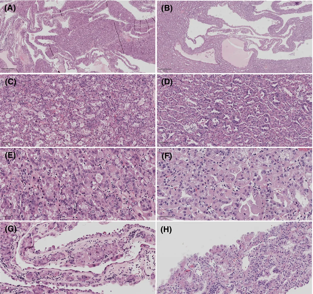
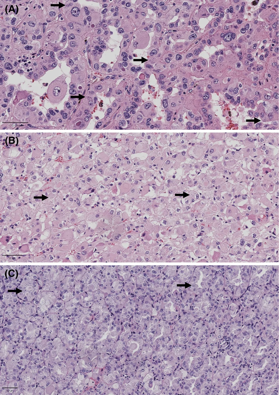
ESC‐RCC 还以实性和囊性结构为特征,实性区域由紧密排列的小管构成(图 1)。ESC‐RCC 恶性细胞的胞质丰富、嗜酸性,伴粗大的嗜碱性点彩,偶呈空泡状;另一些区域的胞质则呈絮状外观(图 2)。ESC‐RCC 的其他特征包括囊腔衬覆细胞呈鞋钉样,以及显著的泡沫样巨噬细胞混杂于肿瘤中或在肿瘤内成簇聚集。
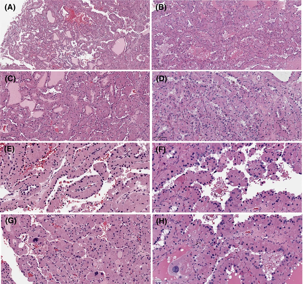
TFEB 扩增型肾细胞癌以实性为主,局部小管状与假乳头状生长交替出现(图 3)。小管通常比 ESC‐RCC 中的小管更大,既无大囊性也无微囊性结构;TFEB 扩增型肿瘤细胞的胞质主要呈致密颗粒状嗜酸性。未见嗜碱性点彩。其他所见包括假乳头状区域内位于顶端的细胞核、细胞外洋红色小球及少见的钙化(图 S1)。
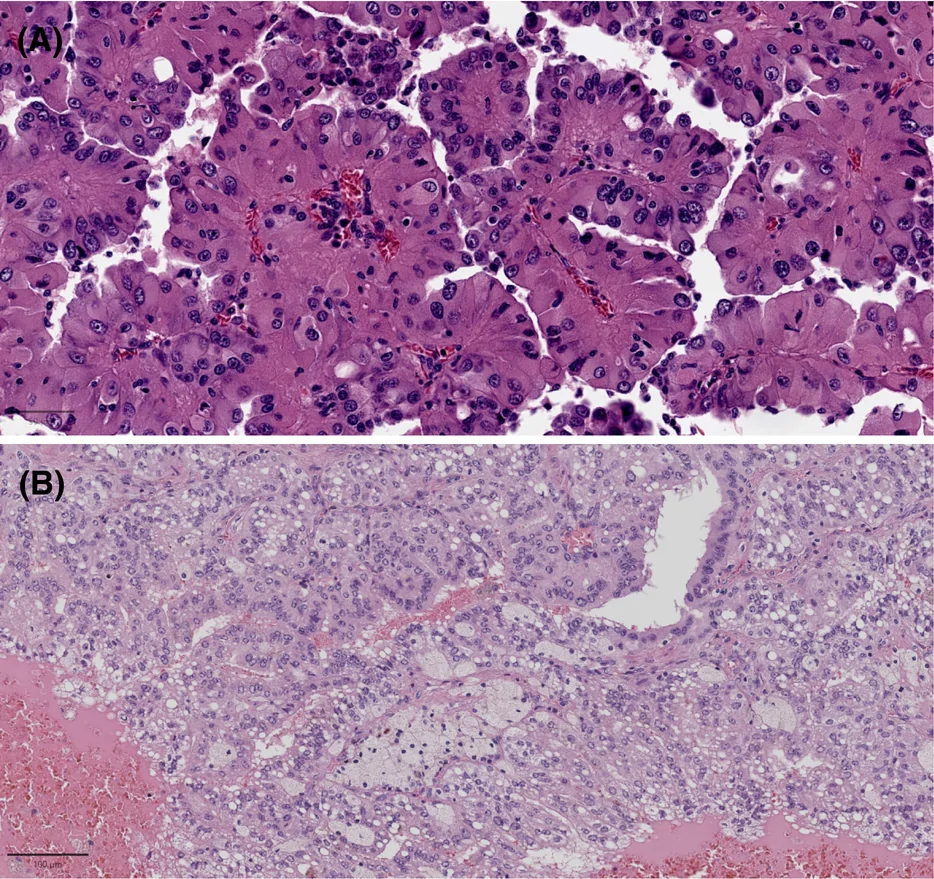
部分肿瘤可见形态学特征重叠。ESC‐RCC 病例 VIII 局部呈假乳头状结构,细胞核位于顶端,类似于更常见于 TFEB 扩增型肾细胞癌的特征(图 4A)。反之,TFEB 扩增型肾细胞癌病例 III 的部分区域可见小管背靠背排列及巨噬细胞簇,类似 ESC‐RCC(图 4B)。
免疫组织化学
采用针对 vimentin、CK7、CK20、P504S、CD10、SDHB、Melan‐A 和 PD‐L1 的抗体进行免疫组织化学(IHC)染色(表 3)。根据呈现相应阳性染色模式的肿瘤细胞百分比进行评分:0%(0 分)、1%–5%(1 分)、5%–50%(2 分)和 >50%(3 分)。阳性细胞 >5% 的肿瘤判定为阳性(对应 IHC 评分 ≥2)。ESC‐RCC 和 TFEB 扩增型肾细胞癌均呈 vimentin 和 P504S 弥漫强阳性,SDHB 表达保留。两类肿瘤均不表达 CK7 或仅局灶表达。CD10 如有染色,则呈局灶性。两组的 PD‐L1 表达均有差异。9 例 ESC‐RCC 中的 7 例(78%)和 6 例 TFEB 扩增型肾细胞癌中的 3 例(50%)可见 CK20 染色(阳性肿瘤细胞 >5%)(p = 0.3287)。9 例 ESC‐RCC 中的 2 例(22%)和 6 例 TFEB 扩增型肾细胞癌中的 4 例(66%)可见 Melan‐A 染色(阳性肿瘤细胞 >5%)(p = 0.1357)(图 S2 和 S3)。
| 病例 | CK20 | MelanA | Vimentin | CK7 | P504S | CD10 | SDHB | PD‐L1 |
|---|---|---|---|---|---|---|---|---|
| ESC‐RCC | ||||||||
| I | 3 | 2 | 3 | 0 | 2 | 0 | ND | 1 |
| IIA | 1 | 0 | 3 | 0 | 3 | 0 | ND | 2 |
| IIB | 1 | 0 | 3 | 0 | 2 | 0 | ND | ND |
| III | 3 | 0 | 3 | 0 | 2 | 0 | 3 | 3 |
| IV | 2 | 0 | 2 | 0 | 3 | 1 | ND | 2 |
| V | 3 | 2 | 3 | 0 | 2 | 0 | 3 | 2 |
| VI | 3 | 0 | ND | 0 | ND | ND | ND | 1 |
| VII | 3 | 0 | ND | 0 | ND | ND | ND | 0 |
| VIII | 2 | 0 | 3 | ND | 3 | ND | ND | 2 |
| TFEB 扩增型肾细胞癌 | ||||||||
| I | 3 | 3 | 3 | 0 | 3 | ND | ND | ND |
| II | 3 | 2 | 3 | 1 | ND | ND | ND | 1 |
| III | 1 | 1 | 3 | 1 | 3 | ND | 3 | 0 |
| IV | 0 | 3 | ND | ND | ND | ND | ND | 3 |
| V | 1 | 3 | ND | ND | ND | ND | ND | 3 |
| VI | 3 | 1 | 3 | 0 | ND | 2 | 3 | 3 |
评分。0%–0% 染色。1%–1% 至 5%。2%–5% 至 50%。3%– >50%。
ND,未进行。
二代测序
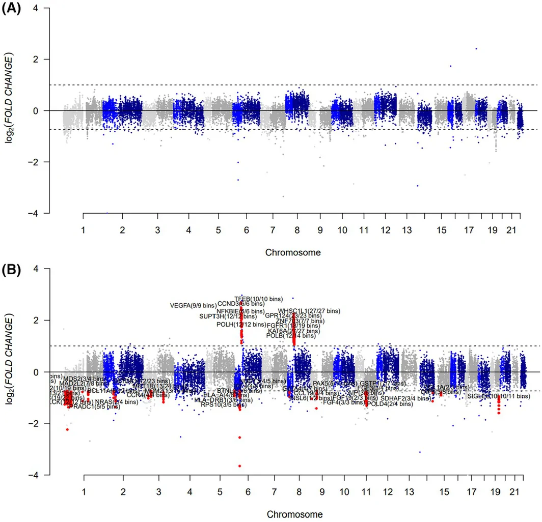
本研究纳入的 9 例 ESC‐RCC 中有 5 例以及全部 6 例 TFEB 扩增型肾细胞癌成功完成二代测序(NGS)(表 4)。所有接受测序的 ESC‐RCC 病例均携带致病性 TSC1 或 TSC2 变异。所有 TFEB 扩增型肾细胞癌病例均显示涉及染色体 6p 并涵盖 TFEB 基因座的局灶性拷贝数增加(图 5)。所有 ESC‐RCC 病例均未检出拷贝数改变。
| 病例 | 包含 TFEB 基因座的 6p 扩增 | 致病性 TSC 变异 | 变异的编码效应 | 编码 DNA 命名 | 蛋白质命名 |
|---|---|---|---|---|---|
| ESC‐RCC | |||||
| I | 否 | TSC2 | 移码 | NM_000548.4:c.276_279dup | p.P94Afs*33 |
| TSC2 | 移码 | NM_000548.4:c.938_939del | p.R313Kfs*24 | ||
| IIA | 否 | TSC1 | 移码 | NM_000368.4:c.2599_2600insCG | p.Q867Pfs*12 |
| III | 否 | TSC2 | 框内缺失 | NM.000548.4:c.5238_5255del | p.H1746_R1751del |
| TSC2 | 剪接 | NM_000548.4:c.975+1G>T | p.? | ||
| V | 否 | TSC2 | 无义 | NM_000548.4:c.1635_1636delinsTT | p.E546* |
| TSC2 | 剪接 | NM_000548.4:c.2356‐1G>C | p.? | ||
| VIII | 否 | TSC1 | 移码 | NM_000368.5:c.976_977del | p.L326Tfs*14 |
| TFEB 扩增型肾细胞癌 | |||||
| I (m) | 是 | 无 | |||
| II | 是 | 无 | |||
| III | 是 | 无 | |||
| IV | 是 | 无 | |||
| V | 是,可疑 | 无 | |||
| VI | 是 | 无 | |||
随访
本研究有 4 名患者失访(n = 4;ESC‐RCC 组有 2 名患者,对应病例 IIA、IIB 和 III;TFEB 扩增型肾细胞癌组有 2 名患者,对应病例 I 和 II)(表 5)。一名失访的 ESC‐RCC 患者有疑似肝转移的记录;值得注意的是,该患者(对应 ESC‐RCC 病例 IIA 和 IIB)在肿瘤切除时已有明确的淋巴结转移。一名失访的 TFEB 扩增型肾细胞癌患者有脑及骨转移的记录(TFEB 扩增型肾细胞癌病例 I),另一名患者已知同侧肾脏存在复发病灶(TFEB 扩增型肾细胞癌病例 II)。
| 病例 | 随访(月) | 目前状态 | 补充信息 |
|---|---|---|---|
| ESC‐RCC | |||
| I | 231 | 存活 | NED |
| IIA | 18 | 失访 | 疑似转移(未证实) |
| IIB | 14 | ||
| III | 64 | 失访 | NED |
| IV | 157 | 死亡 | 死因不明 |
| V | 3 | 死亡 | 死于原发性骨髓纤维化 |
| VI | 49 | 存活 | NED |
| VII | 18 | 死亡 | 死于胆管癌 |
| VIII | 38 | 存活 | NED |
| TFEB 扩增型肾细胞癌 | |||
| I | 205 | 失访 | 脑及骨转移 |
| II | 87 | 失访 | 右肾复发 |
| III | 54 | 存活 | 肺转移 |
| IV | 36 | 存活 | 疑似转移(未证实) |
| V | 17 | 存活 | NED |
| VI | 1 | 存活 | 已安排肾切除术 |
NED,无疾病证据。
在其余未失访的患者中,3 名 ESC‐RCC 患者在分析时已死亡(n = 3;ESC‐RCC 病例 IV、V、VII)。其中 2 名患者死于与 RCC 无关的原因(ESC‐RCC 病例 V 和 VII),另一名患者的死因在我们的记录中未记载(ESC‐RCC 病例 IV)。其余 3 名在分析时仍存活的 ESC‐RCC 患者的随访时间为 38 至 231 个月(n = 3)。这些患者在各自的随访期间均未出现疑似复发或转移的表现。4 名未失访的 TFEB 扩增型肾细胞癌患者的随访时间为 1 至 54 个月(n = 4)。其中 1 名患者已确诊转移(TFEB 扩增型肾细胞癌病例 III),另 1 名患者因有疑似肺转移的记录而正在接受评估(TFEB 扩增型肾细胞癌病例 IV)。其余 2 名未失访的 TFEB 扩增型肾细胞癌患者在本报告时均无疾病证据。
讨论
本研究比较了本机构档案中 9 例 ESC‐RCC 和 6 例 TFEB 扩增型肾细胞癌的临床病理学及分子特征。尽管本研究受限于这些肿瘤的罕见性及由此导致的队列规模较小,但我们病例的临床特征与既往报道一致,并进一步拓展了有关这两类肿瘤的已发表经验。ESC‐RCC 患者通常较年轻(平均年龄 50 岁),以女性为主,多数就诊时为 pT1 期,与报道中该肿瘤预期的惰性临床行为一致。25,26 相比之下,TFEB 扩增型肾细胞癌患者确诊时年龄较大(平均年龄 66 岁),更常以较晚分期肿瘤就诊,且在随访期间更易出现复发或转移,支持既往关于 TFEB 扩增型肾细胞癌是一种侵袭性肿瘤的观察。21,27,28,29
尽管 ESC‐RCC 和 TFEB 扩增型肾细胞癌存在一定的形态学重叠,但各自均呈现一定的特征性组织学模式。ESC‐RCC 通常呈实性和囊性模式,由紧密排列的小管构成,胞质丰富、嗜酸性,伴粗大的嗜碱性点彩或呈絮状外观,囊腔衬覆鞋钉样细胞,并混有泡沫样巨噬细胞。相比之下,TFEB 扩增型肾细胞癌通常以实性生长为主,伴小管状和假乳头状结构,胞质呈致密颗粒状嗜酸性且缺乏嗜碱性点彩,假乳头状区域内细胞核位于顶端,并可见细胞外洋红色小球和偶发钙化。
重要的是,没有任何单一形态学特征对其中任一类肿瘤具有完全特异性。部分肿瘤呈现重叠特征,包括一例 ESC‐RCC 的局灶性假乳头状结构,以及一例 TFEB 扩增型肾细胞癌中局部紧密排列的小管伴巨噬细胞成簇聚集。此外,尽管所有 ESC‐RCC 病例均可见嗜碱性点彩,而所有 TFEB 扩增型肾细胞癌病例均未见,但其他嗜酸性肾肿瘤中也曾报道这一所见,因此应结合整体表现及辅助检查结果加以解读,而不应将其作为孤立的诊断特征。30,31,32,33 同样,既往报道多达 50% 的 ESC‐RCC 可见砂粒体样钙化,但我们的队列中并未发现。27,29
这两类肿瘤的 IHC 染色谱也存在相当程度的重叠。二者均弥漫阳性表达 vimentin 和 P504S,SDHB 表达保留,CK7 多为阴性或仅局灶阳性。尽管 ESC‐RCC 更常呈 CK20 弥漫表达、Melan‐A 阴性或斑片状染色,而 TFEB 扩增型肾细胞癌更常呈相反模式,但这些差异并无统计学意义。因此,CK20 和 Melan‐A 可提供支持性证据,但单独使用不足以可靠地区分这些肿瘤。
两类肿瘤的 PD‐L1 表达均存在差异,既见于不同肿瘤之间,也见于单个病例内部。由于 TFEB 扩增型肾细胞癌具有侵袭性临床行为,且免疫检查点抑制可能发挥作用,其 PD‐L1 表达尤其值得关注。实验研究表明,TFEB 蛋白可促进 PD‐L1 表达,从而促使免疫逃逸及对 mTOR 抑制产生耐药。34 与既往显示 TFEB 扩增型肿瘤表达 PD‐L1 的报道一致,35,36 我们队列中的若干肿瘤呈 PD‐L1 强染色,且与标本存放时间无关,尽管文献报道标本存放时间与 PD‐L1 免疫反应性呈负相关。37,38 此外,一篇已发表的病例报告描述了 TFEB 扩增型肾细胞癌对抗 PD‐L1 治疗具有良好的临床反应,提示免疫检查点抑制可能成为部分患者的治疗策略,但仍需更多临床研究。39
分子分析证实了这两类肿瘤的定义性基因组改变。所有接受测序的 ESC‐RCC 均携带致病性 TSC1 或 TSC2 变异,每例 TFEB 扩增型肾细胞癌均显示累及 TFEB 基因座的染色体 6 短臂局灶性扩增。29 TSC1/2 和 TFEB 编码的蛋白产物参与 PI3K‐AKT–mTOR 通路调控,该通路在肾肿瘤发生过程中反复出现改变及上调。2,40 导致功能丧失的致病性 TSC1/2 变异可引起 mTOR 信号上调,而 TFEB 扩增则导致 mTOR 下游的 TFEB 功能增强。41 这些趋同的分子机制可能部分解释这两类肿瘤之间的形态学重叠。42
传统上,TFEB 扩增通过荧光原位杂交(FISH)确定。近期研究提出,以 TFEB 拷贝数 >5 作为定义扩增的 FISH 阈值,其中 5–10 个拷贝代表低水平扩增,>10 个拷贝代表高水平扩增。36 本研究通过常规杂交捕获 NGS 所产生的拷贝数分析识别 TFEB 扩增。NGS 面板常规用于评估拷贝数增加,43,44,45 此前也曾用于诊断 TFEB 扩增型肾细胞癌。本研究表明,采用 NGS 同时识别 ESC‐RCC 的特征性序列变异和 TFEB 扩增型肾细胞癌的特征性局灶扩增是可行的。
综上,形态学和免疫组织化学对于提出恰当的嗜酸性肾细胞肿瘤鉴别诊断仍至关重要。然而,由于 ESC‐RCC 与 TFEB 扩增型肾细胞癌之间存在显著重叠,常需进行分子特征分析才能确立明确诊断。准确区分这两类肿瘤具有重要的预后意义及潜在治疗意义,因为 ESC‐RCC 通常呈惰性临床病程,而 TFEB 扩增型肾细胞癌与更强的侵袭性行为相关。随着分子检测日益成为肾细胞肿瘤诊断的常规环节,整合临床病理学和分子评估对于准确分类这些少见肿瘤将至关重要。
作者贡献
Tatjana Antic 和 Hayley Zullow 参与了研究构思与设计。所有作者(Hayley Zullow、Melissa Y. Tjota、Jung Woo Kwon 和 Tatjana Antic)共同完成材料准备、数据收集与分析。论文初稿由 Hayley Zullow 撰写,所有作者(Hayley Zullow、Melissa Y. Tjota、Jung Woo Kwon 和 Tatjana Antic)均对论文的先前版本提出意见。所有作者(Hayley Zullow、Melissa Y. Tjota、Jung Woo Kwon 和 Tatjana Antic)均阅读并批准了最终稿。
知情同意
芝加哥大学机构审查委员会予以豁免(IRB‐24‐1223)。
- Two rare eosinophilic renal tumours that are hard to separate on morphology and IHC alone: the authors compared 9 institutional ESC-RCCs (8 patients) with 6 TFEB-amplified RCCs.
- ESC-RCC: younger patients (mean 50 years), female predominance, mostly pT1; solid and cystic architecture, tightly packed tubules, flocculent to coarsely granular cytoplasm with basophilic stippling (9/9), prominent foamy macrophages and hobnail cells lining cysts.
- TFEB-amplified RCC: older patients (mean 66 years), higher stage and more recurrence/metastasis; solid growth with pseudopapillae and loose tubules, dense granular eosinophilic cytoplasm without stippling, apical nuclei in pseudopapillary areas and extracellular magenta globules.
- IHC overlapped: both diffusely vimentin/P504S positive, SDHB retained, CK7 mostly negative; ESC-RCC more often diffuse CK20 with absent/patchy Melan-A and TFEB-amplified RCC the reverse, without statistical significance; PD-L1 variable. Molecular testing is decisive: TSC1/TSC2 pathogenic variants in ESC-RCC and focal 6p amplification involving TFEB in TFEB-amplified RCC.
Scope: PMC open-access full text (CC BY 4.0) — abstract, introduction, methods, results, discussion, author contributions and consent statement, with Figures 1–5 and Tables 1–5. Supporting information, conflict-of-interest and data availability statements, and references are not included (citation numbers are retained).
Abstract
Eosinophilic solid and cystic renal cell carcinoma (ESC‐RCC) and TFEB‐amplified renal cell carcinoma (TFEB‐amplified RCC, a subset of TFEB‐altered RCC) are rare eosinophilic renal cell tumours that can be difficult to distinguish on the basis of morphology and immunohistochemistry alone. In this study, we compared the clinicopathological and molecular features of these entities to identify distinguishing characteristics.
Nine cases of ESC‐RCC and six cases of TFEB‐amplified RCC from our institutional archives were evaluated for clinical, morphological, immunohistochemical and molecular features. Despite morphological overlap, each entity exhibited somewhat distinctive features. ESC‐RCC frequently exhibited solid and cystic architecture with tightly packed tubules, flocculent to coarsely granular cytoplasm with basophilic stippling and prominent admixed macrophages. In comparison, TFEB‐amplified RCC more frequently demonstrated solid growth of pseudopapillae and loose tubules, dense eosinophilic granular cytoplasm without basophilic stippling, focally apical nuclei and extracellular magenta globules. However, neither morphology nor immunohistochemistry was sufficiently specific to completely distinguish these entities. Ancillary molecular testing identified pathogenic TSC1 or TSC2 gene variants in ESC‐RCC and TFEB gene locus amplification in TFEB‐amplified RCC.
Although ESC‐RCC and TFEB‐RCC share overlapping morphological and immunohistochemical features, each demonstrates somewhat characteristic histological findings. Definitive distinction requires molecular testing to identify the underlying molecular alterations. As molecular testing assumes an increasingly important role in RCC diagnosis and classification, recognition of these rare entities and their overlapping and distinguishing features is essential for appropriate testing and accurate diagnosis.
Introduction
Over the past half century, the classification system of renal cell carcinoma (RCC) has changed multiple times to reflect our evolving understanding of renal cell tumour biology. 1 , 2 The most recent World Health Organization (WHO) classification defines six groups of renal cell tumours: clear cell, papillary, oncocytic and chromophobe, collecting duct, molecularly defined and other RCC. Clear cell, papillary and chromophobe RCC account for approximately 95% of renal cell tumour diagnoses. 3 , 4 These more common RCC subtypes have characteristic morphology and immunohistochemical (IHC) staining patterns, and are most often diagnosed without further ancillary testing.
In contrast, rarer RCC subtypes, especially eosinophilic or oncocytic renal tumours, may exhibit significant morphological and immunophenotypic overlap and frequently require advanced testing modalities for definitive diagnosis. 5 , 6 Though ancillary testing is often unavoidable in these cases, identifying specific morphological and IHC features of rare RCC subtypes may guide more effective work‐up and diagnosis of rare renal cell tumours, especially when diagnostic material is limited. Therefore, despite the expanding role of molecular testing in the diagnosis and classification of renal cell tumours, it remains critical to fully characterize and recognize specific features of rare RCC subtypes to improve diagnosis, treatment and outcomes for patients with these tumours. 7 , 8
Eosinophilic solid and cystic renal cell carcinoma (ESC‐RCC) and TFEB‐amplified renal cell carcinoma (TFEB‐amplified RCC, a subset of TFEB‐altered RCC) are rare eosinophilic RCC subtypes that are both associated with TSC/mTOR pathway dysregulation and can be difficult to distinguish on the basis of morphology and immunohistochemistry alone. 9 , 10 , 11 , 12 , 13 Grossly, both entities present as well‐circumscribed tumours with a yellow cut surface with or without areas of haemorrhage and necrosis; histologically, these tumours are composed of large cells with abundant granular eosinophilic cytoplasm, large nuclei and prominent nucleoli. 14 Furthermore, though it has been proposed that these entities can be distinguished by differential IHC staining for Melan‐A and CK20, the reported utility of this strategy is variable. 15 , 16 , 17 , 18 As a result, both ESC‐RCC and TFEB‐amplified RCC may remain in the differential diagnosis for an eosinophilic tumour even after morphological and IHC work‐up. The distinction between these entities is critical, as ESC‐RCC is reported to have a better prognosis than TFEB‐amplified RCC and may have implications on clinical course and treatment strategy. 19 , 20 , 21
Existing comparative studies of ESC‐RCC and TFEB‐amplified RCC remain limited due to the rarity of both entities, and due to the scarcity of papers that compare these entities directly using the same criteria. In this study, we compare the clinical, morphological, IHC and molecular profiles of nine ESC‐RCC cases and six TFEB‐amplified RCC cases from a single institution. In addition to expanding published knowledge of these uncommon renal cell tumours, we demonstrate the use of hybrid‐capture next‐generation sequencing (NGS) to identify the characteristic molecular alterations underlying each entity, including detection of TFEB gene locus amplification through copy‐number analysis. Using this integrated clinicopathological and molecular approach, we aimed to define features that facilitate recognition of these tumours while emphasizing the role of molecular testing in establishing a definitive diagnosis when morphological and IHC findings are equivocal.
Methods
Study Population
This study was approved by the Institutional Review Board at The University of Chicago under protocol IRB‐24‐1223. We retrospectively reviewed The University of Chicago Department of Pathology database from 2005 to 2025 and identified unclassified or unusual eosinophilic renal tumours suspicious for re‐classification as ESC‐RCC or TFEB‐amplified RCC, as well as tumours with established diagnoses of ESC‐RCC or TFEB‐amplified RCC. Seven cases included in this study (ESC‐RCC cases I, IIA, IIB, III, IV, V, and TFEB‐amplified RCC case II) have been reported previously. 13 , 14 ESC‐RCC cases were initially given diagnoses of ESC‐RCC (n = 1), papillary RCC (n = 1) and unclassified RCC (n = 7). TFEB‐amplified RCC cases were initially diagnosed as sarcomatoid RCC (n = 1), renal oncocytoma (n = 1) and unclassified RCC (n = 4). Clinical data were obtained through electronic medical record review.
Pathological Analysis and Immunohistochemical Staining
For each case, the original pathological diagnosis, haematoxylin and eosin (H&E)‐stained slides and IHC stains were reviewed by a genitourinary pathologist at The University of Chicago. IHC stains were performed in the clinical laboratory at The University of Chicago using autoimmunostainers (Leica BOND‐III; Leica Biosystems, Buffalo Grove, IL and BenchMark XT Ventana; Roche, Tucson, AZ) with appropriate controls. The following panel of IHC stains was performed: CK7 (COV‐TL 12/30; Dako, Carpinteria, CA), CK20 (KS20.8; Dako, Carpinteria, CA), Vimentin (V9; Dako, Carpinteria, CA), racemase (P504S, CPK 200 AK, BK, CK; Biocare Medical, Concord, CA), CD10 (56C6; Leica NCL, Buffalo Grove, IL), SDHB (21A11AE7; Abcam, Cambridge, MA), Melan‐A (A103; Dako, Carpinteria, CA) and PD‐L1 (22C3; Agilent, Santa Clara, CA).
Next‐Generation Sequencing
Sufficient material was available for NGS in 5 of 9 ESC‐RCC cases (I, IIA, III, V and VIII) and 6 of 6 TFEB‐amplified RCC cases (I–VI). ESC‐RCC case IIB failed multiple attempts to sequence. ESC‐RCC case IV was not sequenced, as it occurred in a patient with tuberous sclerosis and displayed the typical morphological features and IHC phenotype of ESC‐RCC. From the cases that were sequenced, a representative formalin‐fixed, paraffin‐embedded block was selected for NGS on The University of Chicago Medicine OncoPlus (UCM‐OncoPlus) panel, a hybrid‐capture panel targeting 1005 cancer‐associated genes with 168 clinically reported genes. DNA extraction, DNA quantification, library preparation and NGS were performed as described previously. 22
Data analysis was performed on a HIPAA (Health Insurance Portability and Accountability Act)‐compliant high‐performance computing system (Center for Research Informatics, University of Chicago) using an in‐house developed bioinformatics pipeline. Copy number results were calculated using a combination of CNVkit 14 software 23 and additional in‐house intra‐run normalization to eliminate run‐specific artefacts, by comparison with a pooled cohort of clinical controls. Focal gene‐level and chromosome‐level changes were called using a combination of thresholds and manual interpretation, as described previously. 22 , 23 , 24 The specific thresholds used in this study were log2 (fold change) ≥ 1.5 for amplification and log2 (fold change) between 1.0 and 1.5 for equivocal amplification, consistent with thresholds used previously. 25 Somatic variant calls were inspected using Integrated Genomics Viewer (IGV; Broad Institute, MIT Harvard, Cambridge, MA). Variant review was performed by molecular pathologists and included filters based on population frequencies (the 1000 Genomes Project, https://www.internationalgenome.org/), frequencies in cancer variant databases (COSMIC: Catalogue Of Somatic Mutations In Cancer, https://cancer.sanger.ac.uk/cosmic, and cBioPortal, https://www.cbioportal.org/), and coding effects.
Results
Clinical Demographics
Retrospective review and molecular profiling of previously unclassified or unusual eosinophilic renal cell tumours identified nine ESC‐RCC cases (n = 9) and six TFEB‐amplified RCC cases (n = 6) from our institutional archives (Table 1). Two ESC‐RCC cases (cases IIA and IIB) arose in a single patient who underwent right radical nephrectomy for a right‐sided kidney mass (ESC‐RCC case IIA), and 2 months post‐operatively was found to have a mass in the contralateral kidney (ESC‐RCC case IIB). One of the TFEB‐amplified RCC cases is from a patient who had presented with metastatic disease to the brain; approximately 15 years earlier, the patient had undergone resection of a primary renal mass that was diagnosed as ‘sarcomatoid RCC’. The metastatic lesion (TFEB‐amplified RCC case I) displayed a similar histological appearance to the primary renal tumour and was sequenced for diagnostic purposes. The ESC‐RCC cohort of 9 cases arose in 8 patients (6 female and 2 male) ranging in age from 33 to 68 years at diagnosis (mean: 50 years). The 6 cases of TFEB‐amplified RCC arose in 4 female patients and 2 male patients ranging in age from 53 to 76 years at diagnosis (mean: 66 years). One patient in the ESC‐RCC group had a history of tuberous sclerosis (ESC‐RCC case IV). In the TFEB‐amplified group, one patient had a history of Gaucher's disease, type 1 (TFEB‐amplified RCC case IV) and 1 patient was suspected to have multiple endocrine neoplasia, type 1, though confirmatory testing was not performed (TFEB‐amplified RCC case II). No additional patients were known or suspected to have an underlying hereditary syndrome.
| Cases | Age (years) | Sex | Laterality | Focality | Largest size (cm) | Stage | Original grade | Architecture | Hereditary disease | Original diagnosis |
|---|---|---|---|---|---|---|---|---|---|---|
| ESC‐RCC | ||||||||||
| I* | 52 | F | R | Unifocal | 5.5 | T1bNx | 3 | Solid and cystic | Not tested | Unclassified RCC |
| IIA* | 48 | F | R | Multifocal | 6 | T3bN2 | High | Solid and papillary | Not tested | Papillary RCC, type 2 |
| IIB* | 49 | F | L | Multifocal | 2.5 | T1aN2 | High (3–4) | Solid and papillary | Not tested | Unclassified RCC |
| III* | 33 | F | L | Unifocal | 1.7 | T1aNx | Low | Solid and cystic | Not tested | Unclassified RCC |
| IV* | 47 | F | R | Unifocal | 3 | T1aNx | 3 | Solid and cystic | TSC | Unclassified RCC |
| V* | 65 | F | L | Unifocal | 11.5 | T3aNx | 4 | Solid and cystic | Not tested | Unclassified oncocytic RCC |
| VI | 37 | F | L | Unifocal | 2.2 | T1aNx | NA | Solid and cystic | Not tested | Eosinophilic solid and cystic RCC |
| VII | 68 | M | R | Unifocal | 2 | T1aNx | 2, focal 3 | Solid and cystic | Not tested | Unclassified RCC |
| VIII | 51 | M | L | Unifocal | 4.1 | T3aNx | NA | Solid and cystic | Not tested | Unclassified RCC |
| TFEB‐amplified RCC | ||||||||||
| I | 53 | M | L | Unifocal | 11 | T3aNx | 4 | Solid | Not tested | Sarcomatoid RCC |
| II* | 67 | F | R | Unifocal | 2.9 | T3aNx | NA | Solid and papillary | MEN1 (suspected) | Renal oncocytoma |
| III | 64 | F | L | Unifocal | 7.6 | T3aNx | 3, focal 4 | Solid and papillary | Not tested | Unclassified RCC |
| IV | 65 | F | L | Unifocal | 9.1 | T3aNx | 3 | Solid | Gaucher | Unclassified RCC |
| V | 71 | M | R | Unifocal | 3.7 | T1aNx | 3 | Tubulopapillary | Not tested | Unclassified RCC |
| VI | 76 | F | L | Unifocal | 7.5 | T3aNx | High | Solid and tubulopapillary | Not tested | Unclassified RCC |
Previously published cases.
AJCC Tumour Staging
Upon resection, TFEB‐amplified tumours were larger and more frequently higher in stage than ESC‐RCC tumours (Table 1). ESC‐RCC tumours ranged in size from 1.7 to 11.5 cm in greatest dimension (mean: 4.3 cm). Per the American Joint Committee on Cancer (AJCC), six ESC‐RCC resections revealed pathological stage T1 disease (n = 6) and the remaining three resections revealed pathological stage T3 disease (n = 3). Lymph nodes were recovered in two ESC‐RCC resections corresponding to a single patient (ESC‐RCC cases IIA and IIB); metastatic carcinoma was identified in >1 regional lymph node in each of the resection specimens. Resected TFEB‐amplified tumours ranged in size from 2.9 cm to 11.0 cm in greatest dimension (mean: 7.0 cm). At primary resection, 5 patients with TFEB‐amplified RCC had pathological stage T3 disease (n = 5) and 1 patient had pathological stage T1 disease (n = 1). No lymph nodes were recovered in the resection specimens for TFEB‐amplified RCC.
Morphological Features
Review of H&E slides demonstrated that both entities exhibited shared as well as somewhat distinctive morphological features. Both ESC‐RCC and TFEB‐amplified tumours were at least partially solid and composed of polygonal cells with abundant eosinophilic cytoplasm and focally large, high‐grade nuclei with prominent nucleoli (Table 2).
| Features | ESC‐RCC | TFEB‐amplified RCC |
|---|---|---|
| Architecture | Solid and cystic predominantly tubular, with tightly packed small tubules | Solid pseudopapillary to tubular, with large tubules |
| Cytoplasm | Abundant, eosinophilic, occasionally vacuolated magenta‐blue granules | Abundant, eosinophilic, densely granular |
| Nucleus | Large, pleomorphic, high‐grade nuclei with prominent nucleoli | Large, pleomorphic, high‐grade nuclei with prominent nucleoli focally apical |
| Other | Hobnail cells lining cystic areas foamy macrophages | Extracellular magenta globules calcifications |
ESC‐RCC was further characterized by solid and cystic architecture, with solid areas populated by small, tightly packed tubules (Figure 1). The abundant eosinophilic cytoplasm of malignant cells in ESC‐RCC had coarse basophilic stippling and was occasionally vacuolated; alternatively, the cytoplasm was flocculent‐appearing (Figure 2). Additional features of ESC‐RCC included hob‐nailing of cells lining cystic spaces and conspicuous foamy macrophages admixed with or clustered within the tumour.
TFEB‐amplified RCC was predominantly solid with areas of alternating tubular and pseudopapillary growth (Figure 3). The tubules were generally larger than those observed in ESC‐RCC and neither macrocystic nor microcystic architecture was present; TFEB‐amplified tumour cells predominantly demonstrated dense granular eosinophilic cytoplasm. Basophilic stippling was not identified. Additional findings included apically placed nuclei within pseudopapillary areas, extracellular magenta globules and rare calcifications (Figure S1).
Overlapping morphological features were observed in selected tumours. ESC‐RCC case VIII exhibited focal pseudopapillary architecture with apically positioned nuclei resembling features more associated with TFEB‐amplified RCC (Figure 4A). Conversely, TFEB‐amplified RCC case III contained areas with small back‐to‐back tubules and clusters of macrophages reminiscent of ESC‐RCC (Figure 4B).
Immunohistochemistry
Immunohistochemical (IHC) staining was performed using antibodies against vimentin, CK7, CK20, P504S, CD10, SDHB, Melan‐A and PD‐L1 (Table 3). Staining was scored according to percentage of tumour cells demonstrating the appropriate positive staining pattern as follows: 0% (score 0), 1%–5% (score 1), 5%–50% (score 2) and >50% (score 3). Tumours with >5% positive cells were considered positive (corresponding to IHC score ≥2). Both ESC‐RCC and TFEB‐amplified RCC exhibited strong, diffuse staining for vimentin and P504S with retained SDHB. CK7 expression was absent or only focally present in both entities. CD10 staining was focal when present. PD‐L1 expression was variable in both cohorts. CK20 staining (>5% positive tumour cells) was identified in 7 of 9 ESC‐RCC cases (78%) and in 3 of 6 TFEB‐amplified RCC cases (50%) (p = 0.3287). Melan‐A staining (>5% positive tumour cells) was identified in 2 of 9 ESC‐RCC cases (22%) and in 4 of 6 TFEB‐amplified RCC cases (66%) (p = 0.1357) (Figures S2 and S3).
| Case | CK20 | MelanA | Vimentin | CK7 | P504S | CD10 | SDHB | PD‐L1 |
|---|---|---|---|---|---|---|---|---|
| ESC‐RCC | ||||||||
| I | 3 | 2 | 3 | 0 | 2 | 0 | ND | 1 |
| IIA | 1 | 0 | 3 | 0 | 3 | 0 | ND | 2 |
| IIB | 1 | 0 | 3 | 0 | 2 | 0 | ND | ND |
| III | 3 | 0 | 3 | 0 | 2 | 0 | 3 | 3 |
| IV | 2 | 0 | 2 | 0 | 3 | 1 | ND | 2 |
| V | 3 | 2 | 3 | 0 | 2 | 0 | 3 | 2 |
| VI | 3 | 0 | ND | 0 | ND | ND | ND | 1 |
| VII | 3 | 0 | ND | 0 | ND | ND | ND | 0 |
| VIII | 2 | 0 | 3 | ND | 3 | ND | ND | 2 |
| TFEB‐amplified RCC | ||||||||
| I | 3 | 3 | 3 | 0 | 3 | ND | ND | ND |
| II | 3 | 2 | 3 | 1 | ND | ND | ND | 1 |
| III | 1 | 1 | 3 | 1 | 3 | ND | 3 | 0 |
| IV | 0 | 3 | ND | ND | ND | ND | ND | 3 |
| V | 1 | 3 | ND | ND | ND | ND | ND | 3 |
| VI | 3 | 1 | 3 | 0 | ND | 2 | 3 | 3 |
Scoring. 0%–0% staining. 1%–1% to 5%. 2%–5% to 50%. 3%– >50%.
ND, not done.
Next‐Generation Sequencing
Next‐generation sequencing (NGS) was successfully performed on 5 of 9 ESC‐RCC cases and all 6 TFEB‐amplified RCC cases included in this study (Table 4). All sequenced ESC‐RCC cases harboured pathogenic TSC1 or TSC2 variants. All TFEB‐amplified RCC cases demonstrated focal copy number gain involving chromosome 6p and encompassing the TFEB gene locus (Figure 5). Copy number alterations were not identified in any of the ESC‐RCC cases.
| Cases | 6p amplification with TFEB locus | Pathogenic TSC variant | Variant coding effect | cNomen | pNomen |
|---|---|---|---|---|---|
| ESC‐RCC | |||||
| I | No | TSC2 | Frameshift | NM_000548.4:c.276_279dup | p.P94Afs*33 |
| TSC2 | Frameshift | NM_000548.4:c.938_939del | p.R313Kfs*24 | ||
| IIA | No | TSC1 | Frameshift | NM_000368.4:c.2599_2600insCG | p.Q867Pfs*12 |
| III | No | TSC2 | In‐frame deletion | NM.000548.4:c.5238_5255del | p.H1746_R1751del |
| TSC2 | Splicing | NM_000548.4:c.975+1G>T | p.? | ||
| V | No | TSC2 | Nonsense | NM_000548.4:c.1635_1636delinsTT | p.E546* |
| TSC2 | Splicing | NM_000548.4:c.2356‐1G>C | p.? | ||
| VIII | No | TSC1 | Frameshift | NM_000368.5:c.976_977del | p.L326Tfs*14 |
| TFEB‐amplified RCC | |||||
| I (m) | Yes | None | |||
| II | Yes | None | |||
| III | Yes | None | |||
| IV | Yes | None | |||
| V | Yes‐equivocal | None | |||
| VI | Yes | None | |||
Follow‐Up
Four of the patients in this study were lost to follow‐up (n = 4; 2 patients from the ESC‐RCC group corresponding to cases IIA, IIB and III, and 2 patients from the TFEB‐amplified RCC group corresponding to cases I and II) (Table 5). One of the ESC‐RCC patients lost to follow‐up had documented concern for metastatic disease to the liver; of note, this patient (corresponding to ESC‐RCC cases IIA and IIB) had known lymph node metastases at the time of tumour resection. One patient with TFEB‐amplified RCC lost to follow‐up had documented brain and bony metastases (TFEB‐amplified RCC case I) and the other had known recurrent disease in the ipsilateral kidney (TFEB‐amplified RCC case II).
| Cases | Follow‐up (mo) | Current status | Additional information |
|---|---|---|---|
| ESC‐RCC | |||
| I | 231 | Alive | NED |
| IIA | 18 | Lost to follow‐up | Concern for metastasis (not confirmed) |
| IIB | 14 | ||
| III | 64 | Lost to follow‐up | NED |
| IV | 157 | Deceased | Cause of death unknown |
| V | 3 | Deceased | Secondary to primary myelofibrosis |
| VI | 49 | Alive | NED |
| VII | 18 | Deceased | Secondary to cholangiocarcinoma |
| VIII | 38 | Alive | NED |
| TFEB‐amplified RCC | |||
| I | 205 | Lost to follow‐up | Metastatic disease to brain and bone |
| II | 87 | Lost to follow‐up | Recurrent disease in right kidney |
| III | 54 | Alive | Metastatic disease to lung |
| IV | 36 | Alive | Concern for metastasis (not confirmed) |
| V | 17 | Alive | NED |
| VI | 1 | Alive | Scheduled for nephrectomy |
NED, no evidence of disease.
Of the remaining patients not lost to follow‐up, three ESC‐RCC patients were deceased at the time of analysis (n = 3; ESC‐RCC cases IV, V, VII). Two of these patients died due to causes unrelated to RCC (ESC‐RCC cases V and VII) and the other patient's cause of death was not documented in our records (ESC‐RCC case IV). The remaining three ESC‐RCC patients that were alive at the time of analysis had follow‐up periods ranging from 38 to 231 months (n = 3). These patients did not present with concern for recurrence or metastasis during their respective follow‐up periods. The four TFEB‐amplified RCC patients not lost to follow‐up had follow‐up periods ranging from 1 to 54 months (n = 4). One of these patients had confirmed metastases (TFEB‐amplified RCC case III) and 1 patient was under evaluation due to documented concern for metastatic disease to the lung (TFEB‐amplified RCC case IV). Neither of the remaining two TFEB‐amplified RCC patients not lost to follow‐up had evidence of disease at the time of this report.
Discussion
In this study, we compared the clinicopathological and molecular characteristics of nine ESC‐RCC cases and six TFEB‐amplified RCC cases from our institutional archives. Although this study was limited by the rarity of these entities and the resulting small cohort size, the clinical characteristics of our cases are consistent with prior reports and further expand the published experience with both entities. Patients with ESC‐RCC were generally younger (mean age, 50 years), demonstrated a female predominance and most presented with stage pT1 disease, consistent with the expected indolent clinical behaviour reported for this entity. 25 , 26 In contrast, patients with TFEB‐amplified RCC were older at diagnosis (mean age, 66 years), more frequently presented with more advanced stage tumours and were more likely to develop recurrent or metastatic disease during the follow‐up period, supporting previous observations that TFEB‐amplified RCC is an aggressive neoplasm. 21 , 27 , 28 , 29
Although ESC‐RCC and TFEB‐amplified RCC demonstrated some morphological overlap, each entity demonstrated somewhat characteristic histological patterns. ESC‐RCC typically exhibited a solid and cystic pattern composed of tightly packed tubules, abundant eosinophilic cytoplasm with coarse basophilic stippling or flocculent appearance, hobnail cells lining cystic spaces and admixed foamy macrophages. In contrast, TFEB‐amplified RCC generally demonstrated predominantly solid growth with tubular and pseudopapillary architecture, dense granular eosinophilic cytoplasm lacking basophilic stippling, apically positioned nuclei within pseudopapillary areas, extracellular magenta globules and occasional calcifications.
Importantly, no single morphological feature was entirely specific for either entity. Selected tumours demonstrated overlapping features, including focal pseudopapillary architecture in one ESC‐RCC and focal tightly packed tubules with clustered macrophages in one TFEB‐amplified RCC. Furthermore, while basophilic stippling was present in all ESC‐RCC cases and absent in all TFEB‐amplified RCC cases, this finding has been reported in other eosinophilic renal neoplasms and therefore should be interpreted in the context of the overall and ancillary findings rather than as an isolated diagnostic feature. 30 , 31 , 32 , 33 Likewise, psammomatous calcifications, previously reported in up to 50% of ESC‐RCCs, were not identified in our cohort. 27 , 29
The IHC staining profiles of these entities also demonstrated considerable overlap. Both entities showed diffuse positive expression of vimentin and P504S with retained SDHB expression and were largely negative or only focally positive for CK7. Although ESC‐RCC more frequently exhibited diffuse CK20 expression with absent or patchy Melan‐A staining, TFEB‐amplified RCC more often exhibited the opposite pattern; these differences were not statistically significant. Therefore, CK20 and Melan‐A may provide supportive evidence but are insufficient to reliably distinguish these tumours in isolation.
PD‐L1 expression was variable in both entities, both among tumours and within individual cases. PD‐L1 expression is of particular interest in TFEB‐amplified RCC because of its aggressive clinical behaviour and the potential role of immune checkpoint inhibition. Experimental studies have shown that the TFEB protein promotes PD‐L1 expression, facilitating immune evasion and mTOR inhibition resistance. 34 Consistent with previous reports demonstrating PD‐L1 expression in TFEB‐amplified tumours, 35 , 36 several tumours in our cohort exhibited strong PD‐L1 staining unrelated to the age of the specimen, despite literature reporting a negative correlation between specimen age and PD‐L1 immunoreactivity 37 , 38 Furthermore, a published case report describing a favourable clinical response of TFEB‐amplified RCC to anti‐PD‐L1 therapy suggests that immune checkpoint inhibition may represent a therapeutic strategy for selected patients, though additional clinical studies are needed. 39
Molecular profiling confirmed the defining genomic alterations of both entities. All sequenced ESC‐RCCs harboured pathogenic TSC1 or TSC2 variants and every case of TFEB‐amplified RCC demonstrated focal amplification of the short arm of chromosome 6 involving the TFEB locus. 29 The protein products encoded by TSC1/2 and TFEB participate in regulation of the PI3K‐AKT–mTOR pathway, which is recurrently altered and up‐regulated in renal tumorigenesis. 2 , 40 Pathogenic TSC1/2 variants resulting in loss of function lead to up‐regulation of mTOR signalling, while TFEB amplification results in increased TFEB function downstream of mTOR. 41 These convergent molecular mechanisms may partially explain the morphological overlap between these entities. 42
Traditionally, TFEB amplification has been established using fluorescence in situ hybridization (FISH). Recent studies have proposed FISH thresholds of >5 copies of TFEB to define amplification, with 5–10 copies representing low‐level amplification and >10 copies representing high‐level amplification. 36 In the present study, TFEB amplification was identified using copy‐number analysis generated from routine hybrid capture NGS. NGS panels are routinely used to evaluate copy number gains, 43 , 44 , 45 and have been previously used to diagnose TFEB‐amplified RCC. This study demonstrates the feasibility of using NGS to simultaneously identify sequence variants characteristic of ESC‐RCC and focal amplification characteristic of TFEB‐amplified RCC.
In summary, morphology and immunohistochemistry remain essential for generating an appropriate differential diagnosis in eosinophilic renal cell tumours. However, due to substantial overlap between ESC‐RCC and TFEB‐amplified RCC, molecular characterization is frequently required to establish a definitive diagnosis. Accurate distinction between these entities has important prognostic and potentially therapeutic implications, as ESC‐RCC generally follows an indolent clinical course whereas TFEB‐amplified RCC is associated with more aggressive behaviour. As molecular testing becomes increasingly routine in renal cell tumour diagnosis, integrated clinicopathological and molecular evaluation will be essential for the accurate classification of these uncommon neoplasms.
Author contributions
Tatjana Antic and Hayley Zullow contributed to the study conception and design. Material preparation, data collection and analysis were performed by all authors (Hayley Zullow, Melissa Y. Tjota, Jung Woo Kwon and Tatjana Antic). The first draft of the manuscript was written by Hayley Zullow and all (Hayley Zullow, Melissa Y. Tjota, Jung Woo Kwon and Tatjana Antic) commented on previous versions of the manuscript. All authors (Hayley Zullow, Melissa Y. Tjota, Jung Woo Kwon and Tatjana Antic) read and approved the final manuscript.
Consent
Waived by the University of Chicago Institutional Review Board (IRB‐24‐1223).