- 背景:WHO 1973 三级制 G2 组内异质性大,WHO 2004/2016 二分法(LG/HG)把生物学差异很大的肿瘤归入同一类。作者评估混合分级是否能更好地分层进展风险。
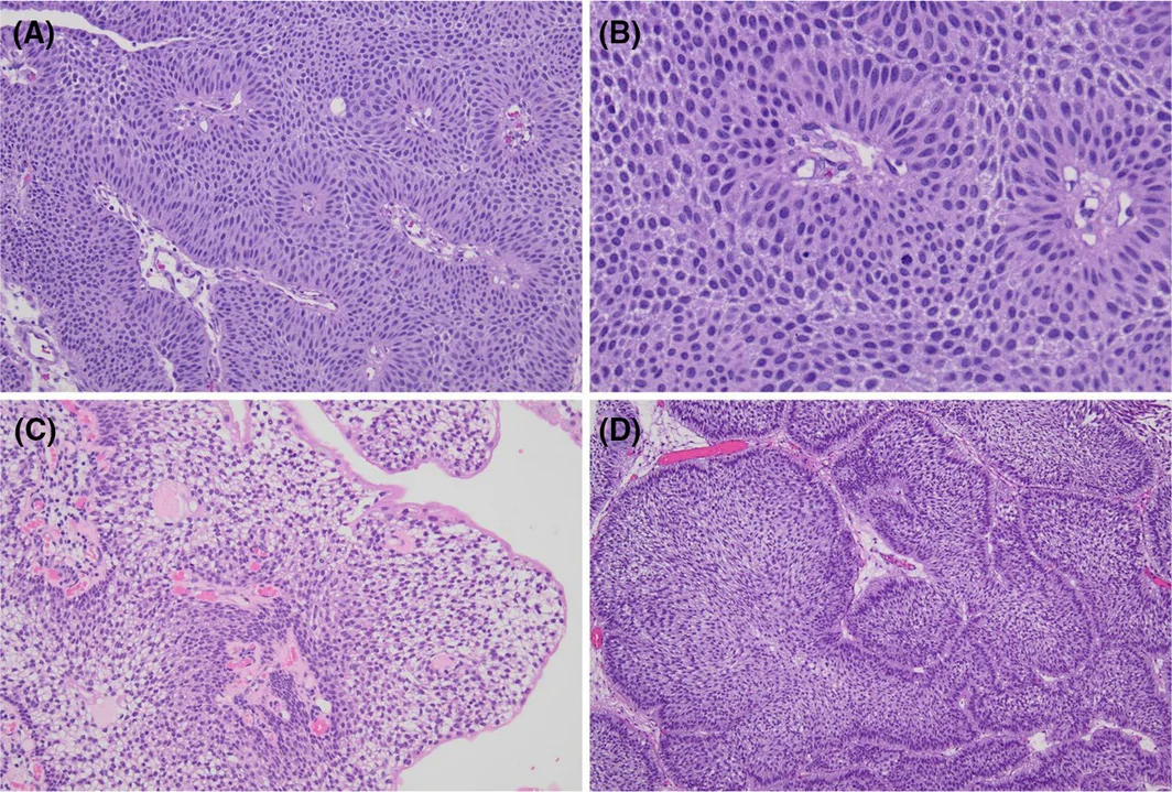
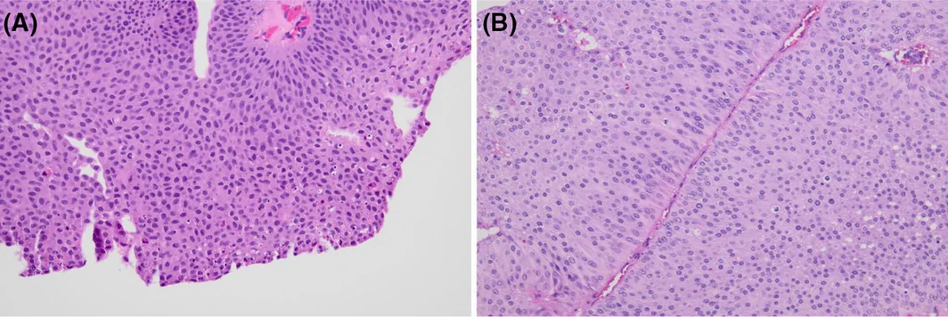
- 方法:2012–2023 年 472 例非浸润性乳头状尿路上皮癌由两位泌尿病理医生复阅,分别按 WHO 2004/2016、混合三级、混合四级及研究性五级框架(I、IIA、IIB、IIIA、IIIB,综合结构、细胞学与增殖特征)分级;图 1–6 为各级别镜下示例,图 3 专门区分 IIA 与 WHO 2022「以低级别为主伴局灶高级别成分」。
- 结果:异型性越高,肿瘤越大、越多灶,进展风险逐级升高,事件集中于 IIIA、IIIB。各系统对复发的区分度均有限(C 指数 0.54–0.56)。
- 预测 T1+ 分期进展:WHO 2004/2016 AUC 0.754,混合三级、四级、五级分别为 0.815、0.828、0.827(均 P < 0.001);混合三级制 AIC/BIC 最优,五级框架 C 指数最高(0.818)。作者认为收益主要来自混合三级制,支持进展风险的组织形态学连续谱。
收录范围:PMC 开放获取全文(CC BY 4.0)——摘要、引言、材料与方法、结果、讨论、结论、作者贡献与知情同意声明,图 1–9、表 1–4 中英对照;利益冲突声明、数据可用性声明与参考文献未收录(正文保留文献序号)。
摘要
组织学分级是非肌层浸润性膀胱癌(NMIBC)进展的关键预测因素。WHO 1973 三级分级系统受限于 G2 类别内部的异质性,而 WHO 2004/2016 二级分类将生物学特征各异的肿瘤归入宽泛的低级别(LG)和高级别(HG)类别,降低了预后区分能力。本研究旨在评估混合分级系统相较于 WHO 2004/2016 的预后预测性能,同时进一步刻画非浸润性乳头状尿路上皮癌的组织形态学连续谱。
由两名泌尿病理医师复核非浸润性乳头状尿路上皮癌病例(2012–2023 年;n = 472),并依据 WHO 2004/2016、混合三级及四级分级系统,以及整合结构、细胞学和增殖特征的研究性五级分级框架(I、IIA、IIB、IIIA、IIIB 级)进行重新分类。主要终点为复发、级别进展及进展至 T1 及以上分期,采用 ROC 分析、Cox 比例风险回归及模型性能指标(AIC、BIC、C‐index)进行评估。
形态学异型性增加与肿瘤体积更大、多灶性更明显及进展风险逐步升高相关,事件集中于 IIIA 级和 IIIB 级。所有系统对复发的区分能力均有限(C‐index 0.54–0.56)。对于进展至 T1 及以上分期,区分能力随分层细化而提高(WHO 2004/2016 的 AUC 为 0.754,混合三级、四级和五级分级系统的 AUC 分别为 0.815、0.828 和 0.827;所有 P < 0.001)。混合 3 级模型的 AIC 和 BIC 最优,而五级模型的 C‐index 最高(0.818)。
与 WHO 2004/2016 相比,混合分级系统改善了非浸润性乳头状尿路上皮癌的进展风险分层,其中大部分获益通过混合 3 级框架即可实现。这些发现支持进展风险具有组织形态学连续谱这一概念,并支持继续完善以生物学为依据的混合分级,以用于 NMIBC 风险分层。
引言
组织学级别是非肌层浸润性膀胱癌(NMIBC)进展的重要预测因素,尽管其预测肿瘤复发的相关性较低。 1 , 2 , 3 , 4 , 5 在过去一个世纪中,人们提出了多种分级系统,以改善 NMIBC 的风险分层并指导临床管理。 4 , 6 , 7 , 8 , 9 , 10 , 11 , 12 , 13 其中,全球应用最广泛的两种系统是世界卫生组织(WHO)1973 三级分类(级别 [G]1、G2 和 G3)及 WHO 2004/2016 分类(乳头状瘤、低度恶性潜能乳头状尿路上皮肿瘤(PUNLMP)、低级别(LG)和高级别(HG))。 14 , 15 , 16 美国泌尿外科学会(AUA)认可 WHO 2004/2016 系统,而欧洲泌尿外科学会(EAU)建议同时使用 WHO 1973 和 WHO 2004/2016 方案进行双重分级。 17 , 18 , 19
WHO 1973 分级系统的局限在于组织学标准定义不明确,导致观察者间变异较大,且大多数肿瘤集中于具有异质性的中间 G2 类别。这一局限促成了 1998 年修订版 WHO/ISUP 分类的制定,该分类引入了 PUNLMP、LG 和 HG 尿路上皮癌类别。 8 该系统随后于 2004 年被 WHO 采纳,并在后续更新中保留。 15 , 16 , 20 与 WHO 1973 相比,WHO 2004/2016 系统提供了更清晰的形态学定义,并提高了可重复性;然而,该系统推出时并无充分证据证明其预后预测性能更优。 21
WHO 1973 和 WHO 2004/2016 系统无法直接比较,因为两者所依据的概念框架根本不同,且级别类别仅部分重叠。评估这些系统的证据表明,两者均未能对复发或进展提供最佳的预后区分能力。 1 , 2 , 22
尽管 WHO 1973 通常对进展风险的分层略优,但 WHO 2004/2016 模型将生物学上异质的肿瘤压缩至过于宽泛的类别中,尤其是在 HG 组内,从而限制了预后区分能力,并掩盖了行为各异、对 BCG 等膀胱内治疗反应不同的肿瘤之间具有临床意义的生物学差异。 22 随着分子、增殖及临床病理数据不断积累,证实传统定义的 HG 肿瘤内部存在显著的生物学多样性,包括进展风险、基因组不稳定性、分子亚型及治疗反应性的差异,这一局限日益受到重视。 23 , 24
越来越多的证据表明,混合分级系统通过整合两种 WHO 分类的优势,可能更好地反映这一组织形态学连续谱。 1 , 25 过去十年间,有研究提出了混合 3 级分级系统,在保留 WHO 2004/2016 LG 类别的同时,依据 WHO 1973 原则,将临床上具有异质性的 HG 组进一步划分为 HG/G2 和 HG/G3。 1 , 4 , 25 , 26 , 27 , 28 重要的是,不应仅将这些混合系统视为替代性分类方案,而应将其视为以生物学为依据的改进,旨在更好地反映乳头状尿路上皮癌内在的形态学连续谱。混合分级方法保留了二级分级(LG 与 HG)在可重复性方面的优势,同时恢复了 HG 类别内部具有临床意义的分层,可能有助于更好地识别进展风险增加的患者,并减少对所有 HG 病变采用统一管理所带来的治疗不足或过度治疗的可能性。
然而,传统分级系统与混合分级系统之间的直接比较验证仍然有限,非浸润性乳头状尿路上皮癌的完整组织形态学连续谱也尚未得到全面刻画。现有分级系统还主要依赖定性形态学描述,对更客观或可半定量的组织学参数整合有限,导致在界定级别类别之间具有生物学意义的过渡时仍存在模糊性。因此,本研究旨在评估混合分级系统相较于 WHO 2004/2016 分类的预后预测性能,同时采用结构化的研究性五级框架,进一步研究非浸润性乳头状尿路上皮癌的组织形态学连续谱。该框架纳入了更精细的结构、细胞学及增殖特征,包括核分裂活性、凋亡活性、核大小、多形性、染色质特征及结构紊乱程度。该框架并非旨在作为常规临床应用的实用分级系统,而是作为研究工具,用于更好地刻画肿瘤异质性、细化现有组织学分级标准,并识别具有生物学和临床意义的分层阈值,从而可能支持将混合分级模型改进为在生物学上更连贯、在临床上更实用的模型。
材料与方法
本研究获克利夫兰诊所基金会机构审查委员会批准。我们对克利夫兰诊所 2012 至 2023 年间本机构所有非浸润性乳头状尿路上皮癌进行了回顾性复核。
队列选择
克利夫兰诊所 PIRO(病理信息检索优化器), 29 是本机构基于网络的平台,可高效进行诊断病理档案的文本检索;我们使用以下关键词检索了 10 年间(2012–2023)的记录:“膀胱”“非浸润性”“尿路上皮”和“癌”。调取所有诊断为非浸润性乳头状尿路上皮癌的经尿道膀胱肿瘤切除术(TURBT)/膀胱活检标本,并由两名泌尿病理医师(J.K.N. 和 R.A.)在对原始诊断及临床结局设盲的情况下复核所有可获得的组织学切片,以确认诊断并对肿瘤进行分级。
临床病理纳入标准与 AUA NMIBC 风险类别一致,即 LG 肿瘤仅限于 AUA 低风险疾病,而 HG 肿瘤包括 AUA 中风险及高风险疾病。 18 具有极高风险特征的患者被排除,包括 BCG 无应答、淋巴血管侵犯、原位癌(CIS)及前列腺部尿道侵犯。此外,含有 WHO 认可的组织学亚型(包括微乳头状、浆细胞样、巢状、肉瘤样及其他亚型)的病例亦被排除,以便在常规型非浸润性乳头状尿路上皮癌中评估分级,避免亚型组织学对预后的混杂影响。由此形成的队列代表了经临床筛选的常规型非浸润性乳头状尿路上皮癌谱系,旨在不受已确立的极高风险特征影响的情况下,促进评估从 LG 到 HG 连续谱中的组织形态学及生物学异质性。我们还排除了再分期标本,其定义为初次诊断后最初 90 天内进行的任何随访 TURBT 标本。
查阅电子病历,获取以下人口学、临床及病理信息:患者初次切除时的年龄、性别、切除日期、肿瘤大小(依据膀胱镜报告)、肿瘤灶数、后续切除的详细信息、复发日期、进展日期及类型(级别或分期),以及转移的发生情况。
病理复核:肿瘤分级的诊断标准
采用结构化五级系统(I、IIA、IIB、IIIA 和 IIIB)(表 1)对肿瘤进行分级,该系统旨在整合并扩展 WHO 1973 和 2004/2016 分类的既有标准,以及既往描述的混合分级方案。 4 , 7 , 14 , 15 , 16 , 30 在切片复核前,通过整合 WHO 及既往发表的混合分级参数,制定各级别的分级标准;研究期间,当其他形态学特征被一致识别为可重复的组织学模式时,也将其纳入。肿瘤分级在概念上被视为一个生物学连续谱,并通过对结构组织、细胞学异型性及增殖活性的综合评价进行评估,旨在反映传统低级别与高级别类别之间的完整形态学谱系。
| 级别 | 结构 | 细胞学 | 增殖 | 其他特征 | 大致对应关系 a |
|---|---|---|---|---|---|
| I 级 经典 LG | 极向及细胞黏附性保留;伞细胞可能保留;基底栅栏状排列、局灶上皮流线状排列 | 细胞核小而均一(约为淋巴细胞大小的 2×);染色质细腻;核膜光滑;核仁不明显;N:C 比值低 | 核分裂象罕见(<2/10 HPF),主要局限于基底层;凋亡罕见 | 可见基底栅栏状排列、核沟及局灶透明胞质 | WHO 2004/2016 LG;WHO 1973 1 级 |
| IIA 级 LG 谱系高端 | 以 LG 结构为主,伴局灶(<5% 肿瘤体积)结构紊乱、轻度极向消失及局灶细胞黏附性丧失;其余背景排列有序 | 局灶核增大(约为淋巴细胞大小的 3–4×);深染;无多形性 | 局灶核分裂活性增加(2–4/10 HPF),包括偶见基底层以上的核分裂;伴凋亡细胞簇 | 伞细胞局灶减少;缺乏 HG 病变的弥漫性异型性及增殖活性;不包括含有任何明确 HG 形态的肿瘤 b | 过渡性的 LG 谱系高端;无直接对应的 WHO 类别 |
| IIB 级 HG 谱系低端 | 以结构紊乱为主,伴乳头融合及分支;细胞黏附性降低;伞细胞显著减少 | 细胞核增大、深染(约为淋巴细胞大小的 3–4×);轻至中度多形性;开始出现核仁 | 核分裂象多见(约 5–7/10 HPF),延伸至基底层以上;凋亡细胞聚集;偶见局灶坏死 | 代表 HG 形态学谱系的低端 | 混合分级 HG‐G2;WHO 2004/2016 HG 谱系低端 |
| IIIA 级 经典 HG | 结构显著紊乱;极向消失;乳头融合和/或局灶实性生长;伞细胞缺失 | 细胞核大且呈多形性(>5× 淋巴细胞大小);弥漫性深染;核仁明显;核沟消失 | 核分裂象多见,包括非典型核分裂象;凋亡多见 | 充分表现出常规型 HG 尿路上皮癌形态 | WHO 2004/2016 HG;WHO 1973 3 级;混合分级 HG‐G3 |
| IIIB 级 HG 伴核间变 | 乳头状/实性生长模式;极向及细胞黏附性完全消失 | 极重度细胞学异型性;显著核间变(细胞核 >4× 相邻肿瘤细胞核大小);高度多形性 | 大量非典型核分裂象;广泛凋亡;坏死常见 | 代表 HG 细胞学异型性的极端 | HG 谱系的间变端;无直接对应的 WHO 类别 |
HG,高级别;HPF,高倍视野;LG,低级别;N:C,核质比;WHO,世界卫生组织。
大致对应关系仅用于概念比较,并不意味着分级系统之间直接等同。
IIA 级不对应于 WHO 2022 中以低级别为主、伴局灶(<5%)高级别成分的乳头状尿路上皮癌类别。任何含有明确常规高级别形态的肿瘤,无论范围大小,均归入高级别谱系(IIB 级或 IIIA 级)。
结构评估包括乳头构型(纤细或融合/分支,可呈外生性或内生性)、细胞黏附程度、极向保留或消失,以及伞细胞的有无。评估的细胞学特征包括核大小(相对于淋巴细胞)、染色质模式、核膜轮廓、核仁显著程度及多形性程度。从混合系统中纳入的其他细胞学特征包括核沟的存在及胞质特征(例如 LG 病变中的透明胞质)。在有代表性的热点区域对增殖活性进行半定量评估,包括核分裂频率、核分裂象分布(基底层或全层)、非典型核分裂象的存在,以及凋亡细胞聚集和肿瘤坏死的存在及范围。通过这种结构化、纲要式方法,肿瘤分级在概念上被视为一个生物学连续谱,使得能够反映并比较完整的形态学变异谱,具体如下:
I 级(经典 LG)(图 1A–D)定义为乳头纤细、极向保留、尿路上皮细胞黏附性良好且伞细胞保留,常伴上皮流线状排列。细胞学异型性极轻,表现为细胞核小而均一,大小约为淋巴细胞的两倍,染色质细腻、核膜光滑、核仁不明显及核质比低。核分裂活性罕见(<2 个核分裂象/10 HPF),主要局限于基底层,仅偶见凋亡。其他支持性特征包括基底栅栏状排列、核沟及局灶透明胞质。
IIA 级(LG 谱系高端)(图 2A,B)由以 LG 为主的肿瘤构成,其中占肿瘤体积 <5% 的局灶区域出现早期偏离经典 LG 形态的结构及细胞学变化。这些病灶在其余排列有序的背景中,呈现局灶结构紊乱、轻度极向消失及局灶细胞黏附性丧失。细胞学异型性包括核增大(约为淋巴细胞大小的 3–4×)、深染,但无多形性。增殖活性局灶增加(2–4 个核分裂象/10 HPF),核分裂象延伸至基底层以上,并伴凋亡细胞簇。伞细胞局灶减少。重要的是,这些局限病灶缺乏 IIB 级和 IIIA 级所特有的弥漫性增殖活性及广泛的细胞学异型性(图 3A,B)。应当指出,IIA 级不对应于 WHO 2022 中以 LG 为主、伴局灶(<5%)HG 成分的乳头状尿路上皮癌类别(图 3C,D)。 20 在我们的框架中,只要存在任何明确的常规 HG 形态,无论范围大小,均归入 HG 谱系(IIB 级或 IIIA 级);其依据是已有证据表明,级别异质性肿瘤代表在生物学上具有进展性的病变。 31 , 32 , 33 因此,IIA 级仅限于缺乏明确 HG 分化、仅表现出常规 LG 与 HG 形态之间有限过渡性特征的肿瘤。
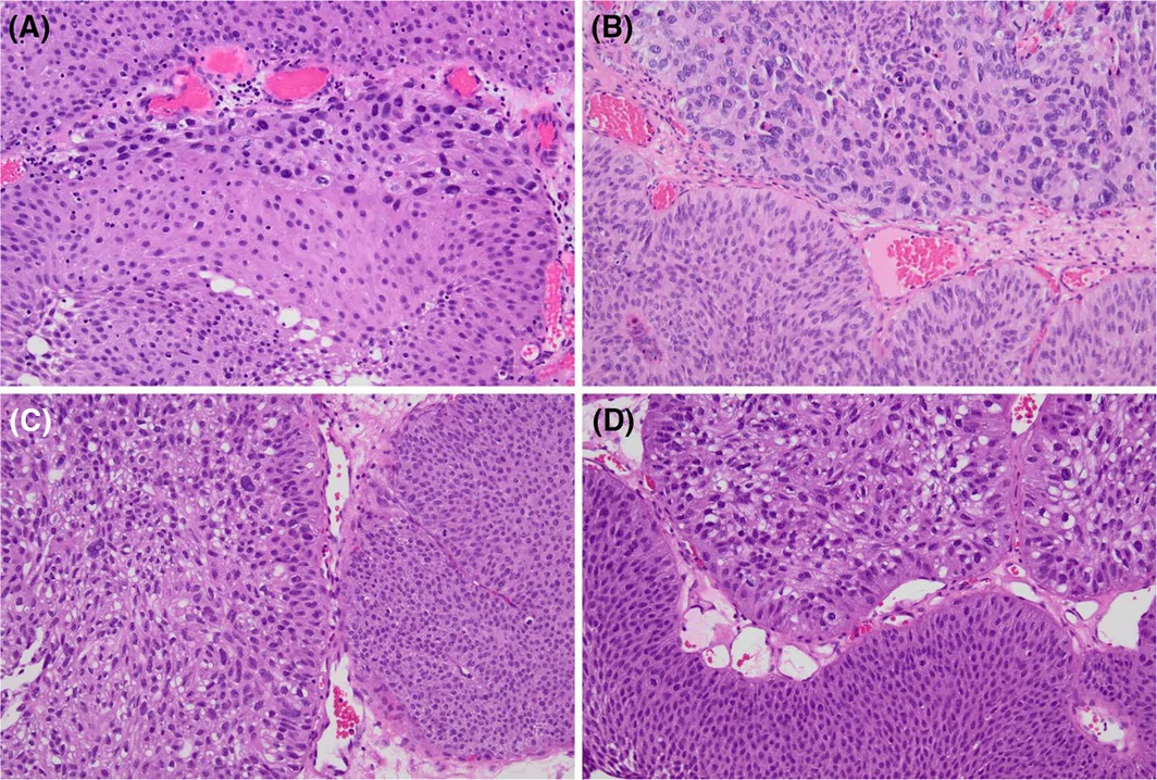
IIB 级(HG 谱系低端)(图 4A–D)表现为以结构紊乱为主,伴乳头融合及分支、细胞黏附性降低及伞细胞显著减少。细胞学异型性更明显,表现为细胞核增大、深染(约为淋巴细胞大小的 3–4×)、轻至中度多形性,并开始出现核仁。核分裂活性频繁(≥3/HPF 或约 5–7 个核分裂象/10 HPF),常伴凋亡细胞聚集,偶见局灶坏死。这些肿瘤代表 HG 形态学谱系的低端。
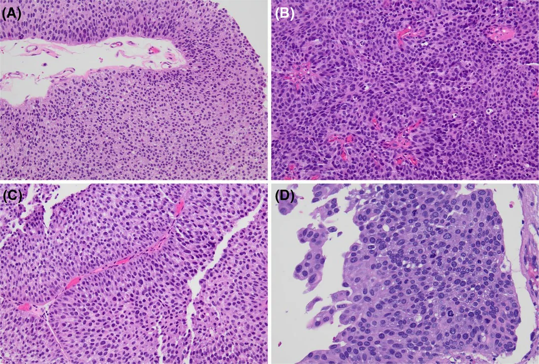
IIIA 级(经典 HG)(图 5A,B)的特征为结构显著紊乱,伴极向消失、乳头融合和/或实性区域,以及伞细胞完全缺失。细胞学异型性严重,包括细胞核大且呈多形性(>5× 淋巴细胞大小)、弥漫性深染、核仁明显及核沟消失。核分裂象多见,包括非典型核分裂象,并伴频繁凋亡。这些特征充分表现出常规型 HG 尿路上皮癌形态。
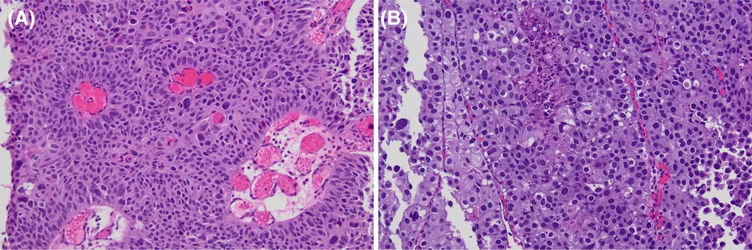
IIIB 级(HG 伴核间变)(图 6A,B)表现出最严重程度的细胞学异型性,包括实性生长、极向及黏附性完全消失,以及显著核间变;核间变定义为细胞核大小超过相邻肿瘤细胞核的四倍。肿瘤表现为高度多形性、非典型核分裂象多见、广泛凋亡,且常伴坏死。此类别代表 HG 细胞学异型性的极端。
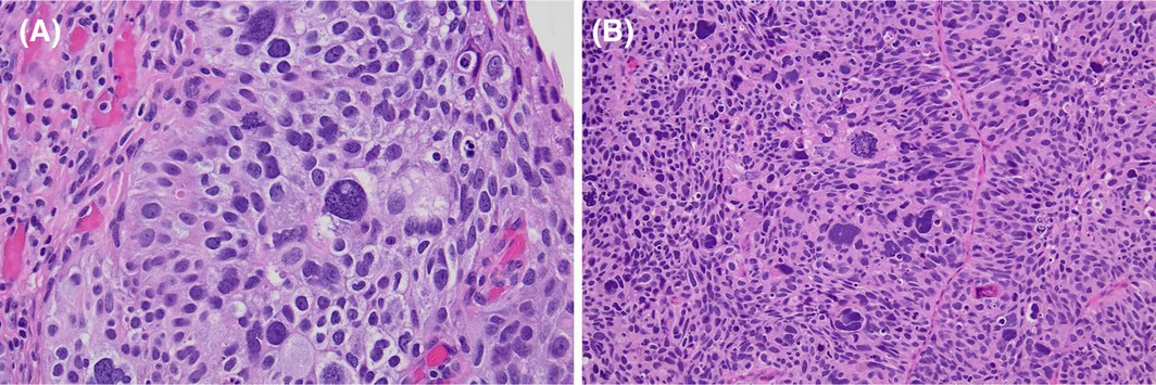
五级框架旨在评估并交叉考察非浸润性乳头状尿路上皮癌生物学谱系中的形态学过渡,而非暗示存在五种临床上不同的管理类别。为与既有分级系统比较,I 级对应 WHO 1973 G1,IIIA 级对应 WHO 1973 G3。中间类别(IIA 和 IIB)代表低级别与高级别疾病之间的过渡性形态学谱系,这得到了既往描述的混合分级模型的支持。在比较分析中,I 级及 IIA 级在 WHO 2004/2016 框架内合并为 LG,IIB 级对应混合三级系统中的 HG‐G2,IIIA/IIIB 级对应 HG‐G3。
所有切片均由两名泌尿病理医师(J.K.N. 和 R.A.)共同复核,复核时不知晓原始病理诊断/级别及临床结局。观察者间一致性分析并非本研究的主要目的。依据为连续形态学分级框架制定的预设研究标准,经双方实时达成一致后确定分级。最终分类依据所识别的最高级别成分(无论范围大小)确定,同时保留连续形态学谱系这一概念框架。
研究结局
本研究的主要终点为复发、级别进展及分期进展。复发定义为初次切除 90 天后疾病的任何再次出现。级别进展定义为级别升高至 HG;分期进展定义为 T 分期从 Ta 升高至 T1 或更高(经组织学证实),包括淋巴结转移或远处转移,但不包括 90 天内再次切除时发现的事件。
至进展时间从 TURBT 日期起计算,未发生进展的患者在末次随访时删失。患者随访至末次临床就诊、进展或死亡。
统计分析
采用描述性统计汇总患者人口学资料及基线临床病理特征。根据情况,采用 Wilcoxon 秩和检验或 Kruskal–Wallis 检验比较连续变量,采用卡方检验比较分类变量,或在单元格期望频数较小时采用 Fisher 精确检验。数据根据情况以均值 ± 标准差(SD)或例数及百分比报告。
采用受试者工作特征(ROC)曲线分析及事件发生时间分析方法,评估所研究分级系统对复发及进展至 T1 及以上分期的预后预测性能。采用 ROC 曲线下面积(AUC)及其 95% 置信区间(CIs)量化区分能力。基于所有分级系统共用的完整病例数据集,采用迭代 10,000 次的自助重抽样法进行 AUC 的两两比较。
采用 Cox 比例风险回归模型,对复发及进展至 T1 及以上分期进行事件发生时间分析。以最低级别类别为参照组,为每个分级系统构建单变量模型。报告风险比(HRs)及其 95% CIs。采用 Schoenfeld 残差评估比例风险假设。
为比较模型的整体性能,计算每个 Cox 模型的似然比(LR)卡方统计量、赤池信息准则(AIC)、贝叶斯信息准则(BIC)及 Harrell 一致性指数(C‐index)及其 95% CIs。较低的 AIC 和 BIC 值被解释为模型拟合与复杂度之间的平衡更佳,而较高的 C‐index 值表示区分能力更强。
所有统计分析均使用 R 软件(版本 4.6.0)完成。所有检验均为双侧检验,P 值 <0.05 被认为具有统计学意义。
结果
检索策略与组织形态学重新分类
PIRO 检索策略最初识别出 1,733 例被诊断为非浸润性乳头状尿路上皮癌的患者,根据原始病理报告,包括 702 例 LG、173 例以 LG 为主伴局灶 HG 以及 858 例 HG 肿瘤(图 7)。应用研究纳入标准后,排除了 494 例 LG、49 例以 LG 为主伴局灶 HG 和 399 例 HG 病例。另外 106 例 LG 和 213 例 HG 病例因无法获得组织学材料而未能进行病理复核。最终研究队列由 472 例具有可供集中病理复核的组织且符合全部研究纳入标准的患者组成,包括 102 例 LG、124 例以 LG 为主伴局灶 HG 和 246 例 HG 肿瘤(图 7)。
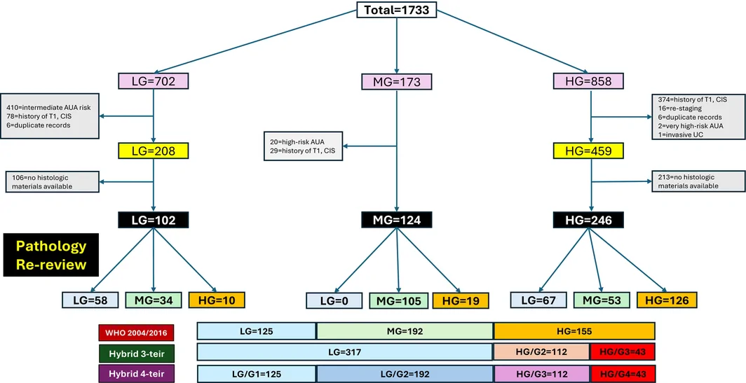
采用预先定义的研究性框架进行联合组织病理学复核后,所有肿瘤均被重新归入五种形态学类别之一,这五种类别代表了结构、细胞学及增殖异型性的连续谱(图 7)。重新分类显示,传统 LG 与 HG 形态之间呈逐步过渡,而非截然分开的二元划分;中间类别涵盖了在极向、黏附性、核异型性、核分裂活性和凋亡负荷方面逐渐出现偏离的肿瘤。
形态学谱系各类别的临床病理特征
表 2 总结了按研究性五级框架分层的人口统计学、临床病理及肿瘤学结局特征。患者总体平均年龄为 71.9 岁,75.8%(358/472)为男性。肿瘤平均大小为 2.8 cm,39.4%(186/472)的肿瘤为多灶性。
| 合计(n = 472) | I (n = 125) | IIA (n = 192) | IIB (n = 112) | IIIA (n = 36) | IIIB (n = 7) | P 值 | |
|---|---|---|---|---|---|---|---|
| 年龄,岁(均值 ± SD) | 71.9 ± 11.7 | 70.8 ± 11.8 | 71.2 ± 11.5 | 73.9 ± 12.2 | 73.0 ± 10.8 | 72.3 ± 11.9 | 0.152 |
| 性别(男性 %) | 358 (75.8%) | 95 (76.0%) | 144 (75.0%) | 88 (78.6%) | 26 (72.2%) | 5 (71.4%) | 0.90 |
| 肿瘤大小,cm(均值 ± SD) | 2.8 ± 1.7 | 2.3 ± 1.2 | 3.0 ± 1.9 | 2.9 ± 1.8 | 3.3 ± 1.7 | 4.3 ± 2.5 | 0.006 |
| 病灶分布(多灶性 %) | 186 (39.4%) | 27 (21.6%) | 83 (43.2%) | 54 (48.2%) | 20 (55.6%) | 2 (28.6%) | <0.001 |
| 随访时间(月,均值 ± SD) | 41.8 ± 28.3 | 40.5 ± 28.4 | 46.0 ± 31.0 | 36.7 ± 24.8 | 38.4 ± 21.6 | 44.5 ± 19.6 | 0.155 |
| 复发,n (%) | 168 (35.6%) | 35 (28.0%) | 66 (34.4%) | 42 (37.5%) | 21 (58.3%) | 4 (57.1%) | 0.011 |
| 复发时间(月,均值 ± SD) | 16.3 ± 17.2 | 18.3 ± 20.5 | 15.8 ± 16.8 | 15.2 ± 14.8 | 13.2 ± 12.3 | 34.9 ± 32.1 | 0.371 |
| 进展(任何类型),n (%) | 51 (10.8%) | 3 (2.4%) | 25 (13.0%) | 6 (5.4%) | 13 (36.1%) | 4 (57.1%) | <0.0001 |
| 级别进展(%) | 23 (4.9%) | 2 (1.6%) | 21 (10.9%) | N/A | N/A | N/A | |
| 进展至 T1 分期(%) | 17 (3.6%) | 1 (0.8%) | 3 (1.6%) | 4 (3.6%) | 7 (19.4%) | 2 (28.6%) | |
| 进展至 T2 分期(%) | 5 (1.1%) | 0 (0%) | 0 (0%) | 2 (1.8%) | 3 (8.3%) | 0 (0%) | |
| 转移性进展(%) | 4 (0.8%) | 0 (0%) | 1 (0.5%) | 0 (0%) | 3 (8.3%) | 0 (0%) | |
| 进展时间(月,均值 ± SD) | 21.5 ± 21.9 | 28.4 ± 20.2 | 19.8 ± 22.5 | 26.8 ± 26.2 | 15.4 ± 13.1 | 56.8 ± 33.9 | 0.423 |
NA,不适用。
总体上,35.6%(168/472)的患者发生复发,7.2%(23/317,排除 HG 肿瘤)出现级别进展,5.5%(26/472)出现分期进展。具体而言,3.6%(17/472)进展至 T1 期,1.1%(5/472)进展至 T2 期,0.8%(4/472)发生转移性进展。平均复发时间和进展时间分别为 16.3 和 21.5 个月。
各形态学类别之间的年龄(范围 70.8–73.9 岁;P = 0.152)或性别分布(男性比例 71.4%–78.6%;P = 0.90)均无统计学显著差异。相比之下,肿瘤大小沿分级连续谱逐步增大,从 I 级的 2.3 cm 增至 IIIB 级的 4.3 cm(P = 0.006)。多灶性同样与形态学异型性的增加密切相关,从 I 级肿瘤的 21.6% 增至 IIIA 级肿瘤的 55.6%(P < 0.001)。
肿瘤学结局也显示出沿形态学谱系逐步变化的生物学梯度。复发率从 I 级肿瘤的 28.0% 逐渐升至 IIIA 级的 58.3% 和 IIIB 级的 57.1%(P = 0.011)。同样,进展风险呈现显著的级别依赖性升高,尤其见于 IIIA 和 IIIB 级(P < 0.0001)。值得注意的是,各种形式的进展,包括级别进展、T1 进展、T2 进展和转移性进展,均不成比例地集中于形态学谱系的高端,尤其是 IIIA 和 IIIB 级(表 2)。尽管各组之间的进展时间无显著差异(P = 0.423),但 IIIA 级肿瘤的进展间隔在数值上较短。
传统与混合分级系统对肿瘤复发的预后预测性能
472 例患者中有 168 例(35.6%)发生肿瘤复发。单变量 Cox 回归显示,在每种分级系统内,随着形态学类别逐步升高,复发风险随之增加(表 3)。在 WHO 2004/2016 分类中,与 LG 肿瘤相比,HG 肿瘤的复发风险增加(HR 1.56,95% CI 1.15–2.13;P = 0.005)。
| 变量/水平 | 复发 | 进展至 T1 及以上分期 | ||
|---|---|---|---|---|
| HR (95% CI) | P 值 | HR (95% CI) | P 值 | |
| WHO 2004/2016 | ||||
| LG | — | — | ||
| HG | 1.56 (1.15, 2.13) | 0.005 | 9.97 (3.74, 26.58) | <0.001 |
| 混合三级 | ||||
| LG | — | — | ||
| HG‐G2 | 1.36 (0.95, 1.96) | 0.092 | 3.94 (1.20, 12.96) | 0.024 |
| HG‐G3 | 2.07 (1.34, 3.22) | 0.001 | 26.09 (9.41, 72.31) | <0.001 |
| 混合四级 | ||||
| LG‐G1 | — | — | ||
| LG‐G2 | 1.22 (0.81, 1.83) | 0.349 | 2.43 (0.27, 21.78) | 0.427 |
| HG‐G3 | 1.54 (0.98, 2.42) | 0.058 | 7.44 (0.89, 61.93) | 0.063 |
| HG‐G4 | 2.35 (1.40, 3.92) | 0.001 | 49.28 (6.49, 373.98) | <0.001 |
| 研究性五级 | ||||
| I | — | — | ||
| IIA | 1.22 (0.81, 1.83) | 0.349 | 2.43 (0.27, 21.76) | 0.427 |
| IIB | 1.54 (0.98, 2.42) | 0.058 | 7.46 (0.90, 62.11) | 0.063 |
| IIIA | 2.53 (1.47, 4.35) | <0.001 | 54.07 (7.04, 415.00) | <0.001 |
| IIIB | 1.70 (0.61, 4.80) | 0.313 | 31.78 (2.88, 350.78) | 0.005 |
分别针对复发(N = 472;168 个事件)和进展至 T1 及以上分期(N = 472;26 个事件)拟合单变量 Cox 比例风险模型。每种分级系统均以最低级别类别作为参照水平。
CI,置信区间;HR,风险比。
在混合三级系统中,HG‐G3 肿瘤的复发风险显著增加(HR 2.07,95% CI 1.34–3.22;P = 0.001),而 HG‐G2 肿瘤的关联较弱(HR 1.36,95% CI 0.95–1.96;P = 0.092)。混合四级和研究性五级系统中也观察到类似趋势,总体上级别逐步升高与复发风险增加相关。
尽管存在上述关联,所有分级系统对复发的总体区分性能均有限(表 4A)。C-index 从 WHO 2004/2016 的 0.544 到混合四级和研究性五级系统的 0.563 不等。随着形态学分层细化,似然比统计量小幅增加,但 AIC 和 BIC 的差异较小。混合三级模型的 AIC 最低(1894.5),提示其在预测复发时,模型拟合度与复杂度之间的平衡最佳。
| A. 复发(N = 472;事件数 = 168) | ||||||
|---|---|---|---|---|---|---|
| 模型 | LR χ2 | Df | LR P 值 | AIC | BIC | C-index(95% CI) |
| WHO 2004/2016 | 7.67 | 1 | 0.006 | 1895.1 | 1898.2 | 0.544 (0.506, 0.582) |
| 混合三级 | 10.31 | 2 | 0.006 | 1894.5 | 1900.7 | 0.549 (0.510, 0.589) |
| 混合四级 | 11.21 | 3 | 0.011 | 1895.6 | 1904.9 | 0.563 (0.520, 0.605) |
| 研究性五级 | 11.78 | 4 | 0.019 | 1897.0 | 1909.5 | 0.563 (0.521, 0.606) |
AIC/BIC 值越低,表示在考虑模型复杂度后模型拟合越好;C-index 值越高,表示区分能力越强。
AIC,赤池信息准则;BIC,贝叶斯信息准则;C-index,Harrell 一致性指数;LR χ2,似然比卡方统计量。
进展至 T1 及以上分期的预测
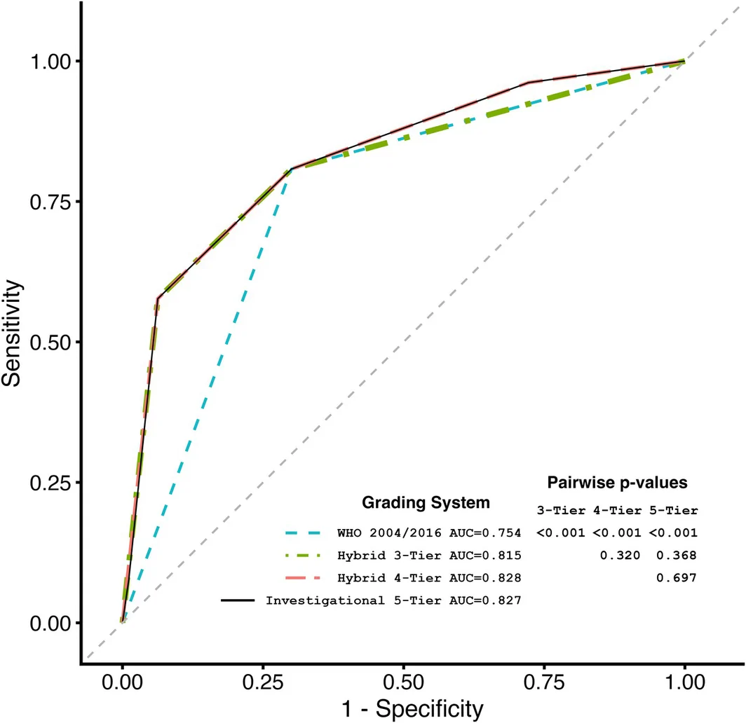
472 例患者中有 26 例(5.5%)进展至 T1 及以上分期。ROC 分析显示,随着形态学分层细化,区分能力提高(图 8)。WHO 2004/2016 系统的 AUC 为 0.754,而混合三级、混合四级及研究性五级系统的 AUC 分别为 0.815、0.828 和 0.827。两两比较显示,与 WHO 2004/2016 相比,所有多级系统的区分能力均显著提高(均为 P < 0.001),而三种多级系统之间的差异无统计学意义。
单变量 Cox 回归分析同样显示,随着形态学类别逐步升高,进展风险逐渐增加(表 3)。在 WHO 2004/2016 系统中,与 LG 肿瘤相比,HG 肿瘤的进展风险显著增加(HR 9.97,95% CI 3.74–26.58;P < 0.001)。
在混合三级系统中,HG‐G2 肿瘤的进展风险增加(HR 3.94,95% CI 1.20–12.96;P = 0.024),而 HG‐G3 肿瘤的风险明显更高(HR 26.09,95% CI 9.41–72.31;P < 0.001)。混合四级和研究性五级系统中也观察到类似的阶梯式升高。研究性五级系统中的 IIIA 级风险最高(HR 54.07,95% CI 7.04–415.00;P < 0.001)。
模型性能分析印证了 ROC 结果(表 4B)。似然比卡方统计量从 WHO 2004/2016 的 28.73 分别增至混合三级、混合四级和研究性五级系统的 46.24、46.98 和 47.53。一致性指数同样从 0.745 分别提高至 0.806、0.814 和 0.818。尽管研究性五级系统的一致性最高,混合三级模型的 AIC(250.6)和 BIC(253.1)最低,表明其在预后预测性能与模型复杂度之间取得了最佳平衡。
讨论
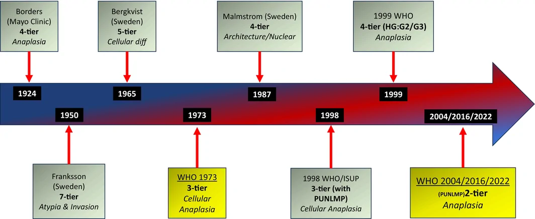
过去一个世纪中,膀胱癌分级系统经历了多次改进,反映了为提高诊断一致性和预后预测准确性而持续作出的努力(图 9)。 4 , 6 , 7 , 8 , 9 , 10 , 11 , 12 , 13 早期系统,如 Broders 12 (1922)和 Franksson 11 (1950)系统,依赖对细胞间变和异型性的主观评估,而后续分类则引入了基于细胞分化和结构紊乱的更为系统化的标准(如 Bergkvist 等 10 ,1965;WHO 1973 14 )。WHO/ISUP 1999 13 和 WHO 2004 15 分类带来了重大的概念转变,纳入了 PUNLMP 类别,并最终确立了广泛采用的 PUNLMP、LG 和 HG 肿瘤分类系统,后续 WHO 更新版(2016、2022)又对其进行了进一步完善。 16 , 20 这些系统旨在规范术语,同时使组织学级别与临床结局更紧密地联系起来。WHO 1973 和 WHO 2004/2016 分级系统在非浸润性乳头状尿路上皮癌的预后分层方面均存在重要但不同的局限性。WHO 1973 能更广泛地反映进展风险,但受限于类别内显著的异质性,尤其是 G2 组。相比之下,WHO 2004 提高了组织学判断的可重复性,但预后分辨能力下降,尤其是在将生物学表现各异的肿瘤归为一组的 HG 类别中。 5 , 34 , 35 , 36
近年来,研究重点转向在保持可重复性的同时优化预后分层。Cheng 等 7 (2012)提出了四级系统,而 van der Kwast 等 4 (2021)提出了其他三级方案,国际泌尿病理学会(ISUP)于 2022 年进一步支持了这些方案, 6 均强调对 HG 病变进行具有临床意义的亚分类。混合三级分级系统结合 WHO 1973 和 WHO 2004 分类的特点,将 WHO 2004 LG 肿瘤(包括 PUNLMP)保留为单一类别,并将 HG 肿瘤细分为 G2 和 G3。 4 尽管本研究仅聚焦于非浸润性乳头状尿路上皮癌,因此本队列未纳入 PUNLMP,但预计 PUNLMP 位于所提出形态学连续谱的最低端,与 I 级最为接近。 21 , 22 我们并不建议替代 WHO 的 PUNLMP 诊断类别,而是将其视为代表该连续谱中生物学侵袭性最低的一端。这一解释也与当前临床指南一致,指南对 PUNLMP 与低级别乳头状尿路上皮癌采用相似的管理方式。 18 , 37
本研究不仅旨在比较当代分级系统,还旨在进一步探究非浸润性乳头状尿路上皮癌背后的组织形态学连续谱。我们的结果支持这样的概念:非浸润性乳头状尿路上皮癌沿着一个逐步发展的生物学谱系存在,其特征是累积的结构紊乱、不断增加的细胞学异型性、极向消失、核多形性、染色质异常、核分裂活性和凋亡变化,而不应将肿瘤级别视为一系列彼此独立的类别状态。这一解释与近期综述及混合分级建议高度一致,后者强调膀胱癌生物学的连续性,而非僵化的二元划分。 3 , 26 , 31 , 38
我们的分析得出了若干重要观察结果。首先,复发和进展与组织学级别的关系存在根本差异。在所有评估的分级系统中,复发预测表现均较差,区分性能普遍有限,ROC 曲线接近随机水平。这些结果与既往文献高度一致,既往研究显示,现有分级系统和已建立的 NMIBC 风险模型对复发的预测性能明显弱于对进展的预测性能。 1 , 17 , 39 Soukup 等在 EAU 关于 WHO 分级系统的系统综述中得出结论:现有分级分类对复发的预测仍不理想,未能证明 WHO 2004/2016 系统明确优于 WHO 1973 系统。 2 同样,Ferro 等显示,在传统与混合分级系统中,复发预测的 C-index 值(0.56–0.61)明显低于进展预测(0.74–0.81),突显了复发与进展风险评估之间的生物学差异。 40 Jahnson 等进一步显示,即使采用联合四级分级系统,对复发区分能力的改善也仅有限。 41
值得注意的是,这些观察结果可能反映了 NMIBC 复发背后本质上的多因素生物学机制。分期进展与肿瘤内在侵袭性和生物学潜能的联系更直接,而复发则受到尿路上皮区域不稳定性、多灶性肿瘤发生、经尿道切除不完全、肿瘤种植和再次播散、膀胱内治疗反应及宿主微环境因素的强烈影响。 17 , 37 Teoh 等总结了导致 NMIBC 复发的若干机制,包括肿瘤切除不完全、隐匿病灶、腔内肿瘤播散和区域癌化。 42 分子和基因组研究进一步支持这一概念。Hafner 等 43 提供了腔内肿瘤播散及区域效应引发寡克隆性肿瘤发生两方面的证据。近期,Strandgaard 等 44 在形态正常的尿路上皮中发现了与后续肿瘤发生相关的驱动突变和免疫耗竭特征,而 Marzouka 等 45 显示,复发肿瘤常携带不符合肿瘤之间直接衍生关系的基因组重排,支持持续演化的区域效应模型,而非简单的局部复发。
因此,单独评估时,组织学级别对复发风险的贡献似乎较为有限。当代临床指南同样强调,NMIBC 的复发风险取决于多个相互作用的临床病理变量,而不仅仅是级别。 18 值得注意的是,尽管复发因其对监测负担、操作相关并发症、患者焦虑和医疗费用的影响而仍具有临床意义,但在日常临床实践中,疾病分期进展具有明显更重要的预后和治疗意义。 5 , 17 , 37 , 39 进展为浸润性疾病仍是癌症特异性病残、死亡、根治性治疗需求及长期肿瘤学结局的主要决定因素。与这一概念一致,我们的分析显示,在所有评估的分级系统中,对进展至 T1 及以上分期的区分能力和生物学分层能力均明显强于对复发的相应能力,支持组织学级别主要反映浸润和生物学进展潜能,而非复发倾向这一概念。
与复发相比,进展至 T1 及以上分期与组织学级别之间的关系明显更强,且在生物学上更为一致。在所有评估的分级系统中,对进展的区分性能始终优于对复发的区分性能,支持组织学分级主要反映浸润和生物学进展潜能,而非仅反映复发倾向这一概念。这些结果与既往研究高度一致,后者表明组织学级别是 NMIBC 进展风险最重要的决定因素之一。Soukup 等显示,在多个队列中,级别始终与进展存在强关联。 2 同样,Chen 等显示,WHO 2004/2016 分类在多变量分析中可显著预测进展,但无法预测复发,进一步强调了这些结局之间的生物学差异。 46 Ferro 等同样显示,在传统与混合分级系统中,进展预测的 C-index 值(0.74–0.81)明显高于复发预测(0.56–0.61),这些结果与本研究的结果非常相近。 40
我们的结果进一步提示,增加形态学细化程度可改善进展至 T1 及以上分期的预后分层。与 WHO 2004/2016 系统相比,混合分级系统显示出更优的区分性能,AUC、似然比统计量和一致性指数均更高。值得注意的是,大部分改善通过混合三级系统即可实现,而进一步细分为四级或五级虽然增加了模型复杂度,却仅带来有限的额外收益。这些观察结果与不断出现的证据一致,支持混合分级方法,即在保留 WHO 1973 系统生物学优势的同时,保持 WHO 2004/2016 框架的临床实用性和可重复性。 1 , 2 , 22 , 25 , 30 , 47
在 EAU 对 5145 例 NMIBC 病例开展的大型个体患者分析中,WHO 1973 和 WHO 2004 均可独立预测进展,其中 WHO 1973 的预后预测性能更强。 1 值得注意的是,混合四级分层的 Harrell 一致性指数优于任一单独系统,并在形态异质性的 WHO 2004 HG 肿瘤内识别出差异显著的进展风险。 1 具体而言,5 年进展率从 LG‐G1(1.4%)和 LG‐G2(3.8%)逐步升至 HG‐G2(7.7%)和 HG‐G3(18.8%),表明传统归为一组的高级别病变内部存在具有生物学意义的进展阈值。 1
Downes 等随后验证了这些观察结果,他们在一个具有长期随访的 609 例双重分级 NMIBC 病例队列中,系统比较了 WHO 1973、WHO 2004 和混合分级系统。 26 与单独采用 WHO 1973 或 WHO 2004 相比,混合三级和四级系统对无进展生存和无事件生存的预后预测性能均更优,一致性指数和 AUC 值均更高。 26 显著的进展差异尤其见于传统 WHO 2004 HG 类别内部,支持 HG 肿瘤之间存在显著的生物学异质性。 26 相比之下,LG 亚组的行为相对相似,因此作者建议保留 WHO 2004 LG 类别,同时将 HG 肿瘤细分为生物学上不同的预后组。 26 这些结果与本研究非常相近,本研究中最大的预后区分同样见于形态学谱系的高端,尤其是在 IIIA 与 IIIB 级之间。
Beijert 等开展的当代最大规模混合分级性能评估研究为这一概念提供了进一步支持,该研究纳入三个独立队列的 9712 例原发性 Ta/T1 NMIBC 病例。 25 值得注意的是,亚组分析显示,仅在 Ta 肿瘤中,混合分级系统仍保持其预后预测价值。 25 作者采用了简化的混合三级模型,保留 WHO 2004 低级别类别,同时将 HG 肿瘤细分为 HG‐G2 和 HG‐G3 类别。在所有队列中,HG‐G3 肿瘤的 5 年进展率(18%–20%)均高于 HG‐G2 肿瘤(7%–9%),且混合分级的预后区分能力优于单独采用 WHO 2004。 25 这些结果支持这样的概念:具有临床意义的生物学异质性集中于传统 HG 类别内,将 HG‐G2 与 HG‐G3 分开可能改善风险分层和治疗决策。
在另一项研究中,Beijert 等在一个来自 17 家机构、包含 5170 例原发性 Ta–T1 膀胱肿瘤的大型多中心队列中,专门考察了 HG‐G3 肿瘤的预后意义。 47 HG‐G3 肿瘤是相对少见的亚群,占所有 Ta–T1 肿瘤的 7.5%,根据更新后的 EAU NMIBC 风险分层系统,其中 42% 被归为中危。 47 虽然 HG‐G3 与 HG‐G2 肿瘤的复发率相似,但进展结局存在明显差异,HG‐G2 和 HG‐G3 肿瘤的 5 年进展率分别为 3.6% 和 13%。 47 值得注意的是,HG‐G3 肿瘤的进展时间明显短于 HG‐G2 肿瘤(P < 0.001),支持 HG‐G3 肿瘤是 NMIBC 内生物学上独特的较高风险亚组这一概念。 47 这些结果支持采用更精细的风险评估方法,而非将所有 HG‐G3 肿瘤一律归为高危,并进一步支持将传统 WHO 2004 高级别类别细化为具有临床意义的预后亚组。
本研究结果也与新近提出的尿路上皮癌发生分子模型一致。低级别乳头状肿瘤常与 FGFR3 改变及相对稳定的腔面型乳头状分子表型相关,而 HG 肿瘤更常出现 TP53 通路改变、染色体不稳定性、增殖活性增加,以及与进展相关的基底型或基因组不稳定型特征。 48 , 49 这些观察结果进一步支持 NMIBC 的形态学进展反映了其背后的生物学连续谱这一概念,并提示混合分级系统可能比单纯二元分类更贴近尿路上皮肿瘤的分子演化过程。
值得注意的是,不应将 IIA 级类别视为等同于 WHO 2022 中以低级别为主、伴局灶(<5%)高级别成分的乳头状尿路上皮癌类别。这些中间型病变似乎代表了在出现明确高级别分化之前,结构和细胞学演化的逐步发展阶段。分子及临床病理研究支持这一解释,这些研究显示,伴局灶高级别成分的肿瘤往往具有与进展相关的增殖和基因组改变,提示明确的高级别形态代表连续生物学谱系中较晚的阶段。 31 , 32 , 33 因此,IIA 级旨在涵盖低级别通路中的早期过渡性变化,而非包含真正高级别形态的级别异质性肿瘤。
另一个需要澄清的问题是 IIIB 级与巨细胞尿路上皮癌的区别。尽管 IIIB 级肿瘤所见的显著核多形性和间变可能与巨细胞尿路上皮癌在形态上有所重叠,但本研究中的所有 IIIB 级肿瘤均为常规型非浸润性乳头状尿路上皮癌。IIIB 级代表高级别非浸润性乳头状尿路上皮癌形态学连续谱的极端,而非独立的诊断实体。相比之下,按定义,巨细胞尿路上皮癌是尿路上皮癌的一种浸润性亚型。 16 本研究进一步拓展了这些观察结果,显示逐步加重的结构紊乱、不断增加的细胞学异型性、极向消失及增殖活性与逐渐升高的浸润性进展风险相关。混合分级系统在传统定义的高级别肿瘤中识别出了生物学上不同的亚群,并较二元 WHO 2004/2016 分类提供了更好的预后区分能力。尽管在混合三级系统基础上进一步细分仅带来有限的模型性能改善,四级及研究性五级框架仍进一步展示了 NMIBC 进展背后的形态学连续谱。这些结果并非旨在界定多个可直接指导临床决策的级别类别,而是提示进展风险沿不良形态学特征谱系逐步富集。总体而言,这些结果支持一个更广泛的概念,即将膀胱癌分级视为生物学进展的连续谱,可能比将其视为严格的二分过程更为准确。
本研究的一项主要优势是由专门从事泌尿病理的病理医师采用预先定义的形态学标准,对所有病例进行集中切片复核,并对结局进行盲法评估。这一方法最大限度地减少了与原始诊断报告有关的变异,确保所有评估系统中分级参数的应用一致,并能够详细描述疾病谱系各处的形态学变异。
除改善预后分层外,所提出的混合分级系统还可能具有重要的临床意义。目前 NMIBC 的管理高度依赖组织学级别,以指导膀胱内治疗、监测强度及是否考虑早期根治性膀胱切除术。 18 , 37 我们的结果提示,进展风险在传统高级别肿瘤中并非均匀分布,而是集中于形态学谱系的高端,尤其是 IIIA 和 IIIB 级。如果得到前瞻性验证,这种细化分层可促进更加个体化的管理。具有较低风险形态的患者可能适合降阶治疗和较低强度的监测,而进展风险最高的患者可能适合强化治疗。在此背景下,风险最高的形态学组可能是新兴一线 BCG 联合策略特别值得关注的人群,例如度伐利尤单抗联合 BCG,该方案在 3 期 POTOMAC 试验中改善了既往未接受 BCG 治疗的高危疾病患者的无病生存, 50 或目前正在接受前瞻性评估的其他强化免疫治疗方法(例如 QUILT‐2.005 中的 nogapendekin alfa inbakicept 联合 BCG);这些患者也可能适合更早考虑根治性膀胱切除术,或参加新型保膀胱策略的临床试验。值得注意的是,由于本研究未评估治疗结局,上述意义应视为用于提出假设,在影响临床实践之前仍需前瞻性验证。
应当承认本研究存在若干局限性。本研究为回顾性研究,来源于单个三级转诊中心,可能引入选择偏倚。正式的观察者间可重复性分析并非本研究的主要目的,因此未予开展;所有病例均由两名泌尿病理医师使用预先定义的分级标准进行集中、盲法共识复核,这可尽量减少但不能消除潜在的系统性分级偏倚。缺乏定量可重复性数据是已认识到的局限性,对于研究性五级框架尤其如此。应当指出,这一挑战并非本框架独有,因为已发表文献显示,现有 WHO 2004/2016 和 WHO 1973 系统的观察者间一致性仅为中等(κ 约为 0.41–0.60),而在专门的可重复性研究中,混合三级分级在国际泌尿病理医师之间取得了明显更高的一致性(κ 0.62–0.68)。 3 , 30 五级框架中更精细的区分是否具有同样的可重复性,仍需正式评估,并是未来研究的优先事项。此外,研究性分级框架在更广泛的临床应用之前,需要在独立队列中进行外部验证和评估。最后,进展事件数量相对有限,导致一些较高级别类别的置信区间较宽,限制了风险估计的精确性。
尽管存在这些局限性,本研究表明,与二元 WHO 2004/2016 分类相比,混合分级系统改善了进展风险分层,同时更好地反映了传统定义的高级别 NMIBC 内部的生物学异质性。研究结果进一步提示,通过对高级别谱系进行选择性细化,即可实现大部分预后预测收益,混合三级系统的优异表现印证了这一点。这些结果支持继续完善分级范式,向以生物学为依据的混合方法发展,使组织形态学与肿瘤行为及具有临床意义的进展风险更紧密地对应。
结论
对于非浸润性乳头状尿路上皮癌的组织学分级,将其理解为生物学和形态学连续谱,比将其理解为严格的二元过程更为恰当。WHO 2004/2016 分类虽然提高了可重复性和临床实用性,但未能充分反映传统归为一组的高级别肿瘤内部的异质性,对进展风险的预后分辨能力有限。
在本队列中,与 WHO 2004/2016 相比,混合分级系统对进展至 T1 及以上分期显示出更优的区分能力,其中混合三级模型在预后预测性能和模型复杂度之间取得了最佳平衡。进一步细分为四级或五级仅带来有限的额外收益。相比之下,所有分级系统对复发的区分能力均有限,这与 NMIBC 复发背后的多因素生物学机制一致,其影响因素超出了肿瘤形态本身。
这些结果支持将高级别谱系的选择性细化作为一种实用且以生物学为依据的方法,以改善 NMIBC 的进展风险分层。未来需要开展结合正式可重复性评估、外部验证及分子相关性研究的多中心研究,以确立最佳分级阈值,并推动在临床实践中整合形态学与生物学风险分层。
作者贡献
Reza Alaghehbandan 构思并设计了本研究。R. Alaghehbandan 和 Jane K. Nguyen 进行了集中组织病理学复核和病例重新分类。Jianbo Li 进行了统计分析。Reza Alaghehbandan 起草了稿件。所有作者均参与数据解释,对稿件的重要学术内容进行了批判性修订,并批准了用于投稿的最终版本。
知情同意
不适用。本研究使用去标识化、回顾性收集的病理和临床数据,根据批准本研究的机构审查委员会的认定,无需获得患者个人对发表的知情同意。本文不包含任何可识别患者身份的信息。
- Background: the WHO 1973 three-tier system suffers from G2 heterogeneity, while the WHO 2004/2016 binary LG/HG scheme lumps biologically diverse tumours together. The authors tested whether hybrid grading better stratifies progression risk.
- Methods: 472 non-invasive papillary urothelial carcinomas (2012–2023) were re-reviewed by two urological pathologists and graded by WHO 2004/2016, hybrid three-tier and four-tier systems, and an investigational five-tier framework (I, IIA, IIB, IIIA, IIIB) integrating architectural, cytological and proliferative features; Figures 1–6 illustrate each grade, with Figure 3 separating IIA from the WHO 2022 “predominantly low grade with focal high-grade component”.
- Results: increasing atypia tracked larger size, multifocality and stepwise higher progression risk, with events concentrated in IIIA and IIIB. Recurrence discrimination was limited for all systems (C-index 0.54–0.56).
- T1+ stage progression: AUC 0.754 for WHO 2004/2016 vs 0.815, 0.828 and 0.827 for hybrid three-, four- and five-tier systems (all P < 0.001); the hybrid 3-tier model had the best AIC/BIC and the five-tier model the highest C-index (0.818). Most benefit comes from the hybrid 3-tier framework, supporting a histomorphological continuum of progression risk.
Scope: PMC open-access full text (CC BY 4.0) — abstract, introduction, materials and methods, results, discussion, conclusions, author contributions and consent statement, with Figures 1–9 and Tables 1–4. Conflict-of-interest and data availability statements and references are not included (citation numbers are retained).
Abstract
Histological grading is a key predictor of progression in non‐muscle‐invasive bladder cancer (NMIBC). The WHO 1973 three‐tier system is limited by heterogeneity within the G2 category, whereas the WHO 2004/2016 binary classification reduces prognostic resolution by grouping biologically diverse tumours into broad low‐grade (LG) and high‐grade (HG) categories. We aimed to evaluate the prognostic performance of hybrid grading systems compared with WHO 2004/2016, while further characterizing the histomorphological continuum of non‐invasive papillary urothelial carcinoma.
Non‐invasive papillary urothelial carcinomas (2012–2023; n = 472) were reviewed and reclassified by two urological pathologists according to WHO 2004/2016, hybrid three‐tier and four‐tier systems, and an investigational five‐tier framework (Grades I, IIA, IIB, IIIA, IIIB) integrating architectural, cytological and proliferative features. Primary endpoints were recurrence, grade progression and T1+ stage progression, assessed using ROC analysis, Cox proportional hazards regression and model‐performance metrics (AIC, BIC, C‐index).
Increasing morphological atypia was associated with larger tumour size, greater multifocality and progressively higher progression risk, with events concentrated in Grades IIIA and IIIB. All systems showed limited recurrence discrimination (C‐index 0.54–0.56). For T1+ progression, discrimination improved with increasing stratification (AUC 0.754 for WHO 2004/2016 vs. 0.815, 0.828 and 0.827 for the hybrid three‐tier, four‐tier and five‐tier systems, respectively; all P < 0.001). The Hybrid 3‐Tier model achieved the most favourable AIC and BIC, while the five‐tier model yielded the highest C‐index (0.818).
Hybrid grading systems improve progression‐risk stratification in non‐invasive papillary urothelial carcinoma compared with WHO 2004/2016, with most benefit achieved by the Hybrid 3‐Tier framework. These findings support the concept of a histomorphological continuum of progression risk and favour continued refinement of biologically informed hybrid grading for NMIBC risk stratification.
Introduction
Histological grade is an important predictor of progression in non‐muscle‐invasive bladder cancer (NMIBC), although it is less relevant for predicting tumour recurrence. 1 , 2 , 3 , 4 , 5 Over the last century, multiple grading systems have been proposed to improve risk stratification and guide clinical management of NMIBC. 4 , 6 , 7 , 8 , 9 , 10 , 11 , 12 , 13 Among these, the two most widely adopted systems worldwide are the World Health Organization (WHO) 1973 three‐tier classification (grade [G]1, G2 and G3) and the WHO 2004/2016 classification (papilloma, papillary urothelial neoplasm of low malignant potential [PUNLMP], low grade [LG] and high grade [HG]). 14 , 15 , 16 The American Urological Association (AUA) endorses the WHO 2004/2016 system, whereas the European Association of Urology (EAU) recommends dual grading using both the WHO 1973 and WHO 2004/2016 schema. 17 , 18 , 19
The WHO 1973 grading system was limited by poorly defined histological criteria, resulting in substantial interobserver variability and accumulation of most tumours within the heterogeneous intermediate G2 category. This limitation prompted development of the revised WHO/ISUP classification in 1998, which introduced the categories of PUNLMP, LG and HG urothelial carcinoma. 8 The system was subsequently adopted by the WHO in 2004 and retained in later updates. 15 , 16 , 20 Compared with WHO 1973, the WHO 2004/2016 system provides clearer morphological definitions and improved reproducibility; however, it was introduced without robust evidence demonstrating superior prognostic performance. 21
The WHO 1973 and WHO 2004/2016 systems are not directly comparable because they rely on fundamentally different conceptual frameworks and partially overlapping grade categories. Evidence evaluating these systems demonstrates that neither provides optimal prognostic discrimination for recurrence or progression. 1 , 2 , 22
While WHO 1973 generally demonstrates slightly improved stratification of progression risk, the WHO 2004/2016 model compresses biologically heterogeneous tumours into overly broad categories, particularly within the HG group, thereby limiting prognostic resolution and obscuring clinically meaningful biological differences among tumours with divergent behaviour and variable response to intravesical therapy such as BCG. 22 This limitation has become increasingly relevant as accumulating molecular, proliferative and clinicopathological data demonstrate substantial biological diversity within conventionally defined HG tumours, including differences in progression risk, genomic instability, molecular subtype and treatment responsiveness. 23 , 24
Increasing evidence suggests that hybrid grading systems may better capture this histomorphological continuum by integrating strengths of both WHO classifications. 1 , 25 Over the last decade, a Hybrid 3‐Tier grading system has been proposed that retains the WHO 2004/2016 LG category while subdividing the clinically heterogeneous HG group into HG/G2 and HG/G3 according to WHO 1973 principles. 1 , 4 , 25 , 26 , 27 , 28 Importantly, these hybrid systems should not be viewed merely as alternative classification schemes, but rather as biologically informed refinements designed to better reflect the underlying morphological continuum of papillary urothelial carcinoma. By preserving the reproducibility advantages of binary grading (LG vs. HG) while restoring clinically meaningful stratification within the HG category, hybrid approaches may improve identification of patients at increased risk for progression and reduce the potential for under‐ or overtreatment associated with uniform management of all HG lesions.
However, direct comparative validation between conventional and hybrid grading systems remains limited, and the full histomorphological continuum of non‐invasive papillary urothelial carcinoma has not been comprehensively characterized. Existing grading systems also rely predominantly on qualitative morphological descriptors with limited integration of more objective or semi‐quantifiable histological parameters, resulting in persistent ambiguity in defining biologically meaningful transitions between grade categories. Therefore, the aim of this study was to evaluate the prognostic performance of hybrid grading systems in comparison with WHO 2004/2016 classification, while further investigating the histomorphological continuum of non‐invasive papillary urothelial carcinoma using a structured five‐tier investigational framework incorporating more refined architectural, cytological and proliferative features, including mitotic activity, apoptotic activity, nuclear size, pleomorphism, chromatin characteristics and degree of architectural disorganization. This framework was not intended as a practical grading system for routine clinical use, but rather as a research tool to better characterize tumour heterogeneity, refine existing histological grading criteria, and identify biologically and clinically meaningful stratification thresholds that may support refinement towards a more biologically coherent and clinically practical hybrid grading model.
Materials and methods
This study was approved by the Institutional Review Board of the Cleveland Clinic Foundation. We performed a retrospective review of all institutional non‐invasive papillary urothelial carcinomas at the Cleveland Clinic between 2012 and 2023.
Cohort Selection
The Cleveland Clinic PIRO (Pathology Information Retrieval Optimizer), 29 an institutional web‐based platform enabling efficient text searches of diagnostic pathology archives, was searched over a 10‐year period (2012–2023) using the following keywords: “bladder”, “non‐invasive”, “urothelial” and “carcinoma”. All transurethral resections (TURBT)/biopsies of bladder diagnosed as non‐invasive papillary urothelial carcinoma were retrieved, and all available histological slides were reviewed by two urological pathologists (J.K.N. and R.A.), with the original diagnoses and clinical outcomes masked, to confirm the diagnosis and grade the tumours.
Clinicopathological inclusion criteria were aligned with AUA NMIBC risk categories, such that LG tumours were limited to AUA low‐risk disease, while HG tumours comprised AUA intermediate‐ and high‐risk disease. 18 Patients with very‐high risk features including BCG unresponsiveness, lymphovascular invasion, carcinoma in situ (CIS) and prostatic urethral invasion were excluded from the study. Further, cases containing recognized WHO histological subtypes (including micropapillary, plasmacytoid, nested, sarcomatoid and other subtypes) were excluded to permit evaluation of grading within conventional non‐invasive papillary urothelial carcinoma without the confounding prognostic effects of subtype histology. The resulting cohort represented a clinically curated spectrum of conventional non‐invasive papillary urothelial carcinoma intended to facilitate evaluation of histomorphological and biological heterogeneity across the LG to HG continuum, independent of established very‐high‐risk features. We also excluded re‐staging specimens, defined as any follow‐up TURBT performed within the first 90 days post the initial diagnosis.
Electronic medical records were reviewed to obtain the following demographic, clinical and pathological information: patient age at initial resection, sex, date of resection, tumour size (based on cystoscopy report), tumour focality, details of follow‐up resections, date of recurrence, date and type of progression (grade vs. stage) and occurrence of metastases.
Pathology Re‐Review: Diagnostic Criteria for Tumour Grading
Tumours were graded using a structured, five‐tier system (I, IIA, IIB, IIIA and IIIB) (Table 1) designed to integrate and extend established criteria from the WHO 1973 and 2004/2016 classifications, as well as previously described hybrid grading schemes. 4 , 7 , 14 , 15 , 16 , 30 Grading criteria for each tier were established based on integration of WHO and previously published hybrid grading parameters before slide review, with additional morphological features incorporated during the study when consistently identified as reproducible histological patterns. Tumour grading was conceptualized as a biological continuum and assessed using a composite evaluation of architectural organization, cytological atypia and proliferative activity, with the aim of capturing the full morphological spectrum between conventional low‐ and high‐grade categories.
| Grade | Architecture | Cytology | Proliferation | Additional features | Approximate correspondence a |
|---|---|---|---|---|---|
| Grade I Classic LG | Preserved polarity and cellular cohesion; possible retained umbrella cells; basal palisading, focal epithelial streaming | Small uniform nuclei (~2× lymphocyte size); fine chromatin; smooth nuclear membranes; inconspicuous nucleoli; low N:C ratio | Rare mitotic figures (<2/10 HPF), mainly confined to basal layer; rare apoptosis | Basal palisading, nuclear grooves and focal clear cytoplasm may be present | WHO 2004/2016 LG; WHO 1973 Grade 1 |
| Grade IIA Upper‐spectrum LG | Predominantly LG architecture with focal (<5% of tumour volume) architectural disarray, mild loss of polarity and focal discohesion; otherwise orderly background | Focal nuclear enlargement (~3–4× lymphocyte size); hyperchromasia; no pleomorphism | Focally increased mitotic activity (2–4/10 HPF), including occasional suprabasal mitoses; associated apoptotic clusters | Umbrella cells focally reduced; lacks diffuse atypia and proliferative activity of HG lesions; excludes tumours with any bona fide HG morphology b | Transitional upper‐spectrum LG; no direct WHO equivalent |
| Grade IIB Lower‐spectrum HG | Predominantly disordered architecture with papillary fusion and branching; reduced cellular cohesion; marked reduction of umbrella cells | Enlarged hyperchromatic nuclei (~3–4× lymphocyte size); mild‐to‐moderate pleomorphism; emerging nucleoli | Frequent mitotic figures (~5–7/10 HPF), extending above the basal layer; apoptotic aggregates; occasional focal necrosis | Represents the lower end of the HG morphological spectrum | Hybrid HG‐G2; lower‐spectrum WHO 2004/2016 HG |
| Grade IIIA Classic HG | Marked architectural disorganization; loss of polarity; fused papillae and/or focal solid growth; umbrella cells absent | Large pleomorphic nuclei (>5× lymphocyte size); diffuse hyperchromasia; prominent nucleoli; loss of nuclear grooves | Frequent mitotic figures, including atypical forms; frequent apoptosis | Fully developed conventional HG urothelial carcinoma morphology | WHO 2004/2016 HG; WHO 1973 Grade 3; Hybrid HG‐G3 |
| Grade IIIB HG with Nuclear Anaplasia | Papillary/solid growth pattern; complete loss of polarity and cellular cohesion | Extreme cytological atypia; marked nuclear anaplasia (nuclei >4× adjacent tumour nuclei); profound pleomorphism | Numerous atypical mitotic figures; extensive apoptosis; necrosis common | Represents the extreme end of HG cytological atypia | Anaplastic end of HG spectrum; no direct WHO equivalent |
HG, high grade; HPF, high‐power field; LG, low grade; N:C, nuclear‐to‐cytoplasmic; WHO, World Health Organization.
Approximate correspondence is provided for conceptual comparison only and does not imply direct equivalence between grading systems.
Grade IIA does not correspond to the WHO 2022 category of predominantly low‐grade papillary urothelial carcinoma with a focal (<5%) high‐grade component. Any tumour containing unequivocal conventional high‐grade morphology, regardless of extent, was assigned to the high‐grade spectrum (Grade IIB or IIIA).
Architectural assessment included papillary configuration (delicate vs. fused/branching, either exophytic or endophytic), degree of cellular cohesion and preservation or loss of polarity, as well as the presence or absence of umbrella cells. Cytological features evaluated included nuclear size (relative to lymphocytes), chromatin pattern, nuclear membrane contour, nucleolar prominence and degree of pleomorphism. Additional cytological features incorporated from hybrid systems included the presence of nuclear grooves and cytoplasmic characteristics (e.g. clear cytoplasm in LG lesions). Proliferative activity was assessed semi‐quantitatively in representative hotspot regions, including mitotic frequency, distribution of mitotic figures (basal vs. full‐thickness), presence of atypical mitotic figures, and the presence and extent of apoptotic aggregates and tumour necrosis. Using this structured, schematic approach, tumour grading was conceptualized as a biological continuum, allowing the full spectrum of morphological variation to be captured and compared as follows:
Grade I (classic LG) (Figure 1A–D) was defined by delicate papillae with preserved polarity, cohesive urothelial cells and retained umbrella cells, often with epithelial streaming. Cytological atypia was minimal, characterized by small uniform nuclei approximately twice the size of lymphocytes, fine chromatin, smooth nuclear membranes, inconspicuous nucleoli and a low nuclear‐to‐cytoplasmic ratio. Mitotic activity was rare (<2 mitotic figures/10 HPF) and mainly confined to the basal layer, with only occasional apoptosis. Additional supportive features included basal palisading, nuclear grooves and focal clear cytoplasm.
Grade IIA (upper‐spectrum LG) (Figure 2A,B) consisted of predominantly LG tumours containing focal areas involving <5% of the tumour volume with early architectural and cytological deviation from classic LG morphology. These foci demonstrated focal architectural disarray, mild loss of polarity and focal discohesion within an otherwise orderly background. Cytological atypia included nuclear enlargement (approximately 3–4× lymphocyte size), hyperchromasia, with no pleomorphism. Proliferative activity was focally increased (2–4 mitotic figures/10 HPF), with mitotic figures extending above the basal layer and associated apoptotic clusters. Umbrella cells were focally reduced. Importantly, these limited foci lacked the diffuse proliferative activity and widespread cytological atypia characteristic of Grades IIB and IIIA (Figure 3A,B). It should be noted that Grade IIA does not correspond to the WHO 2022 category of predominantly LG papillary urothelial carcinoma with a focal (<5%) HG component (Figure 3C,D). 20 In our framework, the presence of any unequivocal conventional HG morphology resulted in assignment to the HG spectrum (Grade IIB or IIIA), irrespective of extent, based on evidence that grade‐heterogeneous tumours represent biologically progressive lesions. 31 , 32 , 33 Accordingly, Grade IIA was restricted to tumours lacking bona fide HG differentiation and exhibiting only limited transitional features between conventional LG and HG morphology.
Grade IIB (lower‐spectrum HG) (Figure 4A–D) demonstrated predominantly disordered architecture with papillary fusion and branching, reduced cellular cohesion and marked reduction of umbrella cells. Cytological atypia was more pronounced, with enlarged hyperchromatic nuclei (approximately 3–4× lymphocyte size), mild–moderate pleomorphism and emerging nucleoli. Mitotic activity was frequent (≥3/HPF or approximately 5–7 mitotic figures/10 HPF), often accompanied by apoptotic aggregates and occasional focal necrosis. These tumours represented the lower spectrum of HG morphology.
Grade IIIA (classic HG) (Figure 5A,B) was characterized by marked architectural disorganization with loss of polarity, fused papillae and/or solid areas, and complete absence of umbrella cells. Cytological atypia was severe, including large pleomorphic nuclei (>5× lymphocyte size), diffuse hyperchromasia, prominent nucleoli and loss of nuclear grooves. Mitotic figures were frequent, including atypical mitotic figures, with frequent apoptosis. These features represented fully developed conventional HG urothelial carcinoma morphology.
Grade IIIB (HG with nuclear anaplasia) (Figure 6A,B) exhibited the most severe degree of cytological atypia, including solid growth, complete loss of polarity and cohesion, and marked nuclear anaplasia defined by nuclei greater than four times the size of adjacent tumour nuclei. Tumours demonstrated profound pleomorphism, frequent atypical mitotic figures, extensive apoptosis and commonly associated necrosis. This category represented the extreme end of HG cytological atypia.
The five‐tier framework was intended to assess and cross‐examine morphological transitions along the biological spectrum of non‐invasive papillary urothelial carcinoma rather than imply the existence of five clinically distinct management categories. For comparison with established grading systems, Grade I corresponded to WHO 1973 G1, while Grade IIIA corresponded to WHO 1973 G3. Intermediate categories (IIA and IIB) represented transitional morphological spectra between low‐ and high‐grade diseases supported by previously described hybrid grading models. For comparative analyses, Grades I and IIA were grouped as LG within the WHO 2004/2016 framework, Grade IIB corresponded to HG‐G2 in the hybrid three‐tier system and Grades IIIA/IIIB corresponded to HG‐G3.
All slides were reviewed jointly by two urological pathologists (J.K.N. and R.A.) without knowledge of original pathology diagnosis/grade or clinical outcomes. Interobserver agreement analysis was not the primary aim of this study. Grading was assigned in real time by mutual agreement using the predefined study criteria developed for the continuous morphological grading framework. Final classification was based on the highest grade component (irrespective of extent) identified while preserving the conceptual framework of a continuous morphological spectrum.
Study Outcomes
The primary endpoints of the study were recurrence, grade progression and stage progression. Recurrence was classified as any disease reappearance, post 90 days initial resection. Grade progression is defined as any increase in grade to HG, stage progression was defined as any increase in T stage from Ta to T1 or higher (histologically confirmed) including lymph node metastasis or distant metastasis, excluding events on re‐resection within 90 days.
Time to progression was measured from the date of TURBT, with patients who did not experience progression censored at their last follow‐up. Patients were followed until their last clinical visit, progression or death.
Statistical Analysis
Patient demographics and baseline clinicopathological characteristics were summarized using descriptive statistics. Continuous variables were compared using the Wilcoxon rank‐sum test or Kruskal–Wallis test, as appropriate, and categorical variables were compared using the chi‐squared test or Fisher exact test when expected cell counts were small. Data were reported as mean ± standard deviation (SD) or counts with percentages, as appropriate.
The prognostic performance of the evaluated grading systems for recurrence and T1+ stage progression was assessed using receiver operating characteristic (ROC) curve analysis and time‐to‐event methods. Discriminative ability was quantified using the area under the ROC curve (AUC) with 95% confidence intervals (CIs). Pairwise comparisons of AUCs were performed using bootstrap resampling with 10,000 iterations based on the common complete‐case dataset used for all grading systems.
Time‐to‐event analyses were performed using Cox proportional hazards regression models for recurrence and T1+ stage progression. Univariable models were constructed for each grading system using the lowest grade category as the reference group. Hazard ratios (HRs) with 95% CIs were reported. The proportional hazards assumption was assessed using Schoenfeld residuals.
To compare overall model‐performance, likelihood‐ratio (LR) chi‐square statistics, Akaike information criterion (AIC), Bayesian information criterion (BIC) and Harrell's concordance index (C‐index) with 95% CIs were calculated for each Cox model. Lower AIC and BIC values were interpreted as indicating a better balance between model fit and complexity, whereas higher C‐index values indicated greater discriminative ability.
All statistical analyses were performed using R software (version 4.6.0). All tests were two‐sided and a P‐value <0.05 was considered statistically significant.
Results
Search Strategy and Histomorphological Reclassification
The PIRO search strategy initially identified 1,733 patients diagnosed with non‐invasive papillary urothelial carcinoma, including 702 LG, 173 predominantly LG with focal HG and 858 HG tumours according to original pathology reports (Figure 7). After application of study inclusion criteria, 494 LG, 49 predominantly LG with focal HG and 399 HG cases were excluded. Histological material was unavailable for pathology re‐review in an additional 106 LG and 213 HG cases. The final study cohort consisted of 472 patients with available tissue for centralized pathology review who met all study inclusion criteria, including 102 LG, 124 predominantly LG with focal HG and 246 HG tumours (Figure 7).
Following joint histopathological review using the predefined investigational framework, all tumours were reassigned into one of five morphological categories representing a continuous spectrum of architectural, cytological and proliferative atypia (Figure 7). Reclassification demonstrated progressive transitions between conventional LG and HG morphology rather than discrete binary separation, with intermediate categories capturing tumours showing incremental deviations in polarity, cohesion, nuclear atypia, mitotic activity and apoptotic burden.
Clinicopathological Characteristics across the Morphological Spectrum
Table 2 summarizes demographic, clinicopathological and oncological outcome characteristics stratified according to the investigational five‐tier framework. The overall mean patient age was 71.9 years, and 75.8% (358/472) of patients were male. Mean tumour size was 2.8 cm, and 39.4% (186/472) of tumours were multifocal.
| Total (n = 472) | I (n = 125) | IIA (n = 192) | IIB (n = 112) | IIIA (n = 36) | IIIB (n = 7) | P‐value | |
|---|---|---|---|---|---|---|---|
| Age, year (mean ± SD) | 71.9 ± 11.7 | 70.8 ± 11.8 | 71.2 ± 11.5 | 73.9 ± 12.2 | 73.0 ± 10.8 | 72.3 ± 11.9 | 0.152 |
| Sex (male %) | 358 (75.8%) | 95 (76.0%) | 144 (75.0%) | 88 (78.6%) | 26 (72.2%) | 5 (71.4%) | 0.90 |
| Tumour size, cm (mean ± SD) | 2.8 ± 1.7 | 2.3 ± 1.2 | 3.0 ± 1.9 | 2.9 ± 1.8 | 3.3 ± 1.7 | 4.3 ± 2.5 | 0.006 |
| Focality (multifocal %) | 186 (39.4%) | 27 (21.6%) | 83 (43.2%) | 54 (48.2%) | 20 (55.6%) | 2 (28.6%) | <0.001 |
| Follow‐up time (months, mean ± SD) | 41.8 ± 28.3 | 40.5 ± 28.4 | 46.0 ± 31.0 | 36.7 ± 24.8 | 38.4 ± 21.6 | 44.5 ± 19.6 | 0.155 |
| Recurrence, n (%) | 168 (35.6%) | 35 (28.0%) | 66 (34.4%) | 42 (37.5%) | 21 (58.3%) | 4 (57.1%) | 0.011 |
| Recurrence time (months, mean ± SD) | 16.3 ± 17.2 | 18.3 ± 20.5 | 15.8 ± 16.8 | 15.2 ± 14.8 | 13.2 ± 12.3 | 34.9 ± 32.1 | 0.371 |
| Progression (any), n (%) | 51 (10.8%) | 3 (2.4%) | 25 (13.0%) | 6 (5.4%) | 13 (36.1%) | 4 (57.1%) | <0.0001 |
| Grade progression (%) | 23 (4.9%) | 2 (1.6%) | 21 (10.9%) | N/A | N/A | N/A | |
| T1 stage progression (%) | 17 (3.6%) | 1 (0.8%) | 3 (1.6%) | 4 (3.6%) | 7 (19.4%) | 2 (28.6%) | |
| T2 stage progression (%) | 5 (1.1%) | 0 (0%) | 0 (0%) | 2 (1.8%) | 3 (8.3%) | 0 (0%) | |
| Metastatic progression (%) | 4 (0.8%) | 0 (0%) | 1 (0.5%) | 0 (0%) | 3 (8.3%) | 0 (0%) | |
| Progression time (months, mean ± SD) | 21.5 ± 21.9 | 28.4 ± 20.2 | 19.8 ± 22.5 | 26.8 ± 26.2 | 15.4 ± 13.1 | 56.8 ± 33.9 | 0.423 |
NA, not applicable.
Overall recurrence occurred in 35.6% (168/472) of patients, whereas grade progression was observed in 7.2% (23/317 excluding HG tumours) and stage progression occurred in 5.5% (26/472). Specifically, progression to T1 disease occurred in 3.6% (17/472), progression to T2 disease in 1.1% (5/472) and metastatic progression in 0.8% (4/472). Mean time to recurrence and progression was 16.3 and 21.5 months, respectively.
No statistically significant differences in age (range 70.8–73.9 years; P = 0.152) or sex distribution (male proportion 71.4%–78.6%; P = 0.90) were observed across the morphological categories. In contrast, tumour size increased progressively across the grading continuum, ranging from 2.3 cm in Grade I to 4.3 cm in Grade IIIB (P = 0.006). Multifocality similarly demonstrated a strong association with increasing morphological atypia, increasing from 21.6% in Grade I tumours to 55.6% in Grade IIIA tumours (P < 0.001).
Oncological outcomes also demonstrated a progressive biological gradient across the morphological spectrum. Recurrence rates increased incrementally from 28.0% in Grade I tumours to 58.3% in Grade IIIA and 57.1% in Grade IIIB tumours (P = 0.011). Similarly, progression risk demonstrated a marked grade‐dependent increase, particularly within Grades IIIA and IIIB (P < 0.0001). Importantly, all forms of progression, including grade progression, T1 progression, T2 progression and metastatic progression, were disproportionately concentrated within the upper morphological spectrum, particularly Grades IIIA and IIIB (Table 2). Although progression time did not differ significantly between groups (P = 0.423), numerically shorter progression intervals were observed in Grade IIIA tumours.
Prognostic Performance of Conventional and Hybrid Grading Systems for Tumour Recurrence
Tumour recurrence occurred in 168 of 472 patients (35.6%). Univariable Cox regression demonstrated increasing recurrence risk across progressively advanced morphological categories within each grading system (Table 3). Within the WHO 2004/2016 classification, HG tumours were associated with increased recurrence risk compared with LG tumours (HR 1.56, 95% CI 1.15–2.13; P = 0.005).
| Variable/level | Recurrence | T1+ progression | ||
|---|---|---|---|---|
| HR (95% CI) | P‐value | HR (95% CI) | P‐value | |
| WHO 2004/2016 | ||||
| LG | — | — | ||
| HG | 1.56 (1.15, 2.13) | 0.005 | 9.97 (3.74, 26.58) | <0.001 |
| Hybrid Three‐Tier | ||||
| LG | — | — | ||
| HG‐G2 | 1.36 (0.95, 1.96) | 0.092 | 3.94 (1.20, 12.96) | 0.024 |
| HG‐G3 | 2.07 (1.34, 3.22) | 0.001 | 26.09 (9.41, 72.31) | <0.001 |
| Hybrid Four‐Tier | ||||
| LG‐G1 | — | — | ||
| LG‐G2 | 1.22 (0.81, 1.83) | 0.349 | 2.43 (0.27, 21.78) | 0.427 |
| HG‐G3 | 1.54 (0.98, 2.42) | 0.058 | 7.44 (0.89, 61.93) | 0.063 |
| HG‐G4 | 2.35 (1.40, 3.92) | 0.001 | 49.28 (6.49, 373.98) | <0.001 |
| Investigational Five‐Tier | ||||
| I | — | — | ||
| IIA | 1.22 (0.81, 1.83) | 0.349 | 2.43 (0.27, 21.76) | 0.427 |
| IIB | 1.54 (0.98, 2.42) | 0.058 | 7.46 (0.90, 62.11) | 0.063 |
| IIIA | 2.53 (1.47, 4.35) | <0.001 | 54.07 (7.04, 415.00) | <0.001 |
| IIIB | 1.70 (0.61, 4.80) | 0.313 | 31.78 (2.88, 350.78) | 0.005 |
Univariable Cox proportional hazards models were fit separately for recurrence (N = 472; 168 events) and T1+ progression (N = 472; 26 events). The lowest grade category served as the reference level for each grading system.
CI, confidence interval; HR, hazard ratio.
Within the Hybrid 3‐Tier system, HG‐G3 tumours demonstrated significantly increased recurrence risk (HR 2.07, 95% CI 1.34–3.22; P = 0.001), whereas HG‐G2 tumours showed a more modest association (HR 1.36, 95% CI 0.95–1.96; P = 0.092). Similar trends were observed in the Hybrid 4‐Tier and Investigational 5‐Tier systems, with progressively higher grades generally associated with increasing recurrence risk.
Despite these associations, overall discriminative performance for recurrence was modest across all grading systems (Table 4A). C‐indices ranged from 0.544 for WHO 2004/2016 to 0.563 for the Hybrid 4‐Tier and Investigational 5‐Tier systems. Likelihood‐ratio statistics increased modestly with greater morphological stratification, but differences in AIC and BIC were small. The Hybrid 3‐Tier model demonstrated the lowest AIC (1894.5), suggesting the most favourable balance between model fit and complexity for predicting recurrence.
| A. Recurrence (N = 472; events = 168) | ||||||
|---|---|---|---|---|---|---|
| Model | LR χ2 | Df | LR P‐value | AIC | BIC | C‐index (95% CI) |
| WHO 2004/2016 | 7.67 | 1 | 0.006 | 1895.1 | 1898.2 | 0.544 (0.506, 0.582) |
| Hybrid 3‐Tier | 10.31 | 2 | 0.006 | 1894.5 | 1900.7 | 0.549 (0.510, 0.589) |
| Hybrid 4‐Tier | 11.21 | 3 | 0.011 | 1895.6 | 1904.9 | 0.563 (0.520, 0.605) |
| Investigational 5‐Tier | 11.78 | 4 | 0.019 | 1897.0 | 1909.5 | 0.563 (0.521, 0.606) |
Lower AIC/BIC values indicate better model fit after accounting for model complexity; higher C‐index values indicate greater discriminative ability.
AIC, Akaike information criterion; BIC, Bayesian information criterion; C‐index, Harrell's concordance index; LR χ2, likelihood‐ratio chi‐square statistic.
Prediction of T1+ Stage Progression
T1+ progression occurred in 26 of 472 patients (5.5%). ROC analysis demonstrated improved discrimination with increasing morphological stratification (Figure 8). The WHO 2004/2016 system achieved an AUC of 0.754, whereas the Hybrid 3‐Tier, Hybrid 4‐Tier and Investigational 5‐Tier systems achieved AUCs of 0.815, 0.828 and 0.827, respectively. Pairwise comparisons demonstrated significantly improved discrimination for all tiered systems compared with WHO 2004/2016 (all P < 0.001), whereas differences among the three tiered systems were not statistically significant.
Univariable Cox regression analysis similarly demonstrated progressively increasing risk of progression across increasingly advanced morphological categories (Table 3). Within the WHO 2004/2016 system, HG tumours were associated with a markedly increased risk of progression compared with LG tumours (HR 9.97, 95% CI 3.74–26.58; P < 0.001).
Within the Hybrid 3‐Tier system, HG‐G2 tumours demonstrated increased progression risk (HR 3.94, 95% CI 1.20–12.96; P = 0.024), whereas HG‐G3 tumours demonstrated substantially higher risk (HR 26.09, 95% CI 9.41–72.31; P < 0.001). Similar stepwise increases were observed within the Hybrid 4‐Tier and Investigational 5‐Tier systems. The highest risk was observed for Grade IIIA within the Investigational 5‐Tier system (HR 54.07, 95% CI 7.04–415.00; P < 0.001).
Model‐performance analyses corroborated the ROC findings (Table 4B). Likelihood ratio chi‐square statistics increased from 28.73 for WHO 2004/2016 to 46.24, 46.98 and 47.53 for the Hybrid 3‐Tier, Hybrid 4‐Tier and Investigational 5‐Tier systems, respectively. Concordance indices similarly improved from 0.745 to 0.806, 0.814 and 0.818. Although the Investigational 5‐Tier system demonstrated the highest concordance, the Hybrid 3‐Tier model achieved the lowest AIC (250.6) and BIC (253.1), indicating the most favourable balance between prognostic performance and model complexity.
Discussion
Bladder cancer grading systems have undergone multiple refinements over the past century, reflecting continuous efforts to improve diagnostic consistency and prognostic accuracy (Figure 9). 4 , 6 , 7 , 8 , 9 , 10 , 11 , 12 , 13 Early systems, such as Broders 12 (1922) and Franksson 11 (1950), relied on subjective assessments of cellular anaplasia and atypia, while subsequent classifications introduced more structured criteria‐based on cellular differentiation and architectural disarray (e.g. Bergkvist et al. 10 1965; WHO 1973 14 ). A major conceptual shift occurred with the WHO/ISUP 1999 13 and WHO 2004 15 classifications, which incorporated the category of PUNLMP and ultimately established the widely used system of PUNLMP, LG and HG tumours, further refined in later WHO updates (2016, 2022). 16 , 20 These systems aimed to standardize terminology while linking histological grade more closely to clinical outcomes. Both WHO 1973 and WHO 2004/2016 grading systems demonstrate important but distinct limitations in the prognostic stratification of non‐invasive papillary urothelial carcinoma. While WHO 1973 provides broader capture of progression risk, it is limited by substantial intra‐category heterogeneity, particularly within the G2 group. In contrast, WHO 2004 offers improved histological reproducibility but shows reduced prognostic resolution, especially within the HG category, where biologically diverse tumours are grouped together. 5 , 34 , 35 , 36
More recently, efforts have focused on optimizing prognostic stratification while maintaining reproducibility. A four‐tier system was proposed by Cheng et al. 7 (2012), while alternative three‐tier approaches were suggested by van der Kwast et al. 4 (2021) and reinforced by the International Society of Urological Pathology (ISUP) in 2022, 6 all emphasizing clinically relevant subclassification of HG disease. The hybrid three‐tier grading system combines features of the WHO 1973 and WHO 2004 classifications by retaining WHO 2004 LG tumours (including PUNLMP) as a single category and subdividing HG tumours into G2 and G3. 4 Although PUNLMP was not included in the present cohort because the study focused exclusively on non‐invasive papillary urothelial carcinoma, PUNLMP would be expected to occupy the lowest end of the proposed morphological continuum, corresponding most closely to Grade I. 21 , 22 We do not propose replacing the WHO diagnostic category of PUNLMP but rather view it as representing the least biologically aggressive end of the continuum. This interpretation is also consistent with current clinical guidelines, in which PUNLMP and low‐grade papillary urothelial carcinoma are managed similarly. 18 , 37
The present study was designed not only to compare contemporary grading systems, but also to further investigate the histomorphological continuum underlying non‐invasive papillary urothelial carcinoma. Rather than viewing tumour grade as a series of discrete categorical states, our findings support the concept that non‐invasive papillary urothelial carcinoma exists along a progressive biological spectrum characterized by cumulative architectural disorganization, increasing cytological atypia, loss of polarity, nuclear pleomorphism, chromatin abnormalities, mitotic activity and apoptotic change. This interpretation aligns closely with recent contemporary reviews and hybrid grading proposals emphasizing the continuous nature of bladder cancer biology rather than rigid binary separation. 3 , 26 , 31 , 38
Several important observations emerged from our analyses. First, recurrence and progression demonstrated fundamentally different relationships with histological grade. Across all evaluated grading systems, recurrence prediction remained poor, with uniformly limited discriminative performance and ROC curves approximating chance levels. These findings are highly consistent with prior literature demonstrating that existing grading systems and established NMIBC risk models show substantially weaker performance for recurrence prediction than for progression prediction. 1 , 17 , 39 Soukup et al. in the EAU systematic review of WHO grading systems, concluded that currently available grading classifications remain suboptimal for recurrence prediction and failed to demonstrate clear superiority of the WHO 2004/2016 system over the WHO 1973 system. 2 Similarly, Ferro et al. demonstrated markedly lower c‐index values for recurrence prediction (0.56–0.61) compared with progression prediction (0.74–0.81) across conventional and hybrid grading systems, highlighting the biological divergence between recurrence and progression risk assessment. 40 Jahnson et al. further showed that even combined four‐tier grading systems provide only modest improvements in recurrence discrimination. 41
Importantly, these observations likely reflect the fundamentally multifactorial biology underlying NMIBC recurrence. Unlike stage progression, which is more directly linked to intrinsic tumour aggressiveness and biological potential, recurrence is strongly influenced by urothelial field instability, multifocal tumourigenesis, incomplete transurethral resection, tumour implantation and re‐seeding, intravesical treatment response and host microenvironmental factors. 17 , 37 Teoh et al. summarized several mechanisms contributing to NMIBC recurrence, including incomplete tumour resection, occult lesions, intraluminal tumour seeding and field cancerization. 42 Molecular and genomic studies further support this concept. Hafner et al. 43 demonstrated evidence for both intraluminal tumour seeding and oligoclonal tumour development arising from field effects. More recently, Strandgaard et al. 44 identified driver mutations and immune exhaustion signatures within morphologically normal urothelium associated with subsequent tumour development, while Marzouka et al. 45 showed that recurrent tumours often harbour genomic rearrangements inconsistent with direct tumour‐to‐tumour derivation, supporting a persistent evolving field effect model rather than simple local recurrence.
Accordingly, histological grade appears to contribute only modestly to recurrence risk when assessed in isolation. Contemporary clinical guidelines similarly emphasize that recurrence risk in NMIBC depends on multiple interacting clinicopathological variables rather than grade alone. 18 Importantly, although recurrence remains clinically relevant because of its impact on surveillance burden, procedural morbidity, patient anxiety and healthcare costs, disease stage progression carries substantially greater prognostic and therapeutic significance in routine clinical practice. 5 , 17 , 37 , 39 Progression to invasive disease remains the principal determinant of cancer‐specific morbidity, mortality, need for radical treatment and long‐term oncological outcome. Consistent with this concept, our analyses demonstrated substantially stronger discrimination and biological stratification for T1+ progression than for recurrence across all evaluated grading systems, supporting the concept that histological grade primarily reflects invasive and biological progression potential rather than recurrence propensity.
In contrast to recurrence, progression to T1+ disease demonstrated a substantially stronger and more biologically coherent relationship with histological grade. Across all evaluated grading systems, discriminatory performance for progression was consistently superior to that observed for recurrence, supporting the concept that histological grading primarily reflects invasive and biological progression potential rather than recurrence propensity alone. These findings are highly concordant with prior studies demonstrating that histological grade is one of the most important determinants of progression risk in NMIBC. Soukup et al. showed that grade remains consistently and strongly associated with progression across multiple cohorts. 2 Similarly, Chen et al. demonstrated that the WHO 2004/2016 classification significantly predicted progression on multivariable analysis while failing to predict recurrence, further emphasizing the biological divergence between these outcomes. 46 Ferro et al. likewise showed substantially higher c‐index values for progression prediction (0.74–0.81) than for recurrence prediction (0.56–0.61) across conventional and hybrid grading systems, findings that closely parallel the results of the present study. 40
Our findings further suggest that increasing morphological refinement improves prognostic stratification for T1+ progression. Compared with the WHO 2004/2016 system, the hybrid grading systems demonstrated superior discriminatory performance, with higher AUCs, likelihood‐ratio statistics and concordance indices. Notably, most of this improvement was achieved with the Hybrid 3‐Tier system, whereas further subdivision into four or five tiers provided only modest incremental gains despite increased model complexity. These observations are consistent with emerging evidence supporting hybrid grading approaches that retain the biological strengths of the WHO 1973 system while preserving the clinical practicality and reproducibility of the WHO 2004/2016 framework. 1 , 2 , 22 , 25 , 30 , 47
In the large EAU individual patient analysis of 5145 NMIBC cases, both WHO 1973 and WHO 2004 independently predicted progression, with WHO 1973 demonstrating stronger prognostic performance. 1 Importantly, hybrid four‐tier stratification improved Harrell's concordance index beyond either system alone and identified substantially different progression risks within morphologically heterogeneous WHO 2004 HG tumours. 1 Specifically, 5‐year progression rates increased progressively from LG‐G1 (1.4%) and LG‐G2 (3.8%) to HG‐G2 (7.7%) and HG‐G3 (18.8%), demonstrating that biologically meaningful progression thresholds exist within conventionally grouped high‐grade disease. 1
These observations were subsequently validated by Downes et al., who systematically compared WHO 1973, WHO 2004 and hybrid grading systems in a cohort of 609 dual‐graded NMIBC cases with long‐term follow‐up. 26 Both hybrid three‐tier and four‐tier systems demonstrated superior prognostic performance for progression‐free and event‐free survival compared with either WHO 1973 or WHO 2004 alone, with higher concordance indices and AUC values. 26 Marked differences in progression were particularly observed within the conventional WHO 2004 HG category, supporting substantial biological heterogeneity among HG tumours. 26 In contrast, LG subgroups showed relatively similar behaviour, leading the authors to propose maintaining the WHO 2004 LG category while subdividing HG tumours into biologically distinct prognostic groups. 26 These findings closely parallel the present study, in which the greatest prognostic separation was likewise observed within the upper morphological spectrum, particularly between Grades IIIA and IIIB.
Further support for this concept was provided by the largest contemporary study by Beijert et al. evaluating hybrid grading performance, which included 9712 primary Ta/T1 NMIBC cases across three independent cohorts. 25 Importantly, subgroup analyses demonstrated that the prognostic value of the hybrid grading system was maintained within Ta tumours alone. 25 The authors employed a simplified hybrid three‐tier model that retained the WHO 2004 low‐grade category while subdividing HG tumours into HG‐G2 and HG‐G3 categories. Across all cohorts, progression rates at 5 years were consistently higher for HG‐G3 tumours (18%–20%) compared with HG‐G2 tumours (7%–9%), with hybrid grading outperforming WHO 2004 alone in prognostic discrimination. 25 These findings support the concept that clinically meaningful biological heterogeneity is concentrated within the conventional HG category and that separating HG‐G2 from HG‐G3 may improve risk stratification and therapeutic decision‐making.
In a separate study, Beijert et al. specifically examined the prognostic significance of HG‐G3 tumours in a large multicentre cohort of 5170 primary Ta–T1 bladder tumours from 17 institutions. 47 HG‐G3 tumours represented a relatively uncommon subset, accounting for 7.5% of all Ta–T1 tumours, and 42% were classified as intermediate risk under the updated EAU NMIBC risk stratification system. 47 While recurrence rates were similar between HG‐G3 and HG‐G2 tumours, progression outcomes differed substantially, with 5‐year progression rates of 3.6% for HG‐G2 and 13% for HG‐G3 tumours. 47 Importantly, time to progression for HG‐G3 tumours was significantly shorter than for HG‐G2 tumours (P < 0.001), supporting the concept that HG‐G3 tumours represent a biologically distinct higher risk subgroup within NMIBC. 47 These findings support a more refined approach to risk assessment rather than uniformly categorizing all HG‐G3 tumours as high risk, and further reinforce refinement of the conventional WHO 2004 high‐grade category into clinically meaningful prognostic subgroups.
The present findings also align with emerging molecular models of urothelial carcinogenesis. Low‐grade papillary tumours are frequently associated with FGFR3 alterations and relatively stable luminal‐papillary molecular phenotypes, whereas HG tumours more commonly demonstrate TP53 pathway alterations, chromosomal instability, increased proliferative activity and progression‐associated basal or genomically unstable features. 48 , 49 These observations further support the concept that morphological progression in NMIBC reflects an underlying biological continuum and suggest that hybrid grading systems may more closely parallel the molecular evolution of urothelial neoplasia than binary classification alone.
Importantly, the Grade IIA category should not be interpreted as equivalent to the WHO 2022 category of predominantly low‐grade papillary urothelial carcinoma with a focal (<5%) high‐grade component. Rather, these intermediate lesions appear to represent progressive stages of architectural and cytological evolution preceding the emergence of unequivocal high‐grade differentiation. This interpretation is supported by molecular and clinicopathological studies demonstrating that tumours with focal high‐grade components often harbour progression‐associated proliferative and genomic alterations, suggesting that overt high‐grade morphology represents a later stage along a continuous biological spectrum. 31 , 32 , 33 Accordingly, Grade IIA was intended to capture early transitional changes within the low‐grade pathway rather than grade‐heterogeneous tumours containing bona fide high‐grade morphology.
An additional point deserving clarification concerns the distinction between Grade IIIB and giant cell urothelial carcinoma. Although the marked nuclear pleomorphism and anaplasia observed in Grade IIIB tumours may show morphological overlap with giant cell urothelial carcinoma, all Grade IIIB tumours in the present study were conventional non‐invasive papillary urothelial carcinomas. Grade IIIB represents the extreme end of the morphological continuum of high‐grade non‐invasive papillary urothelial carcinoma rather than a distinct diagnostic entity. In contrast, giant cell urothelial carcinoma is, by definition, an invasive subtype of urothelial carcinoma. 16 The present study further extends these observations by demonstrating that progressive architectural disorganization, increasing cytological atypia, loss of polarity and proliferative activity are associated with progressively increasing risk of invasive progression. The hybrid grading systems identified biologically distinct subsets within conventionally defined high‐grade tumours and provided improved prognostic discrimination compared with the binary WHO 2004/2016 classification. Although additional subdivision beyond the Hybrid 3‐Tier system yielded only modest improvements in model‐performance, the four‐tier and investigational five‐tier frameworks further illustrated the morphological continuum underlying NMIBC progression. Rather than defining multiple clinically actionable grade categories, these findings suggest that progression risk becomes progressively enriched along a spectrum of adverse morphological features. Overall, the results support the broader concept that bladder cancer grading may be more accurately viewed as a continuum of biological progression than as a strictly dichotomous process.
A major strength of the present study is the centralized slide‐level review of all cases by dedicated urological pathologists using predefined morphological criteria and blinded outcome assessment. This approach minimized variability related to original diagnostic reporting, ensured consistent application of grading parameters across all evaluated systems, and enabled detailed characterization of morphological variation across the disease spectrum.
Beyond improving prognostic stratification, the proposed hybrid grading system may also have important clinical implications. Current management of NMIBC relies heavily on histological grade to guide intravesical therapy, surveillance intensity and consideration of early radical cystectomy. 18 , 37 Our findings suggest that progression risk is not uniformly distributed across conventional high‐grade tumours but is concentrated within the upper end of the morphological spectrum, particularly Grades IIIA and IIIB. If validated prospectively, this refined stratification could facilitate more individualized management. Patients with lower risk morphology may be suitable for treatment de‐escalation and less intensive surveillance, whereas those at highest risk for progression may be candidates for treatment intensification. In this context, the highest risk morphological groups may represent a population of particular interest for emerging first‐line BCG‐based combination strategies, such as durvalumab plus BCG, which improved disease‐free survival in BCG‐naïve high‐risk disease in the phase 3 POTOMAC trial, 50 or other intensified immunotherapy approaches currently under prospective evaluation (e.g. nogapendekin alfa inbakicept plus BCG in QUILT‐2.005), as well as for earlier consideration of radical cystectomy or enrollment in clinical trials of novel bladder‐preserving strategies. Importantly, because treatment outcomes were not evaluated in the present study, these implications should be considered hypothesis‐generating and require prospective validation before influencing clinical practice.
Several limitations should be acknowledged. The study was retrospective and derived from a single tertiary referral centre, introducing the potential for selection bias. Formal interobserver reproducibility analysis was not performed as a primary aim of the current investigation; all cases instead underwent centralized, blinded consensus review by two urological pathologists using predefined grading criteria, which minimizes but does not eliminate potential systematic grading bias. The absence of quantitative reproducibility data are a recognized limitation, particularly for the investigational five‐tier framework. It should be noted that this challenge is not unique to the present framework as published literature demonstrates only moderate interobserver agreement for existing WHO 2004/2016 and WHO 1973 systems (κ approximately 0.41–0.60), whereas hybrid three‐tier grading has achieved substantially higher agreement among international uropathologists (κ 0.62–0.68) in dedicated reproducibility studies. 3 , 30 Whether the more refined distinctions of the five‐tier framework are similarly reproducible requires formal assessment and represents a priority for future investigation. In addition, the investigational grading framework requires external validation and assessment in independent cohorts before broader clinical application. Finally, the number of progression events was relatively limited, resulting in wide confidence intervals for some higher grade categories and limiting the precision of risk estimates.
Despite these limitations, the present study demonstrates that hybrid grading systems improve progression‐risk stratification compared with the binary WHO 2004/2016 classification while better capturing the biological heterogeneity within conventionally defined high‐grade NMIBC. The findings further suggest that much of the prognostic benefit can be achieved through selective refinement of the high‐grade spectrum, as reflected by the strong performance of the Hybrid 3‐Tier system. These results support continued refinement of grading paradigms towards biologically informed hybrid approaches that more closely align histomorphology with tumour behaviour and clinically meaningful progression risk.
Conclusions
Histological grading in non‐invasive papillary urothelial carcinoma is better conceptualized as a biological and morphological continuum than as a strictly binary process. The WHO 2004/2016 classification, while improving reproducibility and clinical practicality, incompletely captures the heterogeneity within conventionally grouped high‐grade tumours and provides limited prognostic resolution for progression risk.
In this cohort, hybrid grading systems demonstrated superior discrimination for T1+ stage progression compared with WHO 2004/2016, with the Hybrid 3‐Tier model achieving the most favourable balance between prognostic performance and model complexity. Further subdivision into four or five tiers provided only modest incremental gains. In contrast, all grading systems showed limited discriminatory ability for recurrence, consistent with the multifactorial biology underlying NMIBC recurrence that extends beyond tumour morphology alone.
These findings support selective refinement of the high‐grade spectrum as a practical and biologically informed approach to improving progression‐risk stratification in NMIBC. Future multicentre studies incorporating formal reproducibility assessment, external validation and molecular correlates are warranted to establish optimal grading thresholds and advance the integration of morphological and biological risk stratification in clinical practice.
Author contributions
Reza Alaghehbandan conceived and designed the study. R. Alaghehbandan and Jane K. Nguyen performed centralized histopathological review and case reclassification. Jianbo Li performed the statistical analysis. Reza Alaghehbandan drafted the manuscript. All authors contributed to data interpretation, critically revised the manuscript for important intellectual content and approved the final version for submission.
Consent
Not applicable. This study used de‐identified, retrospectively collected pathology and clinical data and individual patient consent for publication was not required in accordance with the approving Institutional Review Board's determination. No identifiable patient information is included in this manuscript.