- Acta Neuropathologica OA:主队列 147 例 TMA 免疫组化 + 独立次队列 110 例转录/甲基化/CNA;约 30% 原发瘤可测 Calb 或升高 CALB1,高表达组约 18–19%。
- 主队列校正后高 Calb 蛋白与更短 PFS 相关(HR 2.62);组织学 WHO 2 级、乃至仅因核分裂归 2 级的限制分析中关联仍在;Calr/PR 与 PFS 无显著相关。
- 次队列:CALB1 与 6q 臂缺失、组织学 WHO 2 级、选定外显子/内含子 CpG 低甲基化及甲基化亚类相关;6q 缺失差异表达突出 CALB1、NKX2-5、SOX11。
- 实践提示:Calb IHC(改良 H-score、高表达多弥漫)或可作为整合分子检测前、尤其 WHO 2 级脑膜瘤风险分层的候选辅助标志;可重复性与增量价值待验证。
收录范围:Springer OA 全文(CC BY 4.0)——摘要、引言、材料与方法、结果、讨论与图 1–7 中英对照;原文表 1–5 及补充材料以文内链接指向出版社页面,本站未重制表体;利益冲突、数据可用性、作者贡献细节与参考文献未收录(正文保留文献序号)。
摘要
分子分型已细化脑膜瘤风险分层,但相关转录改变的生物学意义仍不完全清楚。钙结合蛋白 Calbindin D28K(Calb,由 CALB1 编码)具抗凋亡与细胞保护功能。既往无监督转录组分析发现侵袭性细胞特征脑膜瘤中 CALB1 上调,但其生物学与临床意义尚待阐明。我们分析来自 147 例患者的 231 个脑膜瘤组织芯片(TMA)芯块中的 Calb 免疫反应性,并在独立的 110 例原发脑膜瘤队列中评估 CALB1 转录表达、DNA 甲基化与拷贝数改变。约 30% 原发肿瘤检出 Calb 免疫反应性,分子队列中升高的 CALB1 转录水平频率相近。在主队列校正纳入的临床变量后,高 Calb 蛋白表达与更短无进展生存(PFS)相关;该关联在组织学 WHO 2 级脑膜瘤的限制性分析中同样存在。选定外显子与内含子 CALB1 CpG 位点较低甲基化与较高转录表达相关,符合表观遗传参与失控。次队列多变量分析显示 CALB1 转录表达与 6q 臂缺失、组织学 WHO 分级、CALB1 低甲基化及特定甲基化亚类相关。伴 6q 缺失肿瘤的差异表达分析识别出 CALB1 及转录因子(含 NKX2-5、SOX11)表达差异。相较之下,相关钙结合蛋白钙视网膜蛋白(Calr)与孕激素受体(PR)表达在主队列中与 PFS 无显著关联。综合提示 Calb 蛋白表达(尤其在组织学 WHO 2 级脑膜瘤)具潜在预后意义,并在互补队列中将 CALB1 转录表达与选定分子特征相联系。因此,Calb 免疫组化或可在整合分子检测前作为细化该临床异质性肿瘤风险分层的实用工具。
引言
脑膜瘤是最常见的原发性颅内肿瘤,起源于软脑膜蛛网膜帽细胞,组织学亚型与分子改变多样。多数病程相对良好,但现行《中枢神经系统肿瘤 WHO 分类》(CNS WHO)[23] 涵盖从惰性 WHO 1 级到高度侵袭性 WHO 3 级的谱系。除核分裂、脑侵犯与特定形态等经典病理标准外,分子分型已显著细化预后分层。染色体拷贝数改变是肿瘤进展的关键决定因素;据此,cIMPACT-NOW 更新 8 建议对呈 1 级形态但伴 1p 缺失合并 22q 缺失和/或致癌性 NF2 变异的脑膜瘤归为 CNS WHO 2 级 [29],从而将分子风险评估拓展至 2021 年 CNS WHO 标准之外 [23]。6q 与 14q 缺失亦常与不良结局相关 [24]。DNA 甲基化分类与 WHO 分级、染色体畸变整合后可进一步分辨预后,如整合分子评分(IMS)[24]。尽管进展显著,伴这些染色体畸变的侵袭性脑膜瘤下游分子程序仍不完全清楚。
Calb 属 EF-hand 钙结合蛋白家族,在钙稳态中具缓冲与传感样功能 [30]。人类表达主要限于小脑浦肯野神经元 [9]、部分神经元亚群 [9] 及远曲小管与集合管上皮 [6]。除生理作用外,越来越多实验证据提示促瘤特性:抑制 caspase-3 介导凋亡 [5]、促进逃逸细胞衰老 [1]、影响增殖 [15]。与此一致,Calb 见于多种上皮 [1, 7, 16] 与中枢神经系统肿瘤 [21, 25, 34]。脑膜瘤中数据稀少且矛盾:无偏转录组曾将 CALB1 纳入与侵袭性、耐药表型相关的干性基因签名 [18];相应侵袭性生长原代细胞系核 caspase-3 免疫反应性亦较低 [18]。但早期小样本后颅窝脑膜瘤免疫组化未检出 Calb [17]。故其存在、调控机制与临床意义仍未解决。
鉴于其抗凋亡潜能及分子高危脑膜瘤转录改变认识不足,我们假设 CALB1 表达与临床结局及不良分子特征相关。主队列以免疫组化评估 Calb 蛋白表达及其临床病理与预后关联;独立次队列全面分析 CALB1 转录、DNA 甲基化与复发拷贝数改变,以刻画相关生物学并评估预后价值。
材料与方法
人体组织
主队列:231 个 TMA 芯块,对应 147 例患者 179 份标本(UCT Biobank Frankfurt),以高级别脑膜瘤为主,按 2016 年 CNS WHO [22] 分级;临床与随访见表 1。除单/多变量 Cox 与线性回归额外纳入复发瘤外,分析限于原发脑膜瘤。另纳入慕尼黑工业大学病理研究所神经病理部尸检匿名脑膜标本 13 例作起源组织对照。
次队列:110 例公开原发脑膜瘤数据集(原按 2016 WHO [22])。依据拷贝数、NF2 与 TERT 启动子(pTERT)状态,采用基于 2021 CNS WHO [23] 与 cIMPACT-NOW 更新 8 [29] 的改良整合分级:仅原组织学 2 级的 pTERT 突变瘤升为 3 级,1 级形态不单因 pTERT 升级。生存与 WHO 分级表达比较用改良整合分级;临床–分子回归保留原组织学分级。临床见表 2。
两队列互补:主队列研究 Calb 蛋白与临床关联;次队列在同一肿瘤分析 CALB1 转录、甲基化与 CNA,无蛋白数据。
免疫组化
FFPE TMA 切片(5 µm)常规脱蜡水化,pH 6.0 柠檬酸盐缓冲液抗原修复(95 °C),1.5% H2O2 淬灭内源性过氧化物酶,2.5% 正常马血清封闭;一抗(含 avidin)4 °C 过夜,二抗与 biotin 掩蔽 avidin 30 min(抗体见表 3);ABC-HRP 与 DAB 显色,苏木精复染封片。工作浓度经系列稀释确定;小脑(Calb、Calr)与乳腺癌转移灶(PR)作阳性对照,省略一抗作阴性对照。
数字化与评分
Aperio AT2 扫描为 .svs;QuPath 0.5.0 半自动细胞检测与评分 [4]。背景校正与颜色解卷积后识别肿瘤细胞核,按细胞质/核 DAB 三阈值阈值计算 H-score。训练分类器排除红细胞、苏木精结晶等伪影。Calb:阴性 <10,弱阳性 ≥10–<50,中等 ≥50–<100,强阳性 ≥100;PR:阴性 <10,弱 ≥10–<100,强 ≥100;多芯取均值。Calr 因染色弱改为人工分类(阴性/散在或小簇阳性)。
RNA 测序数据
自 GEO 获取 GSE189672 [2] 与 GSE139651 [26] 原始计数,经 edgeR/limma TMM 标准化与批次校正;110 例原发脑膜瘤(GSE189672)为次队列,2 例蛛网膜颗粒细胞模型作 CALB1 参照。回归分析使用未预校正的 TMM log2 CPM,测序批次作协变量。
甲基化数据
使用 GSE189521 [2] 评估全基因组与 CALB1 位点甲基化;并纳入正常肺(GSE173356,低表达对照)及小脑/大脑(GSE154403,高表达对照)。合并 EPIC 与 450K 共有探针(minfi),校正背景、染料偏倚与批次后计算 β 值;Heidelberg Brain Tumor Classifier V12.8 评分与拷贝数图按既述方法生成 [32]。
CNA 由 MethylationEPIC v1.0 经 conumee 推断 [13]:相对 50 例性别匹配拷贝数中性对照标准化,分箱、CBS 分割后按臂受累比例分为局灶(≥5%–≤25%)、节段(>25%–≤95%)与全臂(>95%)增益/缺失;增益兼缺失为 gain/loss,<5% 为平衡。表达回归中将节段与全臂合并为增益或缺失类别;局灶与 gain/loss 因少见且义含糊记为缺失。10、18 号染色体 p/q 合并为染色体水平类别。
统计分析
组间连续变量用双侧 Mann–Whitney U(Holm 校正)。线性回归用 HC1 稳健标准误;主队列结局为 log2 Calb H-score。次队列单因素/临床–分子多变量模型纳入批次、1p/6q/22q、组织学分级、年龄、性别、TERT 启动子、甲基化亚类及三处 CpG(cg26290632、cg12402251、cg18335796)均值等。PFS 用 Kaplan–Meier、log-rank 与 Cox 模型;主队列多变量含年龄、性别、Calb 组、WHO 分级、切除范围、原发/复发;放疗作时间依赖协变量单独评估。6q 缺失差异表达用 DESeq2(含批次),apeglm 收缩 log2FC 可视化;FDR<0.01 且 |收缩 log2FC|>2 高亮。名义或校正 p<0.05 为显著。细节见补充文件 1。
结果
患者资料
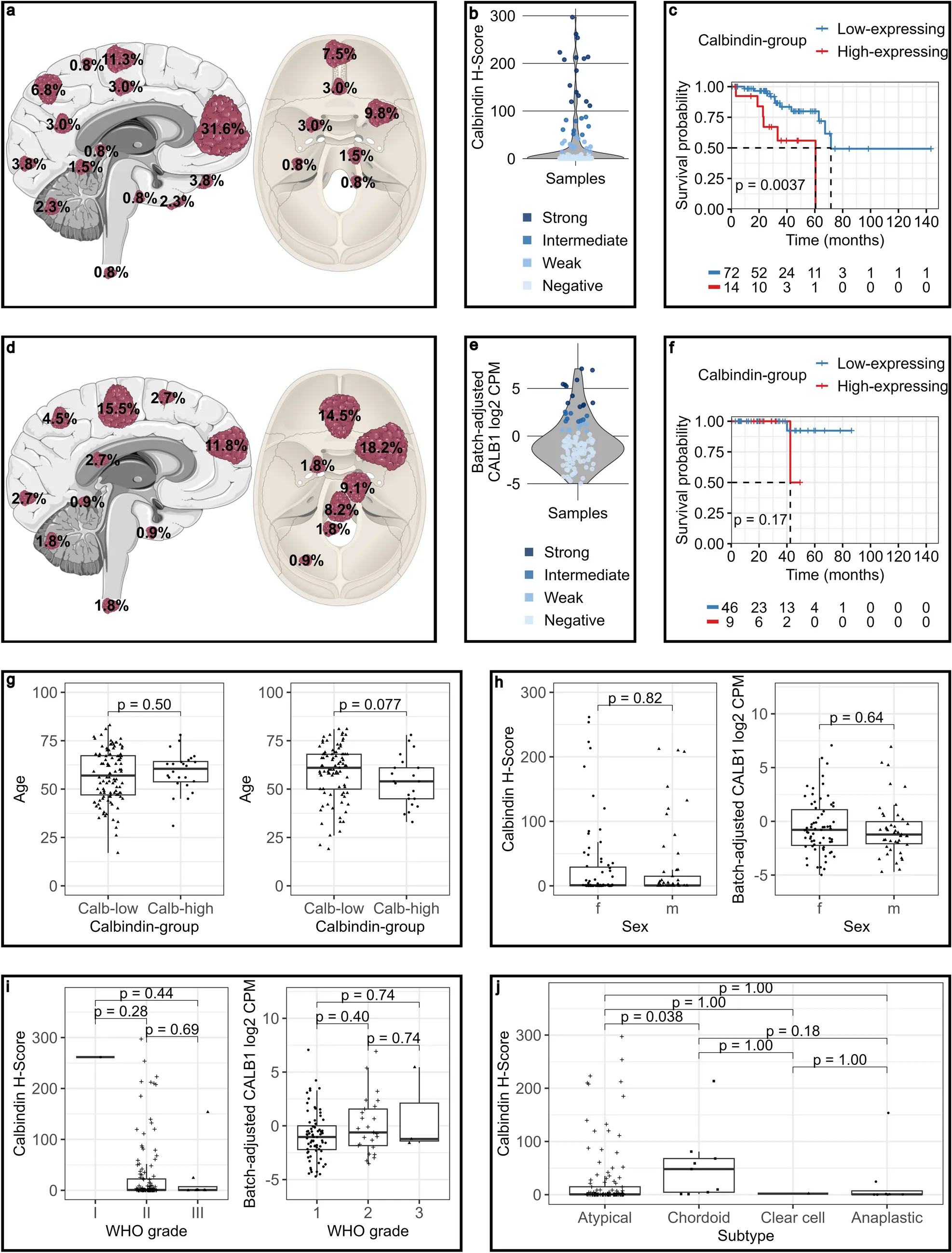
主队列 147 例(原发脑膜瘤 133、非对应复发 14),经有经验神经病理医师按 2016 CNS WHO [22] 复核。次队列 110 例公开原发脑膜瘤(原 2016 WHO [22])。26 例缺 TERT 启动子状态;按方法学改良整合分级,缺 pTERT 不妨碍原组织学 1 级归类,故 100 例可分子分级:WHO 1 级 72(72.0%)、2 级 25(25.0%)、3 级 3(3.0%)。致病性 pTERT(C228T 或 C250T)占 9.1%(含 2 例原 WHO II);纯合 CDKN2A/B 缺失 1 例(原 WHO II)。两队列临床病理见表 1、表 2,常见解剖部位见图 1a, d。
Calbindin 表达
以小脑浦肯野细胞为阳性、省略一抗为阴性对照确认抗体特异性(补充图 1a–d)。H&E 核实肿瘤细胞含量 >90% 后,主队列 132 例原发可评估(1 例技术原因排除)。改良 H-score 下 28.8% 肿瘤检出 Calb 免疫反应性(图 1b);染色强度分布见表 4。
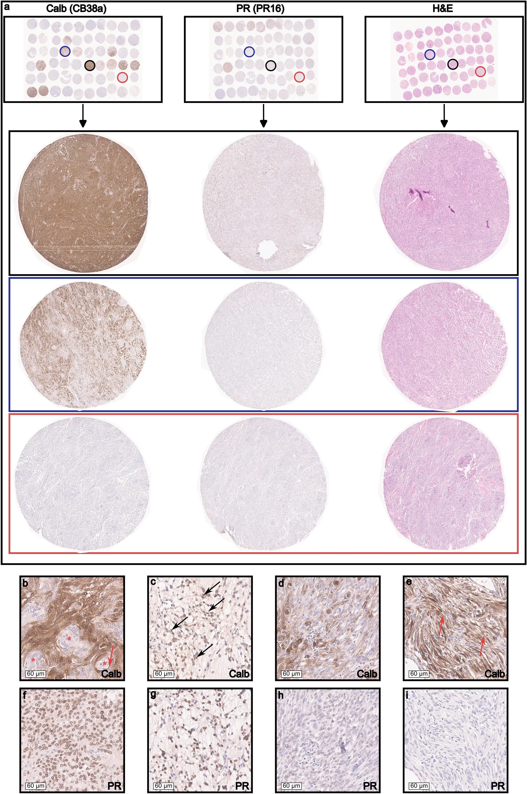
瘤内 Calb 主要见于胞质与胞核,多为弥漫细胞染色;偶仅核或仅胞质而无核标记(图 2b–e)。中至强表达瘤常大片弥漫阳性,其间仅偶见小簇阴性细胞;弱至中等阳性则以灶性细胞簇为主,阴性例最多散在少数阳性细胞。13 例正常脑膜中 9 例无免疫反应性(补充图 1e);余 4 例绝大多数阴性,仅少数弱阳性细胞多位于蛛网膜基底层(补充图 1f)。下游分析将「阴性」与「弱阳性」合并为 Calb-低组(81.8%),「中等」与「强阳性」为 Calb-高组(18.2%)。独立次队列中,相对对照蛛网膜颗粒细胞均值,阳性定义为 ≥2.5 倍至 <5.5 倍,高表达 ≥5.5 倍(中等 ≥5.5–<11.5;强 ≥11.5)。据此 110 例中 8 例(7.3%)弱表达,21 例(19.1%)中等或强转录,与蛋白水平分布相近(图 1e)。
单变量分析中两队列 Calb 表达与性别均无显著关联(女 30.7%/28.4%,男 26.4%/23.3%;图 1h)。Calb-高与 Calb-低中位年龄亦无显著差异(60.5/54 岁 vs 57/61 岁;图 1g)。未见 Calb-高瘤偏好解剖部位(补充图 2f)。核分裂与增殖指数不能区分高低组:低/中/高核分裂计数中 Calb 阳性比例相近(30%、28.7%、25%;补充图 2a);中位 Ki-67 亦相近(6% vs 5% / 3.65% vs 3.5%;补充图 2e)。主队列各 WHO 分级间无显著差异(图 1i)。次队列在临床–分子模型校正后,组织学 WHO 2 级与较高 CALB1 转录相关(p=0.023;补充文件 1)。主队列成对比较中脊索样瘤 Calb H-score 高于不典型瘤(p=0.038;图 1j),多变量模型中亦存(p=0.015;补充文件 1)。
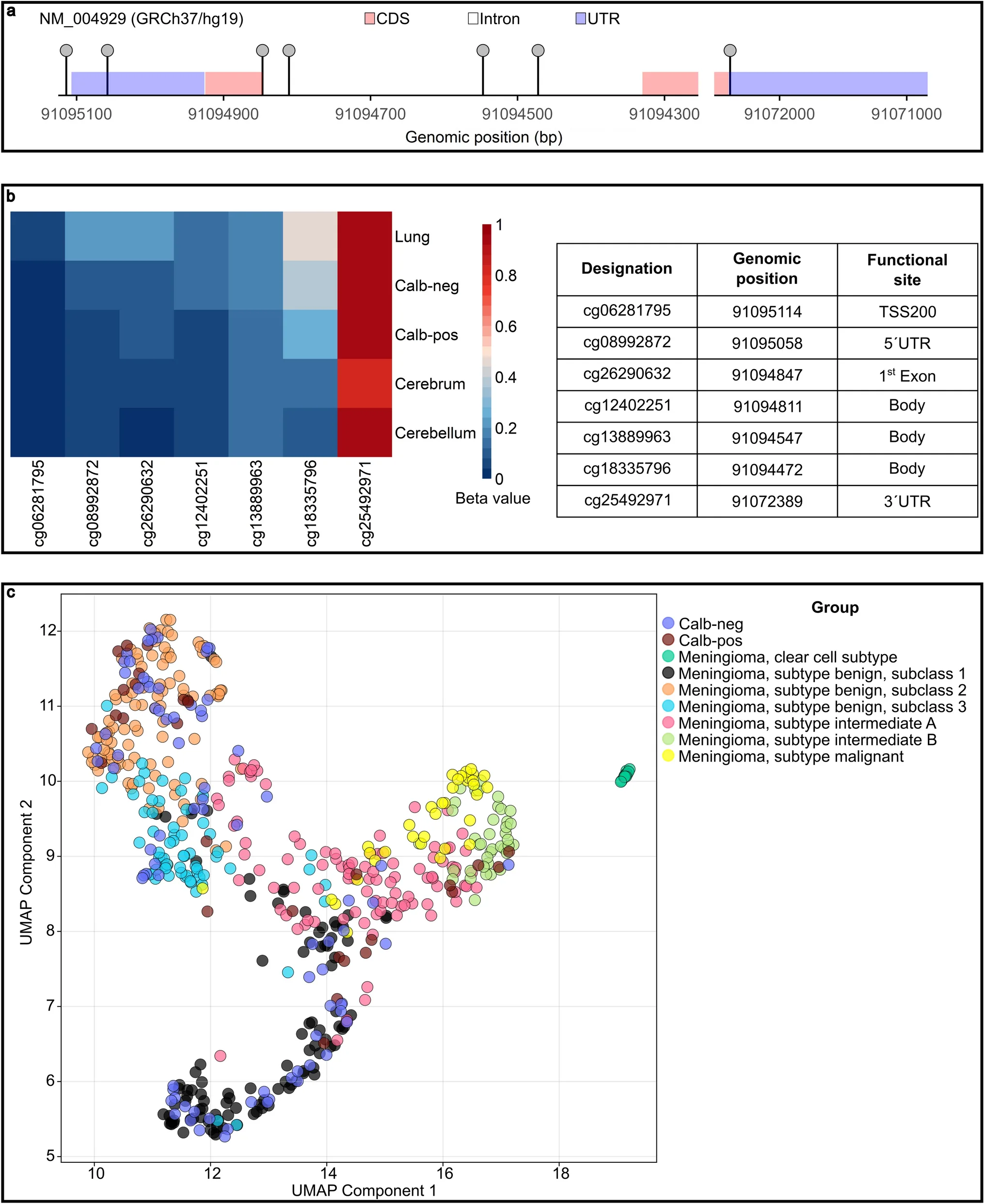
次队列与 Heidelberg Brain Tumor Classifier V12.8 参考集 453 例脑膜瘤的 UMAP 显示 Calb 阳/阴性瘤广泛分布,未形成独立甲基化簇(图 3c、补充图 2c)。相对「Benign 1」,「Benign 3」与较高 CALB1 相关(p<0.05),「Intermediate B」与较低表达相关(p=0.013;补充文件 1)。Calb 与 TERT 启动子突变及 IMS 类别无关(补充图 2b, d)。在主要驱动改变(NF2、SMARCB1、TRAF7、KLF4、AKT1、PIK3CA、SMO、POLR2A、ARID1A;补充图 3)比较中,多重校正后无与 CALB1 关联仍显著。总体而言,Calb 与传统临床病理参数关联有限,但多变量分析显示与组织学 WHO 分级及特定分子特征独立相关。
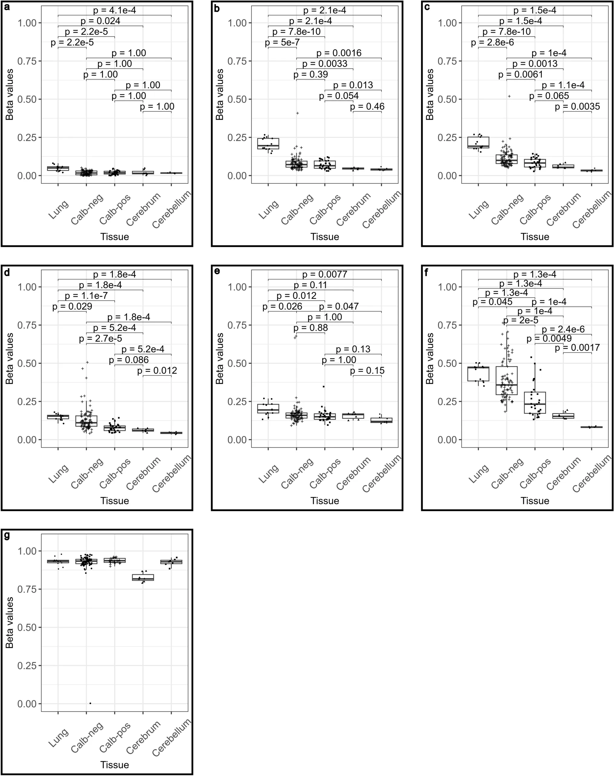
脑膜瘤中 CALB1 调控机制知之甚少。次队列分析 Illumina EPIC 850K 上 12 个 CALB1 相关 CpG 中与 450K 共有的 7 个探针;以肺(低固有表达)、小脑与大脑皮质(高固有表达)为参照比较阳/阴性样本 β 值(图 3b)。三个位于第 1 外显子或内含子的位点(cg26290632、cg12402251、cg18335796;图 4c, d, f)差异显著,其中 cg18335796 相对低甲基化最强(p=2e−5;图 4f)。其余四个(3´UTR、启动子、5´UTR、第 1 内含子:cg25492971、cg06281795、cg08992872、cg13889963)样本间差异轻微(图 4a, b, e, g)。总体脑膜瘤与正常 CNS 组织较肺组织在多数 CALB1 CpG 呈相对低甲基化(图 3a, b)。三位点均值(CpG 评分)较低与临床–分子回归中较高转录相关(p=0.0084;补充文件 1)。
鉴于既往将 CALB1 与雌激素受体信号关联,分析了 ESR1/ESR2 与 6q、14q 及 CALB1 的关系。6q、14q 缺失时 ESR2 较低(p=1.94e−5、p=0.019);14q 缺失时 ESR1 较高(p=0.029),与 6q 无关。ESR1/ESR2 转录均未与 CALB1 显著相关(补充文件 1)。
钙视网膜蛋白表达
亦以 IHC 评估与 Calb 密切相关的 Calr。蛋白表达稀疏、限于少数肿瘤的胞质与核;132 例原发中 12 例(9.1%)见孤立散在、多呈强阳性的肿瘤细胞(补充图 1g),与既往低水平 Calr 描述一致 [31]。因阳性细胞少且灶性,未再分析其他临床病理变量。
孕激素受体表达
为与既定分化标志比较,133 例原发均评估核 PR,阳性 48.1%(表 5)。
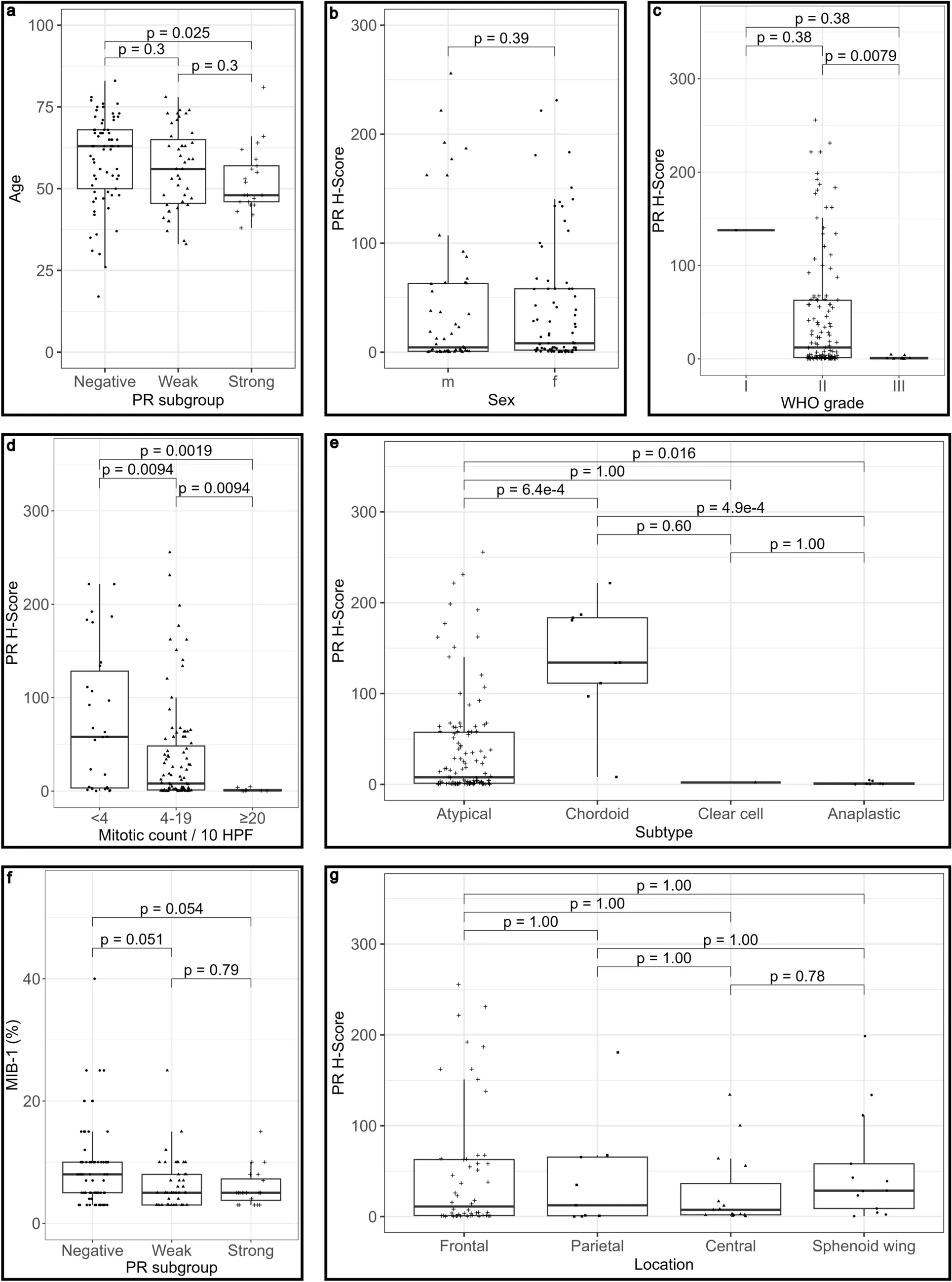
PR 表达随分级升高而降低:WHO III 显著低于 WHO II(p=0.0079;图 5c),脊索样(WHO II)平均 PR H-score 最高(图 5e);并与核分裂活性负相关(图 5d)。强 PR 阳性更多见于较年轻患者(p=0.025;图 5a);性别、MIB-1、部位无差异(图 5b, f, g)。尽管如此,原发全切 WHO II 脑膜瘤中 PR 不能预测 PFS(补充图 4a)。
临床病理因素对脑膜瘤预后的影响
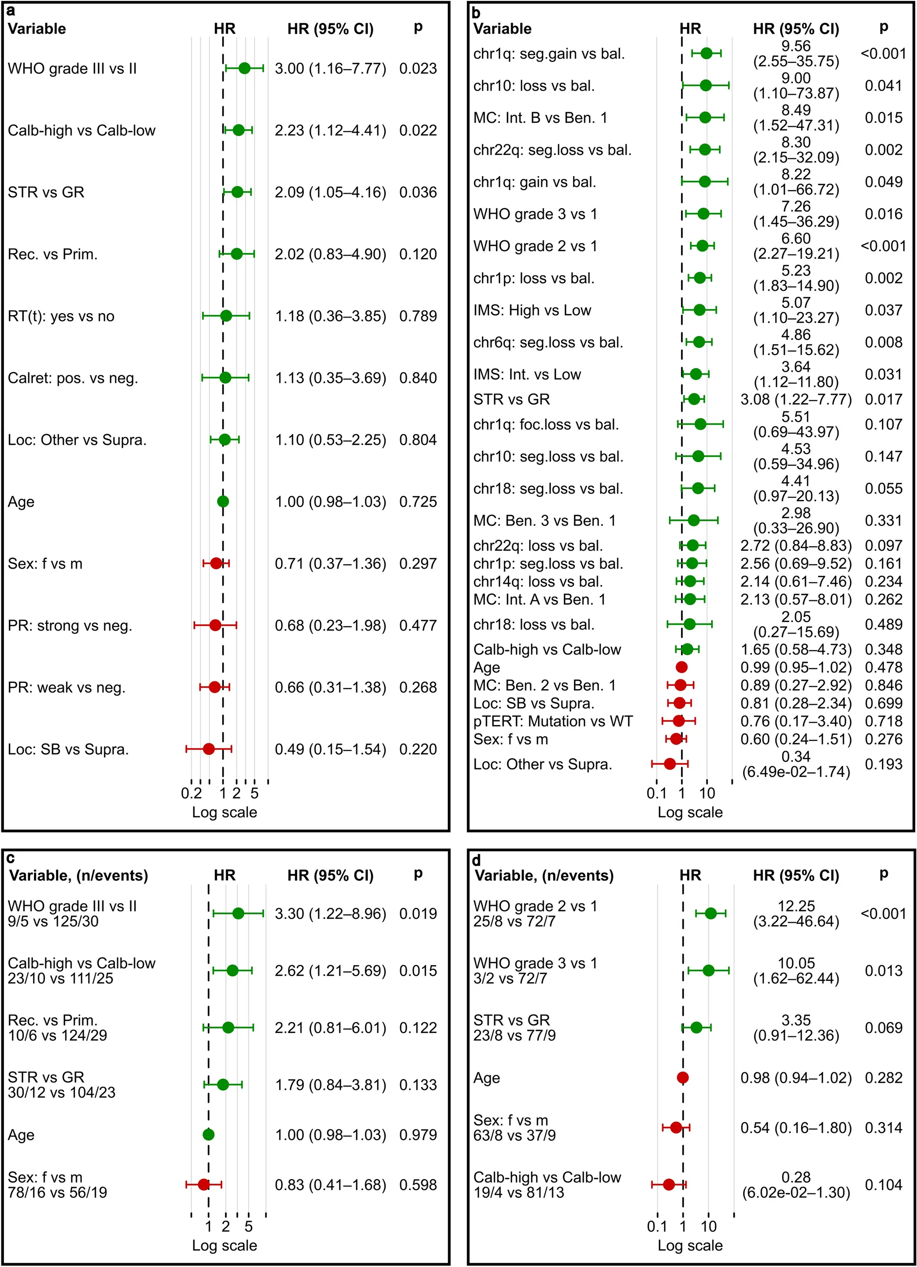
以单/多变量 Cox 评估 Calb、Calr、PR 与既定临床病理参数对 PFS 的影响。主队列中年龄、性别、放疗(时间依赖)、Calr、PR、部位、疾病状态等多与 PFS 无显著相关;高 Calb、次全切与 WHO III 则与更短 PFS 显著相关(图 6a)。校正年龄、性别、WHO 分级、切除范围及原发/复发后,高 Calb 仍与更短 PFS 相关(HR 2.62,95% CI 1.21–5.69;名义 p=0.015;图 6c)。重要的是,限于原发、全切、未放疗、组织学 WHO 2 级时该关联仍存在(图 1c);进一步限于仅因核分裂增多而归 2 级者(排除脊索样、透明细胞及因脑侵犯等非核分裂标准升级者)仍显著(p=0.011;补充图 4b)。
次队列探索性单变量中,WHO 分级、甲基化亚类、若干复发 CNA、IMS 与切除范围与 PFS 名义相关(图 6b)。限制性多变量仅 17 个进展事件,全面建模易过拟合。含年龄、性别、切除范围、改良整合 WHO 分级与 CALB1 表达组的限制模型中,较高 WHO 分级仍与更短 PFS 相关,而 CALB1 未达统计显著(HR 0.28,95% CI 0.06–1.30;名义 p=0.10;图 6d)。限于原发全切 WHO 1 级的 Kaplan–Meier 亦无显著关联,但 Calb-高呈数值上更短 PFS 趋势(p=0.17;图 1f)。
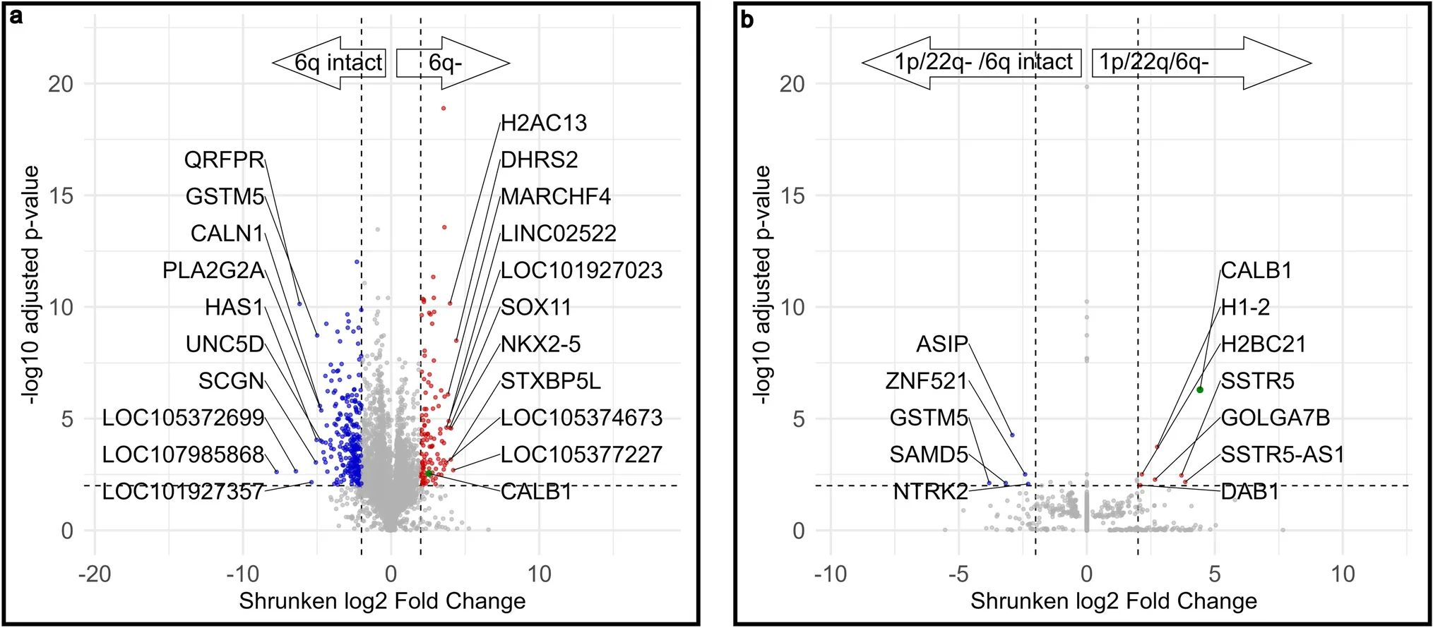
脑膜瘤若干复发染色体畸变与不良结局相关,识别下游转录改变或有助于理解分子高危谱。110 例中 11 例 6q 缺失:局灶 1(0.9%)、节段 8(7.3%)、全臂 2(1.8%;补充文件 2)。10 例节段/全臂纳入差异表达,局灶 1 例因无法分组排除。与其余合格肿瘤比较,节段/全臂 6q 缺失瘤差异表达多基因,含 CALB1 与转录因子 NKX2-5、SOX11(图 7a)。在合并 1p/22q 缺失亚组内,附加 6q 缺失时 CALB1 上调最强且上调基因中 FDR 校正 p 最低(图 7b)。染色体聚焦多变量线性模型(批次及 1p、1q、6q、10、14q、18、22q;n=95)中,6q 缺失在校正后仍与较高 CALB1 相关(名义 p=0.037;补充文件 1)。另一含批次、1p/6q/22q、组织学 WHO、年龄、性别、TERT 启动子、甲基化亚类与 CALB1 CpG 评分的临床–分子模型(n=80)中,6q 缺失亦与较高表达相关(p=0.023;补充文件 1)。
讨论
脑膜瘤中 Calbindin D28K 表达与临床结局
本研究在两个独立队列中证实部分脑膜瘤表达 Calb 蛋白及升高的 CALB1 转录:约 30% 原发瘤可检出免疫反应性或升高转录,高表达组约 18–19%。正常脑膜细胞缺如或仅弱而稀疏。主队列校正纳入临床病理变量后,高 Calb 蛋白与更短 PFS 相关;组织学 WHO 2 级限制分析亦然。次队列限制性多变量中 CALB1 转录与 PFS 无显著关联;WHO 1 级亚组数值趋势须因样本量与进展事件有限而审慎解读。结果支持将 Calb 免疫组化作为风险分层候选辅助标志(尤其组织学 WHO 2 级)进一步研究。改良 H-score 提供系统半定量评估,加之 Calb-高瘤多为弥漫染色,或利于肉眼判读,并支持在分子检测前评估其作为快速、潜在低成本辅助标志的价值;可重复性与增量预后价值须验证后方可临床应用。
Calb 既往见于髓母细胞瘤 [17, 25]、胚胎发育不良性神经上皮肿瘤 [34]、室管膜下巨细胞星形细胞瘤 [14, 21] 与肺癌 [1, 7, 16] 等。本研究扩展至脑膜瘤,与 Khan 等转录组发现一致 [18],而与早期小样本免疫组化阴性观察不同 [17]。相关 Calr 少见且多限于散在细胞,与既往一致 [31]。次队列多变量临床–分子分析显示较高 CALB1 与 6q 缺失、组织学 WHO 2 级、甲基化亚类「Benign 3」及 CALB1 相关 CpG 较低甲基化相关,「Intermediate B」呈反向关联。脊索样瘤 Calb 尤高,但排除脊索样后预后关联仍在,提示不限于该亚型;是否反映脊索样分化程序仍属推测。总体将 CALB1 与特定染色体及表观特征相联系,超越常规组织病理;其为主动驱动抑或下游标志尚待解决。
其他细胞系统实验提示 Calb 可通过抑制 caspase-3 [3, 5, 19](钙及 pH 依赖机制均有提出 [5, 19])、激活 PI3K/AKT [33] 及促进逃逸衰老 [1] 促进存活。本研究中 CALB1 与核分裂或 Ki-67 升高无关,不支持以增殖为主的表型。既往侵袭性耐药原代脑膜瘤细胞系核 caspase-3 减低且转录组提示 CALB1 过表达 [18],支持其或通过增强存活与抗凋亡而非增殖促进肿瘤持续。跨瘤种预后高度情境依赖:髓母细胞瘤 [25] 与一则 NSCLC 报告 [16] 中高表达不良,肺鳞癌 [1] 与腺癌 [7] 则有不一甚至有利关联。
PR 作为既定分化标志,与既往一致:与 WHO 分级负相关并总体预后偏向良好 [27]。单变量 Cox 与 Kaplan–Meier 中 PR 与 PFS 无显著关联,符合其在既定病理参数之外预后价值有限的报道 [28]。
表观遗传调控与潜在上游机制
数据支持 DNA 甲基化可能参与 CALB1 调控:内含子与第 1 外显子 CpG 低甲基化与表达升高相关。此外,调控还曾关联 ΔFosB、维甲酸、维生素 D3、内源性逆转录病毒及雌激素信号 [1, 12, 35–37]。ESR1/ESR2 分别位于 6q、14q。分析中 6q 缺失伴较低 ESR2 而无显著 ESR1 改变;14q 缺失伴较低 ESR2、较高 ESR1。但批次校正线性或秩相关分析中两受体转录均未与 CALB1 显著相关,不支持雌激素受体丰度与 CALB1 上调的简单关系。
临床–分子多变量中,校正临床病理、甲基化亚类、选定 CNA 及三处 CpG 衍生评分后,6q 缺失仍与较高 CALB1 相关;染色体聚焦模型校正更多复发 CNA 后亦支持。机制未明,需功能研究判定 CALB1 是否贡献相关表型抑或主要为下游标志。
局限性与临床意义
局限包括:瘤内异质性可使 TMA 抽样偏倚;次队列可在同瘤分析转录、甲基化与 CNA,但 Calb IHC 在独立队列进行,缺乏蛋白–转录–CNA 整合,无法直接验证 6q 缺失与蛋白表达关系,需同瘤三水平研究。6q–CALB1 关联在校正复发 CNA 及限于 1p/22q 共缺失后仍存,但共现与 6q 缺失例数有限,特异性结论受限。
次队列进展事件少亦限制多变量生存建模并增加过拟合风险,故未确立超越既定临床病理与分子危险因素的增量预后价值。最终需功能研究判定 CALB1 是否促进进展抑或仅为相关分子程序的标志。
尽管如此,主队列校正后高 Calb 蛋白与更短 PFS 相关,组织学 WHO 2 级限制分析亦支持;次队列同样本分子分析将 CALB1 转录与 6q 缺失及特定表观特征相联系。综合支持进一步评估 Calb IHC 作为风险分层候选辅助标志(尤其组织学 WHO 2 级),并为探究 CALB1 调控与细胞存活关系提供依据,但未确立因果机制。
- Acta Neuropathologica OA: primary IHC cohort (147 pts, TMA) plus independent molecular cohort (110) with transcript/methylation/CNA; ~30% of primaries show Calb or elevated CALB1, high-expression ~18–19%.
- High Calb protein associated with shorter PFS after adjustment (HR 2.62), including in restricted histological WHO grade 2 analyses; Calr/PR not linked to PFS.
- Secondary cohort links CALB1 to 6q loss, WHO grade 2, selected CpG hypomethylation and methylation subclasses; DE with 6q loss highlights CALB1, NKX2-5, SOX11.
- Practical angle: Calb IHC may be a candidate adjunct for risk stratification—especially WHO grade 2—before integrative molecular testing; validation still required.
Scope: Springer open-access full text (CC BY 4.0)—Abstract, Introduction, Materials and methods, Results, Discussion and Figures 1–7 in parallel ZH/EN. Tables 1–5 and supplements are linked to the publisher page and not rebuilt here; conflicts of interest, data-availability statements, author-contribution details and the reference list are omitted (in-text citation numbers retained).
Abstract
Molecular profiling has refined risk stratification in meningioma, but the biological relevance of associated transcriptional changes remains incompletely understood. Calbindin D28K (Calb), encoded by CALB1, is a calcium-binding protein with reported anti-apoptotic and cytoprotective functions. Previous unsupervised transcriptomic analysis identified CALB1 upregulation in meningiomas with aggressive cellular features, but its biological and clinical relevance remains unresolved. We analysed Calb immunoreactivity in 231 meningioma tissue microarray (TMA) cores from 147 patients and evaluated CALB1 transcript expression, DNA methylation and copy-number alterations in an independent cohort of 110 primary meningiomas. Calb immunoreactivity was detected in approximately 30% of primary tumours, and elevated CALB1 transcript levels were observed at a similar frequency in the molecular cohort. High Calb protein expression was associated with shorter progression-free survival (PFS) after adjustment for the included clinical variables in the primary cohort. This association was also observed in restricted analyses of histological WHO grade 2 meningiomas. Lower methylation of selected exonic and intronic CALB1 CpG sites correlated with a higher transcript expression, consistent with an epigenetic contribution to deregulation. Multivariable analyses identified associations of CALB1 transcript expression with chromosome arm 6q loss, histological WHO grade, CALB1 hypo-methylation and specific methylation subclasses in the secondary cohort. Differential expression analysis of tumours harbouring 6q loss identified expression differences in CALB1 and transcription factors, including NKX2-5 and SOX11. In contrast, expression of a related calcium-binding protein Calretinin (Calr) and the Progesterone Receptor (PR) was not significantly associated with PFS in the primary cohort. Together, these findings support the potential prognostic relevance of Calb protein expression, particularly in histological WHO grade 2 meningiomas, and associate CALB1 transcript expression with selected molecular features in complementary cohorts. Calb immunohistochemistry may therefore provide a practical tool for refining risk stratification in this clinically heterogeneous tumour type prior to integrative molecular testing.
Introduction
Meningiomas are the most common primary intracranial neoplasms and arise from arachnoid cap cells of the leptomeninges. They comprise a biologically heterogeneous group of tumours characterised by diverse histological subtypes and molecular alterations. Whilst most meningiomas follow a relatively favourable clinical course, the current World Health Organization Classification of Tumors of the Central Nervous System (CNS WHO) [23] recognises a spectrum ranging from indolent WHO grade 1 lesions to highly aggressive WHO grade 3 tumours. In addition to classical histopathological criteria—including mitotic activity, brain invasion and specific morphological features—molecular profiling has substantially refined prognostic stratification. Chromosomal copy-number alterations have emerged as key determinants of tumour progression. By this rationale, cIMPACT-NOW update 8 recommends assigning CNS WHO grade 2 to meningiomas with grade 1 morphology that harbour chromosome arm 1p deletion together with 22q deletion and/or an oncogenic NF2 variant [29]. These recommendations extend molecular risk assessment beyond the criteria of the 2021 CNS WHO classification [23]. Losses of 6q and 14q have also been frequently associated with adverse clinical outcomes [24]. In addition, DNA methylation-based classification schemes provide further prognostic resolution when integrated with WHO grade and chromosomal aberrations, as exemplified by the integrated molecular score (IMS) [24]. Despite these advances, the downstream molecular programmes associated with aggressive behaviour of meningiomas harbouring these chromosomal aberrations remain incompletely understood.
Calb is a member of the EF-hand calcium-binding protein family with established buffering and sensor-like functions in calcium homeostasis [30]. In humans, its expression is largely restricted to cerebellar Purkinje neurons [9], selected neuronal subpopulations [9] and epithelial cells of the distal nephron and collecting duct [6]. Beyond its physiological role, increasing experimental evidence suggests tumour-promoting properties. Calb has been shown to inhibit caspase-3-mediated apoptosis [5], facilitate escape from cellular senescence [1] and influence proliferation [15]. Consistent with these findings, Calb expression has been reported in several epithelial [1, 7, 16] and CNS neoplastic entities [21, 25, 34]. In meningioma, however, data on Calb remain scarce and contradictory. CALB1 was previously identified in unbiased transcriptomic analyses of meningioma as part of a stemness-associated gene signature linked to aggressive and drug-resistant tumour phenotypes [18]. Corresponding primary meningioma cell lines with aggressive growth characteristics also exhibited lower nuclear caspase-3 immunoreactivity [18]. However, early immunohistochemical analyses in a small posterior fossa meningioma cohort did not detect Calb immunoreactivity [17]. Thus, the presence, regulatory mechanisms, and clinical relevance of Calb in meningioma remain unresolved.
Given its anti-apoptotic potential and the incomplete understanding of transcriptional changes associated with molecularly defined high-risk meningiomas, we hypothesised that CALB1 expression is associated with clinical outcome and adverse molecular features. We investigated Calb protein expression and its clinicopathological and prognostic associations in a primary immunohistochemical cohort. In an independent secondary cohort, CALB1 transcript expression, DNA methylation and recurrent copy-number alterations were comprehensively analysed. These complementary analyses aimed to characterise CALB1-associated biology and evaluate its potential prognostic relevance.
Materials and methods
Human tissues
To analyse the expression of Calb in meningioma and compare it to clinical data, a primary cohort of 231 TMA cores, corresponding to 179 samples of 147 patients, was assembled. This extensive and already comprehensively clinically documented cohort of predominantly high-grade meningiomas was selected for a focussed evaluation of the association between clinicopathological parameters, Calb expression in the tumours, and recurrence during follow-up. The samples were provided by the UCT Biobank Frankfurt. All clinical and follow-up data are depicted in Table 1. Whilst no molecular data were available, samples were graded according to the 2016 version of the CNS WHO classification [22], which was the valid version at the time of study initiation. In the primary cohort, analyses were restricted to primary meningiomas, except for the univariable and multivariable Cox and linear regression analyses, which additionally included recurrent tumours. In addition, to compare meningiomas with their tissue of origin, 13 completely and irreversibly anonymised meningeal samples from routine autopsies, conducted at the Department of Neuropathology, Institute of Pathology, Technical University of Munich, were included as reference.
A secondary cohort comprising 110 publicly available primary meningioma datasets with associated clinical information was included to enable molecular comparisons across histological WHO grades 1 and 2. All cases were originally classified according to the 2016 version of the CNS WHO classification [22]. Using available copy-number profiles and NF2 as well as TERT promoter mutation status (pTERT), we applied a modified integrated grading scheme based on the 2021 CNS WHO framework [23] and the cIMPACT-NOW update 8 recommendations [29]. Informed by recent findings on the context-dependent prognostic relevance of pTERT mutations [10], pTERT-mutant tumours were upgraded to grade 3 only when their original histological grade was 2. Tumors with grade 1 morphology were not upgraded solely on the basis of a pTERT mutation. The modified integrated grading scheme was used for the secondary-cohort survival analyses and the WHO grade expression comparisons, whereas original histological WHO grades were retained for the clinical–molecular regression analyses. Clinical characteristics and follow-up data are summarised in Table 2.
The two cohorts were complementary. The primary cohort was used to investigate Calb protein expression and its clinical associations. In the secondary cohort, CALB1 transcript expression, DNA methylation and CNA status were analysed in the same tumours. Calb protein expression was not available for these molecularly characterised tumours.
Immunohistochemistry
5 µm-thick, ready-to-use sections of formalin-fixed, paraffin-embedded (FFPE) TMA samples were deparaffinised and rehydrated, using a series of xylene, ethanol dilutions and DM water, followed by antigen retrieval in pH 6.0 citrate buffer (95 °C) and quenching of endogenous peroxidase with 1.5% hydrogen peroxide solution. Nonspecific antibody binding sites were blocked, using blocking buffer with 2.5% normal horse serum (Vector Laboratories, USA). Incubation with primary antibody solutions, containing avidin (Vector Laboratories, USA), was performed overnight at 4 °C, with subsequent binding of secondary antibodies and masking of avidin with biotin (Vector Laboratories, USA) for 30 min. All primary and secondary antibodies are listed in Table 3. Visualisation of antigen–antibody complexes was done by incubation with ABC-reagent (Vector Laboratories, USA), containing horseradish peroxidase (HRP) for 30 min, followed by 3,3´-diaminobenzidine (DAB, Vector Laboratories, USA). After counterstaining with haematoxylin for 5 min and blueing, slides were dehydrated using ethanol dilutions and xylene and finally mounted.
Final concentrations of all antibodies were determined by serial dilutions prior to the start of experiments (Table 3). Slides of cerebellum (Calb, Calr) and breast cancer metastasis (PR) were used as positive controls in each run. Negative controls were prepared by omitting the primary antibody.
Digitisation and evaluation
All stained slides were scanned using an Aperio slide scanner (Aperio AT2, Leica Biosystems, Germany) and converted to.svs files. Calb and PR status were assessed using a semi-automated, quantitative cell detection and scoring-algorithm implemented in the open-source software QuPath 0.5.0, as previously described for PR status [4]. Following background correction and colour deconvolution to separate Haematoxylin and DAB channels, tumour cell nuclei were identified using the Positive Cell Detection command with customised parameter settings. Tumour cell nuclei identification was based on optical density within the Haematoxylin channel. The H-scores were calculated by applying three intensity thresholds for cellular (Calb) or nuclear (PR) DAB intensity. Afterwards, multiple regions of interest (ROIs) from several Calb- and PR-stained TMA slides were selected to train a cell classifier to exclude detections corresponding to faulty results from erythrocytes, haematoxylin crystals and other artefacts. After application of the complete workflow to all TMA samples, an individual grading system based on H-score calculation was applied for Calb (negative: <10, weak positive: ≥10 to <50, intermediate positive: ≥50 to <100, strong positive: ≥100) and for PR (negative: <10, weak positive: ≥10 to <100, strong positive: ≥100). In tumour cases with multiple cores, the mean H-score was calculated. Calr immunohistochemistry, however, was manually evaluated in categories due to its generally weak staining results (negative: no positive tumour cells, positive: singular or singular groups of positive tumour cells).
RNA sequencing data
Raw count matrices from two publicly available meningioma RNA sequencing (RNA-seq) datasets (GSE189672 [2] and GSE139651 [26]) were obtained from the Gene Expression Omnibus (GEO) database of the National Center for Biotechnology Information (NCBI) [8], together with the corresponding metadata. RNA-seq data were processed in R (version 4.2.1) using edgeR (version 3.38.4) and limma (version 3.52.4) packages. Raw counts were restricted to genes present in both datasets, filtered for lowly expressed genes and normalised for library size using the trimmed mean of M-values (TMM) method. Log2 CPM values were batch-corrected using limma’s removeBatchEffect() function whilst preserving tumour versus arachnoid reference status. Corrected CALB1 expression values were used for visualisation and expression-category assignment. Data from 110 primary meningioma samples (GSE189672 [2]) were then used for the secondary cohort, whereas 2 arachnoid granulation cell model samples (GSE139651 [26]) served as reference for CALB1 expression levels. For regression analyses, TMM-normalised log2 CPM values were generated separately from all 110 secondary-cohort tumours, without prior batch correction; sequencing batch was included directly as a model covariate.
Methylation data
To assess global DNA methylation patterns and CpG sites within the CALB1 locus, publicly available methylation BeadChip samples from the NCBI GEO repository GSE189521 [2] were used.
To compare CALB1 methylation between meningiomas and tissues with intrinsically either a high or low Calb expression, .idat files from 13 normal lung samples (GSE173356 [20]) as well as seven normal cerebellum and seven normal cerebrum samples (GSE154403 [11]) were incorporated into the analysis.
Two different array versions were used within the data processing (Illumina EPIC and 450 K methylation array). Common probes were identified according to the manufacturer’s annotations, allowing both array types to be merged using the minfi package (version 1.43.1) in R (version 4.2.2). For comparison of different samples of several batches, a background and dye-bias correction, followed by log2-transformation of the values was performed. In the next step, batch effects related to array type and tissue preservation were addressed using the limma package, with methylated and unmethylated signals corrected separately. Beta values were subsequently calculated according to
where M and U denote the re-transformed methylated and unmethylated signal intensities, and α represents an offset of 100. Copy-number variation plots and methylation-based classifier scores of the Heidelberg Brain Tumor Classifier V12.8 were generated as previously described [32].
Chromosomal copy-number alterations were inferred from Illumina Infinium MethylationEPIC v1.0 BeadChip data using the conumee R package as previously described [13]. All secondary-cohort tumour samples were profiled on this platform. Raw.idat files were imported using minfi and processed with preprocessRaw, without background correction, dye-bias correction or control-probe normalisation at this preprocessing stage. Query samples were subsequently normalised against a linear combination of 50 sex-matched copy-number-neutral control samples. Log2 ratios between observed and fitted intensities were aggregated into 26,446 genomic bins (GRCh37/hg19), baseline-centred using conumee and segmented by circular binary segmentation with alpha = 0.001, 50,000 permutations, a minimum of five bins per segment, undo.splits = "sdundo" and undo.SD = 2.2. Thresholds were applied to individual segments: segments with a mean log2 ratio ≥ +0.10 were called gains, and those with a mean log2 ratio ≤ −0.10 were called losses. Chromosomal arms were categorised according to the fraction of their segment-covered length affected by copy-number alterations. Segments spanning a centromere were split at the p/q boundary, and the resulting portions were assigned to the respective arms. For each alteration direction, changes affecting ≥5% and ≤25% of the assessed arm length were classified as focal, changes affecting >25% and ≤95% as segmental, and changes affecting >95% as complete arm-level gains or losses. Arms harbouring both gains and losses, each affecting ≥5% of the assessed arm length, were classified as gain/loss. Arms with <5% altered genomic content were considered balanced. Arm-level extent categories were adapted from the framework described by Hielscher et al. [13], with an additional distinction between focal and larger segmental alterations for descriptive reporting. For linear expression regression analyses, segmental and complete arm-level alterations were merged into single gain or loss categories, respectively. Focal alterations and gain/loss classifications were coded as missing owing to their low frequency and ambiguous interpretation. To reduce model complexity, p- and q-arm CNA calls for chromosomes 10 and 18 were combined into composite chromosome-level categories. When one arm was balanced, focal or segmental alterations of the other arm retained their category, whereas complete arm-level gains or losses were classified as segmental chromosome-level gains or losses, respectively. Concordant arm-level categories were retained, same-direction combinations of segmental and complete alterations were classified as segmental, and opposite-direction alterations were classified as gain/loss.
Statistical analysis
Continuous clinicopathological parameters were compared between expression groups using two-sided Mann–Whitney U tests. The same test was used for Calb and PR H-scores across clinicopathological groups in the primary cohort and for CpG beta values across tissue/expression groups in the secondary cohort. Pairwise comparisons were Holm-adjusted within each factor or CpG site.
Linear regression analyses used heteroscedasticity-consistent HC1 robust standard errors and model-specific complete cases. In the primary cohort, the outcome was log2-transformed Calb H-score; the multivariable model included age, sex, primary versus recurrent disease status, and histological grade/subtype. In the primary cohort, the single histological WHO grade 1 tumour and the single clear cell meningioma were excluded from linear regression models including histological grade/subtype because each category contained only one case. Secondary-cohort regression analyses used TMM-normalised log2 CPM generated from all 110 tumours, without prior batch correction, and included sequencing batch as a covariate. Batch-corrected values were used only for visualisation and expression-category assignment. Single-factor models included one variable of interest and batch. The clinical–molecular multivariable model included batch, 1p, 6q and 22q status, original histological WHO grade, age, sex, TERT promoter status, methylation subclass, and the mean beta value of cg26290632, cg12402251 and cg18335796. The single CDKN2A/B homozygously deleted tumour was excluded from these clinical–molecular models but retained for normalisation and other analyses. A chromosome-focussed model included batch and CNA categories for 1p, 1q, 6q, chromosome 10, 14q, chromosome 18 and 22q. Its targeted 6q-loss versus balanced contrast used a nominal p value; other CNA coefficients were reported descriptively.
Secondary-cohort pairwise contrasts were calculated using emmeans and Holm-adjusted within each factor. Mutation-status comparisons were jointly Holm-adjusted across NF2, SMARCB1, TRAF7, KLF4, AKT1, PIK3CA, SMO, POLR2A and TERT promoter status. Models restricted to a single batch omitted the batch term. ARID1A was displayed descriptively and excluded from inference because only one mutant tumour occurred within the batch containing both status groups.
Exploratory coefficient tests were Benjamini–Hochberg FDR-adjusted separately within the primary-cohort univariable and multivariable analyses, secondary-cohort single-factor and multivariable clinical–molecular analyses, and single-factor CNA analyses. Intercept and batch coefficients were excluded. Additional batch-adjusted models examined CNA-ESR1/ESR2 and ESR-CALB1 associations. Rank-based sensitivity analyses ranked both CALB1 and the ESR predictor. FDR correction was applied separately across the CNA-ESR coefficient tests, two linear ESR-CALB1 tests, and two rank-based tests. Linear-model estimates, confidence intervals, reference categories and sample sizes are provided in Supplementary File 1.
PFS was analysed using Kaplan–Meier estimates, log-rank tests, and univariable and multivariable Cox proportional hazards models. The primary multivariable model included age, sex, Calb expression group, WHO grade, extent of resection, and primary versus recurrent disease status. Radiotherapy was evaluated separately as a time-dependent covariate in a univariable model. The restricted secondary model included age, sex, CALB1 expression group, extent of resection, and modified integrated WHO grade. Cox models used model-specific complete cases. Hazard ratios are reported with 95% confidence intervals and nominal two-sided Wald p values.
DESeq2 analyses used raw RNA sequencing counts and included sequencing batch. Tumors with segmental or whole-arm 6q loss were compared with all remaining eligible tumours and, separately, within the 1p/22q-codeleted subgroup. Focal or mixed alterations precluding group assignment were excluded. Wald-test p values were Benjamini–Hochberg-adjusted within each genome-wide comparison. apeglm-shrunken log2 fold changes [38] were used for visualisation and ranking; volcano plots highlighted adjusted p<0.01 and absolute shrunken log2 fold change >2. Differential expression analyses used R 4.2.1 and DESeq2 1.36.0. Other statistical analyses used R 4.5.0 with ggplot2 4.0.3, survminer 0.5.0, broom, dplyr, lmtest, sandwich, emmeans, edgeR, limma, and survival. Nominal or adjusted p<0.05 defined statistical significance, as applicable. Final layouts were edited using Inkscape 1.4.2.
Results
Patient data
The primary cohort comprised 147 patients, including 133 primary meningiomas and 14 non-corresponding recurrent tumours. All cases were reviewed by experienced neuropathologists and classified according to the 2016 CNS WHO version [22]. The secondary cohort consisted of 110 publicly available primary meningiomas, originally diagnosed according to the 2016 WHO criteria [22]. TERT promoter mutation status was unavailable for 26 tumours. Under the modified integrated grading scheme described in Methods, missing pTERT status did not preclude classification of tumours with original histological WHO grade 1. Molecular grading was therefore possible for 100 tumours. This resulted in 72 tumours (72.0%) classified as WHO grade 1, 25 tumours (25.0%) as WHO grade 2, and 3 tumours (3.0%) as WHO grade 3. Pathogenic pTERT mutations (C228T or C250T) were identified in 9.1% of the cases, including 2 tumours previously classified as WHO grade II. Homozygous CDKN2A/B deletion was detected in 1 tumour (previously WHO grade II). Detailed clinicopathological characteristics of both cohorts are summarised in Tables 1 and 2, common anatomical locations are depicted in Fig. 1a, d.
Calbindin expression
Antibody specificity was confirmed using cerebellar Purkinje cells as positive and omission of the primary antibody as negative controls (Supplementary Fig. 1a–d). After verification of tumour cell content >90% on H&E staining, 132 primary meningiomas from the primary cohort were evaluable; one case was excluded for technical reasons. Using a modified H-score system, Calb immunoreactivity was detected in 28.8% of tumours (Fig. 1b). The distribution of staining intensities is summarised in Table 4.
Intratumoural Calb immunoreactivity was predominantly observed in both the cytoplasm and nucleus, most commonly showing a diffuse cellular staining pattern. Rarely immunoreactivity was restricted exclusively to the nucleus or the cytoplasm without nuclear labelling (Fig. 2b–e). Tumors with intermediate to strong Calb expression generally exhibited diffuse staining throughout large tumour areas, with only occasional small, interspersed clusters of negative cells. In contrast, weak-to-intermediate positive meningiomas showed predominantly focal staining limited to clusters of tumour cells, whereas negative cases contained a few scattered positive cells at most. Normal meningeal cells did not show any immunoreactivity in 9 of 13 cases as demonstrated in Supplementary Fig. 1e. In the remaining 4 samples, the vast majority of meningeal cells was negative, with a small number of weak positive cells, predominantly located in the basal layers of the arachnoid mater (Supplementary Fig. 1f). For downstream analyses, “negative” and “weak positive” samples were combined into a Calb-low group (81.8%), whereas “intermediate positive” and “strong positive” samples defined the Calb-high group (18.2%). In the independent secondary cohort, positivity was defined as ≥2.5-fold to <5.5-fold relative to the mean CALB1 expression of the control arachnoid granulation cell samples, with a high Calb-expression determined as ≥5.5x-fold (intermediate: ≥5.5-fold to <11.5-fold; strong: ≥11.5-fold, respectively). Using these thresholds, 8 of 110 cases (7.3%) showed weak CALB1 expression, whereas 21 cases (19.1%) demonstrated intermediate or strong CALB1 transcript levels, closely mirroring the distribution observed at the protein level (Fig. 1e).
Systematic evaluation of clinicopathological parameters of the primary cases revealed no significant association between Calb expression and patient sex in either cohort in univariable analysis, with positive tumours detected in 30.7% and 28.4% of female and 26.4% and 23.3% of male patients, respectively (Fig. 1h). Likewise, median age did not differ significantly between patients with Calb-high (60.5/54 years) and Calb-low tumours (57/61 years; Fig. 1g). No preferential neuroanatomical localisation of Calb-high tumours was observed (Supplementary Fig. 2f). Mitotic activity and proliferative index failed to discriminate between Calb-low and -high tumours, with comparable proportions of Calb-positive cases amongst tumours with low (30%), intermediate (28.7%), and high (25%) mitotic counts (Supplementary Fig. 2a). Accordingly, median Ki-67 indices were similar between Calb-low and -high tumours (6% vs. 5% / 3.65% vs. 3.5%; Supplementary Fig. 2e). Comparison across WHO grades in the primary cohort revealed no significant differences as shown in Fig. 1i. In the secondary cohort, histological WHO grade 2 was associated with a higher CALB1 transcript expression after adjustment for the covariates in the clinical–molecular model (p=0.023; Supplementary File 1). In the primary cohort, chordoid tumours showed higher Calb H-scores than atypical tumours in pairwise analysis (p=0.038; Fig. 1j). This association was also observed in the multivariable model (p=0.015; Supplementary File 1).
UMAP-based dimensionality reduction of the secondary cohort together with 453 meningioma samples from the Heidelberg Brain Tumor Classifier V12.8 reference set demonstrated a broad distribution of Calb-positive and -negative tumours without formation of a distinct methylation cluster (Fig. 3c and Supplementary Fig. 2c). Relative to methylation subclass “Benign 1”, “Benign 3” was associated with a higher CALB1 transcript expression (p<0.05), whereas “Intermediate B” was associated with a lower expression (p=0.013; Supplementary File 1) in the multivariable analysis. Calb expression was neither associated with TERT promoter mutation status nor IMS-category (Supplementary Fig. 2b, d). CALB1 expression was further analysed across the major molecular driver alterations associated with meningioma subgroups (NF2, SMARCB1, TRAF7, KLF4, AKT1, PIK3CA, SMO, POLR2A and ARID1A; Supplementary Fig. 3). Amongst evaluable driver-mutation comparisons, no association with CALB1 expression remained statistically significant after adjustment for multiple testing. Taken together, Calb expression showed only limited associations with conventional clinicopathological parameters, whereas multivariable analyses demonstrated independent associations with histologically defined WHO grade and distinct molecular features.
To date, little is known about the regulatory mechanisms controlling CALB1, especially in meningioma. To investigate potential epigenetic mechanisms underlying CALB1 upregulation, DNA methylation patterns of the secondary cohort were analysed. Of 12 CALB1-associated CpG sites represented on the Illumina EPIC 850 K array, 7 probes were shared with the 450 K array and included in the analysis. Beta values were compared between positive and negative samples, using lung tissue (low intrinsic expression), cerebellar and cerebral cortex tissue (high intrinsic expression) as references. Probe designations, chromosomal positions and functional sites—according to the manufacturer’s annotation files—can be seen in Fig. 3b. Significant differences between positive and negative meningiomas were observed for three CpG sites (cg26290632, cg12402251, cg18335796; Fig. 4c, d, f), located within the 1st exon or intron of CALB1, with cg18335796 showing the strongest relative hypo-methylation (p=2e−5; Fig. 4f). The remaining four CpG sites (cg25492971, cg06281795, cg08992872 and cg13889963), located within the 3´UTR, promoter region, 5´UTR and 1st intron, exhibited only minor differences between all samples (Fig. 4a, b, e, g). Overall, meningiomas and normal CNS-tissue displayed relative hypo-methylation across most CALB1 CpG sites compared with lung tissue (Fig. 3a, b). Lower values of the CALB1 CpG scores (mean of the three sites cg26290632, cg12402251, cg18335796) were associated with a higher transcript expression in the clinical–molecular regression model (p=0.0084; Supplementary File 1).
As CALB1 expression has previously been linked to oestrogen receptor signalling, ESR1 and ESR2 expression was analysed in relation to chromosome arm 6q and 14q status, as well as CALB1 expression. ESR2 expression was lower in tumours with 6q loss and 14q loss (p=1.94e−5 and p=0.019, respectively). ESR1 expression was higher in tumours with 14q loss (p=0.029), whereas no association with 6q loss was observed. Neither ESR1 nor ESR2 transcript expression was significantly associated with CALB1 transcript expression in the corresponding analyses (Supplementary File 1).
Calretinin expression
Calr, a calcium-binding protein closely related to Calb, was also assessed via IHC. Calr protein expression was sparse and restricted to a small subset of tumours, involving both cytoplasmic and nuclear compartments. Twelve of 132 primary cases (9.1%) displayed isolated scattered, predominantly strong positive tumour cells (Supplementary Fig. 1 g), consistent with previous findings describing a low-level Calr expression in meningiomas [31]. Owing to the limited number and focal distribution of positive cells, no further clinicopathological variables were investigated.
Progesterone receptor expression
To contextualise and compare Calb expression with an established immunohistochemical differentiation marker in meningioma, nuclear PR expression was assessed in all 133 primary meningiomas, resulting in 48.1% positive samples, as demonstrated in Table 5.
Increasing PR expression was inversely correlated with grading, with WHO grade III meningiomas exhibiting significantly lower PR scores than WHO grade II tumours (p=0.0079; Fig. 5c), whilst chordoid meningiomas (WHO grade II) had the highest mean PR H-score (Fig. 5e). This association was further supported by an inverse correlation between PR expression and mitotic activity, as shown in Fig. 5d. Strong PR-positive meningiomas occurred more frequently in younger patients (p=0.025; Fig. 5a), whereas no differences were detected with respect to patient sex, MIB-1 index and location (Fig. 5b, f, g). Despite these associations, PR expression did not predict PFS in primary gross-totally resected WHO grade II meningiomas (Supplementary Fig. 4a).
Influence of clinicopathological factors on prognosis of meningioma
The influence of Calb, Calr and PR expression, together with established clinicopathological parameters, on PFS was assessed using univariable and multivariable Cox proportional hazards models. In the primary cohort, most variables were not significantly associated with PFS, including age, sex, radiotherapy modelled as a time-dependent covariate, Calr and PR expression, tumour location and disease status. In contrast, a high Calb expression, subtotal resection and WHO grade III tumours were significantly associated with shorter PFS (Fig. 6a). In the primary cohort, a high Calb expression remained associated with shorter PFS after adjustment for age, sex, WHO grade, extent of resection, and primary versus recurrent disease status (HR 2.62, 95% CI 1.21–5.69; nominal p=0.015; Fig. 6c) in this cohort of predominantly high-grade meningiomas. Importantly, the association between a high Calb expression and a shorter PFS was also observed in analyses restricted to primary, gross-totally resected, non-irradiated, histologically WHO grade 2 meningiomas (Fig. 1c). This association persisted after further restricting the analysis to meningiomas classified exclusively by increased mitotic activity, thereby excluding chordoid and clear cell subtypes as well as tumours upgraded based on brain invasion or other non-mitotic criteria (p=0.011; Supplementary Fig. 4b).
In the secondary cohort, WHO grade, methylation subclass, several recurrent CNAs, IMS category, and extent of resection showed associations with PFS at nominal p<0.05 in exploratory single-variable analyses (Fig. 6b). As the restricted multivariable model included only 17 progression events, comprehensive multivariable survival modelling was not feasible without substantial risk of overfitting. In the restricted multivariable model, which included age, sex, extent of resection, modified integrated WHO grade, and CALB1 expression group, a higher WHO grade remained associated with shorter PFS, whereas CALB1 expression was not statistically significant (HR 0.28, 95% CI 0.06–1.30; nominal p=0.10; Fig. 6d). Likewise, Kaplan–Meier analysis restricted to primary, gross-totally resected WHO grade 1 meningiomas demonstrated no significant association between CALB1 expression and PFS, although Calb-high tumours showed a numerical trend towards shorter PFS (p=0.17; Fig. 1f).
As several recurrent chromosomal aberrations in meningioma are associated with adverse clinical outcome, identifying downstream transcriptional alterations may provide insights into the biological mechanisms underlying these molecular high-risk profiles. Chromosome arm 6q losses were identified in 11 of 110 tumours, including focal (n=1, 0.9%), segmental (n=8, 7.3%) and whole-arm losses (n=2, 1.8%; Supplementary File 2). The ten tumours with segmental or whole-arm 6q loss were included in the differential expression analyses; the tumour with focal 6q loss was excluded because it could not be assigned to either comparison group. A comparison of tumours harbouring segmental or whole-arm 6q loss with all remaining eligible tumours revealed differential expression of multiple genes, including CALB1 and the transcription factors NKX2-5 and SOX11 (Fig. 7a). Within tumours with combined 1p/22q loss, CALB1 showed the strongest upregulation and the lowest FDR-adjusted p value amongst upregulated genes in tumours with additional 6q loss (Fig. 7b). A chromosome-focussed multivariable linear regression model included sequencing batch and CNA categories for 1p, 1q, 6q, chromosome 10, 14q, chromosome 18 and 22q (n=95). In the focussed 6q-loss contrast, 6q loss remained associated with a higher CALB1 transcript expression after adjustment for these covariates (nominal p=0.037; Supplementary File 1). In a separate clinical–molecular model including batch, 1p, 6q and 22q status, histological WHO grade, age, sex, TERT promoter status, methylation subclass and the CALB1 CpG score (n=80), 6q loss was also associated with a higher CALB1 expression (p=0.023; Supplementary File 1).
Discussion
Calbindin D28K expression and clinical outcome in meningioma
In the present study, we demonstrate Calb protein expression and elevated CALB1 transcript levels in distinct subsets of meningiomas assessed in two independent cohorts. Approximately 30% of primary tumours showed detectable Calb immunoreactivity or elevated CALB1 transcript levels, whereas the high-expression groups comprised approximately 18–19%. Normal meningeal cells showed absent or predominantly weak and sparse Calb immunoreactivity. In the primary cohort, a high Calb protein expression was associated with shorter PFS after adjustment for the included clinicopathological variables. This association was also observed in analyses restricted to histologically WHO grade 2 meningiomas. In contrast, CALB1 transcript expression was not significantly associated with PFS in the restricted multivariable model of the secondary cohort. A numerical trend towards shorter PFS in its WHO grade 1 subgroup should be interpreted cautiously given the limited sample size and number of progression events. These findings support further investigation of Calb immunohistochemistry as a candidate adjunct marker for risk stratification, particularly within histologically WHO grade 2 meningiomas. Immunoreactivity was assessed using a modified H-score, providing a systematic semiquantitative assessment. Together with the predominantly diffuse staining pattern of Calb-high tumours, this may facilitate visual assessment and supports further evaluation of Calb immunohistochemistry as a rapid, potentially cost-effective adjunct marker for risk stratification in histologically WHO grade 2 meningiomas prior to molecular testing. Its reproducibility and incremental prognostic value require validation before clinical implementation.
Calb expression has previously been reported in several neoplastic entities, including medulloblastoma [17, 25], dysembryoplastic neuroepithelial tumour [34], subependymal giant cell astrocytoma [14, 21] and lung cancer [1, 7, 16]. Our findings extend these observations to meningioma and are consistent with the transcriptomic findings of Khan et al. [18], contrasting with earlier immunohistochemical observations in a small meningioma cohort [17]. Expression of the related calcium-binding protein Calr was rare and largely limited to scattered tumour cells, consistent with previous reports [31]. In the secondary cohort, multivariable clinical–molecular analysis identified associations of a higher CALB1 expression with chromosome arm 6q loss, histological WHO grade 2, methylation subclass “Benign 3”, and lower methylation of CALB1-associated CpG sites, whereas methylation subclass “Intermediate B” showed an inverse association. Although chordoid meningiomas exhibited particularly a high Calb expression, the prognostic association persisted in a restricted subgroup excluding chordoid tumours, suggesting that it was not confined to this histological subtype. A possible relationship between Calb expression and a differentiation programme characteristic of chordoid meningiomas remains speculative. Together, these findings link CALB1 expression to specific chromosomal and epigenetic features beyond conventional histopathological characteristics. Whether CALB1 actively contributes to this programme or primarily represents a downstream marker remains unresolved.
Experimental studies in other cellular systems suggest that Calb can promote cellular survival by inhibiting caspase-3 activity [3, 5, 19], with both calcium- and pH-dependent mechanisms being proposed [5, 19]. In addition, Calb has been reported to reduce apoptosis through activation of the PI3K/AKT signalling pathway [33] and to facilitate escape from cellular senescence [1]. Notably, CALB1 expression in the present study was not associated with increased mitotic activity or Ki-67 index, arguing against a predominantly proliferation-driven phenotype. Instead, previous studies have demonstrated reduced nuclear caspase-3 expression in an aggressive, drug-resistant subgroup of primary meningioma cell lines alongside transcriptomic evidence implicating CALB1 overexpression [18], supporting the hypothesis that CALB1 expression may promote tumour persistence through enhanced cellular survival and resistance to apoptosis rather than increased proliferation. Whether CALB1 acts as a functional driver of these processes or represents a marker of this biological programme requires further investigation. The prognostic relevance of Calb expression appears to be highly context-dependent across tumour entities. A high expression has been associated with unfavourable outcomes in medulloblastoma [25] and in one report of non-small cell lung cancer [16], whereas variable or even favourable associations also have been reported in lung squamous cell carcinoma [1] and adenocarcinoma [7].
PR expression, an established differentiation marker in meningioma, was consistent with previous reports, showing an inverse correlation with WHO grade and generally favourable prognostic implications [27]. PR expression was not significantly associated with PFS in the univariable Cox and Kaplan–Meier analyses, consistent with previous studies reporting limited prognostic value beyond established histopathological parameters [28].
Epigenetic regulation and potential upstream mechanisms
Our data support a possible contribution of DNA methylation to CALB1 regulation. Hypomethylation of intronic and 1st exonic CpG sites correlated with increasing CALB1 expression, indicating an epigenetic contribution to its regulation. Besides DNA methylation, CALB1 regulation has been linked to ΔFosB, retinoic acid, vitamin D3, endogenous retroviruses and oestrogen signalling [1, 12, 35,36,37]. ESR1 and ESR2 are located on chromosome arms 6q and 14q, respectively. In our analyses, 6q loss was associated with a lower ESR2 expression but not with significantly altered ESR1 expression. Loss of 14q was associated with a lower ESR2 and a higher ESR1 expression. However, neither receptor transcript showed a significant association with CALB1 expression in batch-adjusted linear or rank-based analyses. These findings do not support a simple relationship between oestrogen receptor transcript abundance and CALB1 upregulation.
In the clinical–molecular multivariable model, chromosome arm 6q loss remained associated with a higher CALB1 expression after adjustment for the included clinicopathological variables, methylation subclass, selected CNAs, and a methylation score derived from the CALB1-associated CpG sites cg26290632, cg12402251, and cg18335796. A separate chromosome-focussed model also supported this association after adjustment for additional recurrent CNAs. Together, these findings associate CALB1 expression in meningioma with specific epigenetic and chromosomal features. The molecular mechanisms underlying these associations remain unresolved. Functional studies are required to determine whether CALB1 contributes to the associated biological phenotype or primarily represents a downstream marker.
Limitations and clinical implications
This study has several limitations. Intra-tumoural heterogeneity may introduce sampling bias in tissue microarrays. In the secondary cohort, CALB1 transcript expression, DNA methylation, and CNA status were assessed in the same tumours, enabling direct transcript-level analyses of their associations. However, Calb immunohistochemistry was performed in an independent cohort. The absence of integrated protein, transcript, and CNA assessment therefore precludes direct validation of the relationship between chromosome arm 6q loss and Calb protein expression. Future studies assessing all three levels in the same tumours are required. The association between 6q loss and CALB1 transcript expression persisted after adjustment for recurrent CNAs and in analyses restricted to tumours with 1p/22q codeletion. However, the co-occurrence of these alterations and the limited number of 6q-loss tumours constrain conclusions regarding specificity for 6q loss.
The limited number of progression events in the secondary cohort also constrained multivariable survival modelling and increased the risk of overfitting. The present study therefore does not establish incremental prognostic value beyond established clinicopathological and molecular risk factors. Finally, functional studies are needed to determine whether CALB1 contributes to tumour progression or represents a marker of the associated molecular programmes.
Despite these limitations, a high Calb protein expression was associated with shorter PFS in the primary cohort after adjustment for the included clinicopathological variables, with supporting findings in analyses restricted to histologically WHO grade 2 meningiomas. Complementary same-sample molecular analyses in the secondary cohort linked CALB1 transcript expression to chromosome arm 6q loss and specific epigenetic features. Together, these findings support further evaluation of Calb immunohistochemistry as a candidate adjunct marker for risk stratification, particularly in histologically WHO grade 2 meningiomas. The molecular findings also provide a rationale for investigating a possible relationship between CALB1 regulation and cellular survival in meningioma, without establishing a causal mechanism.